Iranian Journal of Basic Medical Sciences
ijbms.mums.ac.ir
Antioxidant and antiapoptotic effects of erdosteine in a rat
model of ovarian ischemia-reperfusion injury
Vedat Ugurel
1, Ahmet Cagatay Cicek
2, Mustafa Cemek
3,
Selim Demirtas
2, A Tuba Kocaman
3,
Turan Karaca
2*
1 University of Trakya, Faculty of Medicine, Department of Obstetrics and Gynecology, 22030, Balkan Campus, Edirne, Turkey 2 University of Trakya, Faculty of Medicine, Department of Histology and Embryology, 22030, Balkan Campus, Edirne, Turkey
3 Yildiz Technical University, Biochemistry Division, Department of Bioengineering, Faculty of Chemical and Metallurgical Engineering, Istanbul, Turkey
A R T I C L E I N F O A B S T R A C T
Article type: Original article
Objective(s): To evaluate the protective effect of erdosteine, an antiapoptotic and antioxidant agent, on torsion–detorsion evoked histopathological changes in experimental ovarian ischemia-reperfusion (IR) injury.
Materials and Methods: Eighteen female Wistar albino rats were used in control, IR, and IR+Edosteine (IR-E) groups, (n=6 in each). The IR-E group received the erdosteine for seven days before the induction of torsion/retorsion, (10 mg/kg/days). The IR and IR-E groups were exposed to right unilateral adnexal torsion for 3 hr. Three hours later, re-laparotomy was performed, and the right ovaries were surgically excised. Oxidant and antioxidants levels were determined in serum. The ovarian tissue samples were received and fixed with % neutral buffered formalin. The sections were stained with H&E, anti-PCNA, and TUNEL.
Results: The IR group were showed severe acute inflammation, polynuclear leukocytes and macrophages, stromal oedema and haemorrhage. Treatment with erdosteine in rats significantly retained degenerative changes in the ovary PCNA (+) cell numbers were significantly decreased in the IR and IR-E groups unlike the control group. However, its numbers were significantly increased in the IR-E group unlike the IR group. TUNEL (+) cell numbers were significantly increased in the IR group unlike the control and the IR-E groups. In erdosteine treated group, TUNEL (+) cells were detected significantly less than the IR group (P<0.05).
Conclusion: In conclusion, erdosteine maybe a protective agent for ovarian damage and decreasing lipid peroxidation products and leukocytes aggregation after adnexal torsion in animals.
Article history: Received: Jun 22, 2015 Accepted: Nov 5, 2015
Keywords: Antioxidant Erdosteine
Ischemia-reperfusion Oxidant
Ovary Rat
►
Please cite this article as:Ugurel V, Cagatay Cicek A, Cemek M, Demirtas S, Kocaman AT, Karaca T. Antioxidant and antiapoptotic effects of erdosteine in a rat model of ovarian ischemia-reperfusion injury. Iran J Basic Med Sci 2017; 20:53-58; http://dx.doi.org/10.22038/ijbms.2017.8093
Introduction
Adnexal torsion is an infrequent gynaecologic emergency with a prevalence of 2.7%, which denotes the bending of the ovary and fallopian tube around the broad ligament. Because 70% of the cases are women of reproductive age, the early diagnosis and treatment is necessary for the preservation of the affected ovary, and hence that of fertility (1).
Symptoms are nonspecific, and the diagnosis is not always made until the surgical exploration of the adnexa. It is not always possible to determine the extent of necrosis intraoperatively, and subsequent pregnancies are possible even in patients with morphologically necrotic appearing ovaries; so, conservative surgical management with detorsion of the pedicle is preferred over adnexectomy (2).
Adnexal torsion related pathology can be divided
into two phases: a) The time elapsed until the detorsion operation, as the ischemia period, b) The creation of reactive oxygen species (ROS) after detorsion, as the reperfusion period (3).
While exhaustion of cellular energy reservoirs leads
to dysfunction of Na+/K+-ATP pumps in the cell
membranes and results in swelling of the cell during ischemia period, increased production of hydroxyl free radicals and hydrogen peroxide by conversion of hypoxanthine to xanthine, through xanthine oxidase enzyme, occurs only after adequate oxygenation by reperfusion of the ischemic tissue. Thus, reperfusion followed by ischemia causes more severe tissue damage. Ovarian torsion followed by detorsion leads to biochemical and histological changes in ovarian tissue. Hypoxia by weakening oxidative phosphorylation incites catabolism of ATP with accumulation of ADP (4).
In healthy conditions, ROS levels are retained under control by enzymes such as catalase (CAT) and glutathione peroxidase (GPx) (5). However, in case of oxidative stress, it is likely that uncontrolled production of ROS damages the cell structure and several biomolecules such as protein, DNA, RNA, and lipids (6).
Current research interest is directed to the agent that possibly prevents the ischemia- reperfusion
(IR) related cellular damage by antioxidant
effects. Erdosteine
[S-(2-(N-3-(2-oxo-tetrahydro-thienyl) acetamido) thioglycolic acid], originally a mucolytic drug, contains two sulfhydryl groups which are released after hepatic first-pass metabolism to be active and to act as free oxygen radical scavengers that these groups calculus for the antioxidant activity of erdosteine (7).
The aim of this experimental study is to investigate the protective effect of erdosteine on rat ovarian IR injury model, by determining the levels of antioxidant enzymes (CAT and glutathione (GSH)), antioxidant
vitamins ascorbic acid, -carotene, retinol, and
-tocopherol), lipid peroxidation products
(malondialdehyde (MDA)) in serum, and changes in the numbers of apoptotic and proliferative cells.
Materials and Methods
Animals
In the current study, we used a total of 18 female
Wistar albino rats, which were 9–10 weeks old and
200–230 g. The experiment was conducted in the
Experimental Research Center of Trakya University. The animals were preserved under specific pathogen free and optimum laboratory conditions. All animals were fed a standard laboratory diet and had access
to tap water ad libitum. All animals were treated
humanely and in compliance with the recommenda-tions of the Animal Ethical Committee of Trakya University (Permission No: TUHDYEK-2014/37).
Animals were randomly separated into three
groups: the control (sham operation, n=6),
the IR (torsion-retorsion, n=6), and the IR-E (torsion/retorsion plus erdosteine, n=6). The IR-E group was treated with erdosteine for seven days (orally, 10 mg/kg/days) before the induction of torsion/retorsion.
The rats were anesthetized with 75 mg/kg
ketamine hydrochloride Ketalar, Eczacıbası,
Turkey), and IP injection of xylazine hydrochloride (10 mg/kg, Rompun, Bayer, Leverkusen, Germany).
Rats were placed in a dorsal recumbent position, the incision area was cleaned and dressed. A 2.5 cm midline incision was performed for laparotomy, the uterine horns and adnexa were located in the control group. Rats, except the control group, were exposed to right unilateral adnexal torsion for 3 hr. The bended adnexa were fixed to abdominal muscles by a 3/0 silk suture in the IR and the IR-E groups. The skin was sutured with 5/0 silk. Through this
procedure, the right ovary, its vessels, and the right cornu of the uterus were rotated by 360 ° in a clockwise direction. Three hours later, re-laparotomy was performed, and the right ovaries were surgically excised. Subsequently, the rats were scarified by a high dose of anaesthetic (8).
Chemicals
Methylene blue, hydrogen peroxide, reduced
glutathione, thiobarbituric acid, phosphate
buffer, butylated hydroxytoluene, trichloroacetic acid, 5,5-dithiobis-(2-nitrobenzoic acid) [EDTA], disodium
hydrogen phosphate, hexane, ethanol,
phenyl-endiamine, sodium nitrite, ethylenediamine
dihydrochloride, and sodium nitrate were purchased from Sigma Aldrich (MA, USA). The chemicals and reagents used in this study were of analytical grade. Ultra-distilled water was used as the solvent.
Biochemical analyses
Fasting blood samples were drawn into heparinized and heparin-free tubes during routine blood sampling for biochemical analyses. After
immediate centrifugation (4000g for 7 min at +4 °C),
the serum was stored in a polystyrene plastic tube at
–80 °C, until the time of analysis. The red blood cells
were washed with isotonic saline (0.89% NaCl), and further processed for the preparation of hemolysate. Whole blood was collected into heparinized tubes and whole blood MDA and GSH levels were studied on the same day of admission.
Whole blood MDA levels were determined using
the method described by Jain et al (9) and based
on the thiobarbituric acid reactivity. The optical
density was measured at 532–600 nm in a
spectrophotometer (Jenway 6305 UV/VIS). The results were calculated by the absorbance coefficient of this complex as nmol/ml. Blood GSH concentration was measured using the method described by
Beutler et al (10). The results were expressed in
mg/dl.
Ascorbic acid (vitamin C) serum concentration was measured by spectrophotometric method (12).
The levels of -carotene at 425 nm and retinol
(vitamin A) at 325 nm were detected after the reaction of serum:ethanol:hexane at the ratio of
1:1:3, respectively (13 . The level of -tocopherol
(vitamin E) was determined with 2,4,6-tripridyl-s-triazin and ferric chloride, after the extraction (14).
CAT activity was calculated according to the constant rate of hydrogen peroxide decomposition, by catalase enzyme at 240 nm in erythrocytes (15).
Histopathological examination
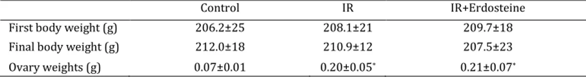
Table 1. Body and ovary weights of groups
*: P<0.05; Compared with Control group
PCNA immunohistochemistry
Ovaries were removed and fixed in 10% neutral buffered formalin solution, and following the routine laboratory methods, blocked in paraffin. Immunocytochemical stains were performed based on the avidin-biotin complex (ABC) technique
described by Oguz et al (16). The samples were
incubated with specific monoclonal antiproliferative cell nuclear antigen (anti-PCNA, 1:100, ab2426-1; Abcam, USA).
Terminal dUTP nick-end labeling (TUNEL)
staining
Apoptotic cells were determined by the terminal deoxynucleotidyl transferase-mediated dUTP nick-end labeling (TUNEL) technique using an apoptosis detection kit (Calbiochem, San Diego, CA, USA), as previously reported (16).
To determine the numerical distribution of PCNA and TUNEL (+) cells in the ovaries samples stained with anti-PCNA antibody and TUNEL kit, cells were examined under light microscopy (400X). In each section, the numbers of positive cells in 10 different enlarged areas selected at random, were counted in random high-power sections using a light
microscope (Olympus BX51, Japan) and
incorporating a software analysis system (Argenit Kameram, ver. 2.11.5.1, Istanbul, Turkey). Finally, all the counts were converted to number of PCNA and
TUNEL (+) cells per mm2 area.
Statistical analyses
A statistical comparison of differences between experimental groups was performed by means of
analysis of variance (ANOVA) and Tukeys Post hoc test,
and a value of P<0.05 was considered statistically
significant. All values were expressed as mean standard deviation (SD), and statistical tests were performed using SPSS version 12.0 PL for Windows.
Results
Biochemical findings
The oxidant and antioxidant levels in the study and control groups are summarized in Table 1. The highest MDA level was observed in the IR group and
the lowest in the control group (P<0.01). Comparison
among the groups revealed that the MDA level in the IR group was higher than those in the control and IR-
E groups (P<0.01). The GSH and CAT levels in the IR
group were lower than in the control and IR-E
groups (P<0.01 and P<0.05, respectively). We also
detected the lowest level of ascorbic acid, -carotene,
and -tocopherol in the IR group, and the highest
level in the control group (P<0.05).
Histopathological findings
The body and ovaries weight changes and histopathological results are summarized in Table 1 and Figure 1, respectively. In the control group, the ovaries have normal appearance of the cortex and the medulla. The IR group showed severe acute
inflammation, polynuclear leukocytes, and
macrophages, as well as degenerative cells, stromal oedema, and haemorrhage. Erdosteine treatment, significantly retained degenerative changes in the ovary. Histopathological changes were significantly decreased in IR-E group compared with IR group. The IR and IR-E groups showed significant increase in ovaries weight compared with the control group
(P <0.05; Table 1).
Immunohistochemical and TUNEL findings
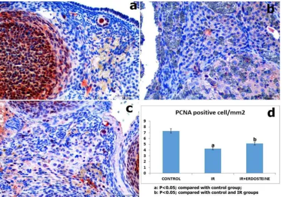
Results of PCNA staining of the ovary are summarised in Figure 2. PCNA (+) cells were detected in the oocytes, follicle epithelium, theca follicle, and stromal cells of all groups. PCNA (+) cell numbers were significantly decreased in the IR and the IR-E groups compared with control group. However, PCNA (+) cell numbers were significantly increased in the IR-E group compared with the IR group (Figure 2).
Ovaries tissues were rarely stained for TUNEL (+) cells in the control rats. TUNEL (+) cell numbers were significantly increased in the IR group compared with the control and the IR-E groups. In the erdosteine
Figure 1. (a) Control group, healthy appearance ovary, (b)
Ischemia–reperfusion group, (c) Ischemia–reperfusion plus erdosteine group. Treatment with erdosteine significantly ameliorated the histological structure. (Hematoxylin and eosine staining, Scale bar =10 μm)
Control IR IR+Erdosteine
First body weight (g) 206.2±25 208.1±21 209.7±18
Final body weight (g) 212.0±18 210.9±12 207.5±23
Figure 2. PCNA staining. (a) Control group, (b) Ischemia–reperfusion group; decreased PCNA (+) cells were seen in the ovary,
(c) Ischemia–reperfusion plus erdosteine-treated group; (Immunoperoxidase, hematoxylin counterstain; Scale bar =5 μm), (d) PCNA (+) cells counts per mm2 in groups
treated group, TUNEL (+) cells were detected
significantly less than the IR group (P <0.05) (Figure 3).
Discussion
The previous studies suggested that ovarian torsion/detorsion (IR injury) causes morphological and
biochemical variation in ovarian tissue (18). The aim of this study was to investigate the antioxidant effect as well as DNA protective effect of erdosteine on torsion/detorsion induced ovarian injury in rats. To our knowledge, this is the first study demonstrating the
protective effect of erdosteine onDNA damage, and
Figure 3. TUNEL staining. (a) Control group, (b) Ischemia–reperfusion group; increased TUNEL (+) cells were seen in the ovary,
proliferation in ovarian torsion/detorsion rat experimental model by TUNEL and PCNA test, respectively. In the present study we demonstrated that treatment with erdosteine prominently decreases
the ovarian damage of rats subjected to IR injury. IR damage contains ROS, production of proteolytic
enzymes, and polymorphonuclear leucocytes
chemotaxis/degranulation (19). Erdosteine has been used to protect against IR injury in diverse organs (4, 19, 20). Erdosteine is likely protects against ovarian torsion/detorsion by avoiding scavenging of free radicals, release of oxidant free radicals, and through prevention of proinflammatory processes. ROS are believed to cause cellular injury and subsequent necrosis by different mechanisms, including oxidation of cellular proteins and peroxidation of cellular membrane lipids (21, 22). In the present study, our results suggested that IR causes an important increase in the MDA level of ovarian tissue; similar findings have been reported in earlier studies (23, 24).
It has been recently reported that 150 mg/kg erdosteine is effective in preventing local tissue injury following testicular torsion (18). In another
study, Yurdakul et al (2010) showed that erdosteine
treatment decreased the decreased levels of MDA and xanthine oxidase (25). It has been also suggested that multiple doses of erdosteine before application have protective effects against radiocontrast-induced nephrotoxicity (19).
During lipid peroxidation, MDA is formed and increased in the case of tissue ischemia. Our adnexal ovary rat model showed that serum MDA level was significantly increased in IR cases; these results suggested that erdosteine treatment regulates serum MDA levels. It has been previously showed that sildenafil treatment reduced the myeloperoxidase activity; however, the enzyme activity was lower in the IR+sildenafil treatment group compared to the IR group (24).
It has been recently suggested that erdosteine treatment increases serum antioxidant enzymes, SOD, CAT, and GSH-Px in renal IR injured rats (26). In
the present study, SOD, CAT, GSH, -carotene,
retionol, and -tocopherol activities were decreased
in the IR group; however, serum MDA level was significantly increased in the IR group. While the
activities of SOD, CAT, GS(, -carotene, retinol, and
-tocopherol were increased in the IR+E group, ascorbic acid activity remained stable. These antioxidant products protect tissues from the harmful effects of ROS by blocking free radical production and easing the neutralization of the formed free radicals (24, 27). A previous study showed, erdosteine treatment increased SOD activities and GSH levels in radio contrast media-induced liver toxicity in rats (28).
In the presented study, we observed increased number of TUNEL (+) cells, but decreased number of PCNA (+) cells in the IR group. The previous studies suggested that the number of apoptotic cells was increased significantly in ovarian and testis IR models (8, 29). Another study showed that apoptotic cell counts were increased significantly in ovaries after IR (30). However, an experimental IR study of
Gencer et al (2014) revealed that the TUNEL and
caspase-3 positive cell number was increased after ischemia but decreased significantly in the quercetine treatment group (8). Similarly, our results showed that erdosteine treatment decreased apoptotic cell number in rat ovarian I/R model.
Conclusion
The present results suggested that erdosteine decreased oxidative stress caused by IR, thereby; it may decrease free radical induced damages, such as germinal cell death. Erdosteine treatment also reduced IR-induced histological injury in the ovaries. These effects may be due to decreased MDA levels and increased antioxidant enzymes (CAT and GSH)
and antioxidant vitamins ascorbic acid, -carotene,
-tocopherol, and retinol) activities. Further studies are needed to clarify whether erdosteine has a direct
protective effect on ovarian injury caused by IR or not.
Conflict of interest
The authors declared that they have no competing interests.
Acknowledgment
The project was not financially supported by any funding agency.
References
1. Hibbard LT. Adnexal torsion. Am J Obstet Gynecol
1985; 152:456–461.
2. Cohen SB, Oelsner G, Seidman DS, Admon D,
Mashiach S, Goldenberg M. Laparoscopic detorsion
allows sparing of the twisted ischemic adnexa. J Am Assoc Gynecol Laparosc 1999; 6:139- 43.
3. Ozkisacik S, Yazici M, Gursoy H, Culhaci N. Does
gradual detorsion protect the ovary against ischemia-reperfusion injury in rats? Pediatr Surg Int 2014;
30:437-440.
4. Sirmali M, Uz E, Sirmali R, Kilbaş A, Yilmaz (R,
Ağaçkiran Y,et al. The effects of erdosteine on lung injury induced by the ischemia-reperfusion of the hind-limbs in rats. J Surg Res 2008; 145:303-307.
5. Fardoun RZ. The use of vitamin E in type 2 diabetes
mellitus. Clin Exp Hypertens 2007; 29:135-148.
6. Eppihimer MJ, Granger DN. Ischemia/ reperfusion-
induced leukocyte-endothelial interactions in
postcapillary venules. Shock 1997; 8:16–25.
7. Sirmali M, Uz E, Sirmali R, Kilbaş A, Yilmaz
ischemia-reperfusion-induced lung oxidative stress and plasma copper and zinc levels in a rat hind limb model. Biol Trace Elem Res 2007; 118:43-52.
8. Gencer M, Karaca T, Güngör AN, (acıvelioğlu SÖ,
Demirtaş S, Turkon (, et al. The protective effect of quercetin on IMA levels and apoptosis in experimental ovarian ischemia-reperfusion injury. Eur J Obstet Gynecol Reprod Biol 2014; 177:135-140.
9. Jain SK, McVie R, Duett J, Herbst JJ. Erythrocyte
membrane lipid peroxidation and glycocylated
hemoglobin in diabetes. Diabetes 1989; 38:1539–1543.
10.Beutler E, Duron O, Kelly BM. Improved method
for the determination of blood glutathione. J Lab Clin Med. 1963; 61: 882-888.
11.Griffith OW. Determination of glutathione and
glutathione disulfide using glutathione reductase and
2-vinylpyridine. Anal Biochem 1980; 106:207–212.
12.Omaye ST, Turnbul JD, Savberlich HE. Ascorbic
acid analysis II. Determination after derivatisation with 2.2. dinitrophenylhidrazine. Selected methods for determination of ascorbic acid in animal cells tissues and fluids. In: McCormick DB, Wright LD.editors. Methods in Enzymology. New York, USA:
Academic Press; 1979.p.62:7–8.
13.Suzuki I, Katoh N. A simple and cheap method for
measuring serum vitamin A in cattle using
spectrophototmeter. Nihon Juigaku Zasshi 1990;
52:1281–1283.
14.Martinek R. Method for determination of vitamin
E (total tocopherol) in serum. Clin Chem 1964;
10:1078–1086.
15.Aebi H. Catalase in vitro. Methods Enzymol 1984;
105:121–126.
16.Oguz S, Kanter M, Erboga M, Ibis C. Protective
effect of Urtica dioica on liver damage induced by biliary obstruction in rats. Toxicol Ind Health 2013; 29:838-845.
17.Ayvaz S, Aksu B, Karaca T, Cemek M,
Tarladacalisir Y-T, Ayaz A, et al. Effects of methylene
blue in acute lung injury induced by blunt chest trauma. Hippokratia 2014; 18:50-56.
18.Dokuyucu R, Karateke A, Gokce H, Kurt RK, Ozcan
O, Ozturk S, et al. Antioxidant effect of erdosteine
and lipoic acid in ovarian ischemia-reperfusion injury.
Eur J Obstet Gynecol Reprod Biol 2014; 183:23-27.
19.Yesilyurt A, Erden IA, Bilgic I, Erden G, Albayrak A.
The protective effect of erdosteine on radiocontrast induced nephrotoxicity in rats. Environ Toxicol 2011;
26:395–402.
20.Ozerol E, Bilgic S, Iraz M, Cigli A, Ilhan A, Akyol O.
The protective effect of erdosteine on short-term global brain ischemia/reperfusion injury in rats. Prog Neuropsychopharmacol Biol Psychiatry 2009; 33:20-24.
21.Tunc T, Uysal B, Atabek C, Kesik V, Caliskan B,
Oztas E, et al. A. Erdosteine and ebselen as
useful agents in intestinal ischemia/reperfusion injury. J Surg Res 2009; 155:210-216.
22.Mallick IH, Yang W, Winslet MC, Seifalian AM.
Ischemia-reperfusion injury of the intestine and protective strategies against injury. Dig Dis Sci 2004; 49:1359.
23.Caglayan EK, Caglayan K, Göcmen AY, Cinar
H, Seckin L, Seckin S, et al. Protective effect of ethyl
pyruvate on ischemia-reperfusion injury in rat ovary: biochemical and histopathological evaluation. Eur J Obstet Gynecol Reprod Biol 2014; 182:154-159.
24.Celik M, Aksoy AN, Aksoy H, Aksoy Y, Halici Z.
Sildenafil reduces ischemia-reperfusion injury in
rat ovary: biochemical and histopathological
evaluation. Gynecol Obstet Invest 2014; 78:162-167.
25.Yurdakul T, Kulaksizoglu H, Pişkin MM, Avunduk
MC, Ertemli E, Gokçe G, et al. Combination antioxidant
effect of -tocoferol and erdosteine in ischemia-reperfusion injury in rat model. Int Urol Nephrol 2010; 42:647-655.
26.Sirmali R, Armağan A, Öktem F, Uz E, Kirbas
A, Dönmez S, et al. Protective effects of erdosteine,
vitamin E, and vitamin C on renal injury induced by the ischemia-reperfusion of the hind limbs in rats. Turk J Med Sci 2015; 45:33-37.
27.Carden DL, Granger DN. Pathophysiology of
ischaemia-reperfusion injury. J Pathol 2000; 190: 255–
266.
28.Yesildağ A, Ozden A, Yilmaz HR, Uz E, Ağackiran
Y, Yesildağ M, et al. Erdosteine modulates radiocontrast-induced hepatotoxicity in rat. Cell Biochem Funct 2009;
27:142–147.
29.Minutoli L, Irrera N, Squadrito F, Marini H, Nicotina
PA, Arena S, et al. Effects of ischaemic post-conditioning
on the early and late testicular damage after experi-mental testisischaemia-reperfusion. Andrology 2014; 2:76-82.
30.Sapmaz-Metin M, Topcu-Tarladacalisir Y, Uz YH,
Inan M, Omurlu IK, Cerkezkayabekir A, et al. Vitamin
E modulates apoptosis and c-jun N-terminal kinase
activation in ovarian torsion–detorsion injury. Exp