Reveal Novel Genetic Variants and Putative Causative
Genes in Congenital Hyperinsulinism
Maria Carla Proverbio1, Eleonora Mangano2, Alessandra Gessi3, Roberta Bordoni2, Roberta Spinelli2, Rosanna Asselta4, Paola Sogno Valin5, Stefania Di Candia5, Ilaria Zamproni6, Cecilia Diceglie6, Stefano Mora6, Manuela Caruso-Nicoletti7, Alessandro Salvatoni8, Gianluca De Bellis2,
Cristina Battaglia2,4*
1Dipartimento di Fisiopatologia e dei Trapianti (DePT), Universita` degli Studi di Milano, Milan, Italy,2Institute of Biomedical Technologies (ITB), CNR, Segrate, Milan, Italy, 3Scuola di Dottorato di Medicina Molecolare, Universita` degli Studi di Milano, Milan, Italy,4Dipartimento di Biotecnologie Mediche e Medicina Traslazionale (BIOMETRA), Universita` degli Studi di Milano, Milan, Italy,5Department of Pediatrics, San Raffaele Scientific Institute, Milan, Italy,6Laboratory of Pediatric Endocrinology, Division of Metabolic and Cardiovascular Sciences, San Raffaele Scientific Institute, Milan, Italy,7Dipartimento di Scienze Mediche e Pediatriche, Universita` di Catania, Catania, Italy, 8Department of Clinical and Experimental Medicine, Pediatric Unit, Insubria University, Varese, Italy
Abstract
Congenital hyperinsulinism of infancy (CHI) is a rare disorder characterized by severe hypoglycemia due to inappropriate insulin secretion. The genetic causes of CHI have been found in genes regulating insulin secretion from pancreaticb-cells; recessive inactivating mutations in theABCC8andKCNJ11genes represent the most common events. Despite the advances in understanding the molecular pathogenesis of CHI, specific genetic determinants in about 50 % of the CHI patients remain unknown, suggesting additional locus heterogeneity. In order to search for novel loci contributing to the pathogenesis of CHI, we combined a family-based association study, using the transmission disequilibrium test on 17 CHI patients lacking mutations inABCC8/KCNJ11,with a whole-exome sequencing analysis performed on 10 probands. This strategy allowed the identification of the potential causative mutations in genes implicated in the regulation of insulin secretion such as transmembrane proteins (CACNA1A, KCNH6, KCNJ10, NOTCH2, RYR3, SCN8A, TRPV3, TRPC5), cytosolic (ACACB, CAMK2D, CDKAL1, GNAS, NOS2, PDE4C, PIK3R3) and mitochondrial enzymes (PC, SLC24A6), and in four genes (CSMD1,SLC37A3,SULF1,
TLL1) suggested by TDT family-based association study. Moreover, the exome-sequencing approach resulted to be an efficient diagnostic tool for CHI, allowing the identification of mutations in three causative CHI genes (ABCC8,GLUD1, and HNF1A)in four out of 10 patients. Overall, the present study should be considered as a starting point to design further investigations: our results might indeed contribute to meta-analysis studies, aimed at the identification/confirmation of novel causative or modifier genes.
Citation:Proverbio MC, Mangano E, Gessi A, Bordoni R, Spinelli R, et al. (2013) Whole Genome SNP Genotyping and Exome Sequencing Reveal Novel Genetic Variants and Putative Causative Genes in Congenital Hyperinsulinism. PLoS ONE 8(7): e68740. doi:10.1371/journal.pone.0068740
Editor:Degui Zhi, University of Alabama at Birmingham, United States of America
ReceivedJanuary 7, 2013;AcceptedMay 31, 2013;PublishedJuly 15, 2013
Copyright:ß2013 Proverbio et al. This is an open-access article distributed under the terms of the Creative Commons Attribution License, which permits unrestricted use, distribution, and reproduction in any medium, provided the original author and source are credited.
Funding:The present study was supported by grants of MIUR PRIN2008WY2TY9. The funders had no role in study design, data collection and analysis, decision to publish, or preparation of the manuscript.
Competing Interests:The authors have declared that no competing interests exist.
* E-mail: [email protected]
Introduction
Congenital hyperinsulinism (CHI), previously known as persis-tent hyperinsulinemic hypoglycemia of infancy (PHHI, MIM256450), is characterized by severe hypoglycemia due to inappropriate insulin secretion from pancreaticb-cells. If improp-erly managed, hypoglycemia can cause brain damage, learning disability, and even death [1]. This condition affects at least 1/ 50,000 children of European descent, and it has been reported in nearly all major ethnic groups [2].
Histologically, CHI can be associated either with diffuse insulin secretion or with focal adenomatous hyperplasia. These two forms share a similar clinical presentation, but result from different molecular mechanisms. Recently, a positron emission tomography scan using Fluorine-18 L-3,4-dihydroxyphenylalanine (18-fluoro DOPA-TC-PET-scan) has been used to distinguish focal from
diffuse forms. Diffuse CHI (Di-CHI) is characterized by autosomal recessive or (less frequently) dominant inheritance, whereas focal CHI (Fo-CHI) is due to a germline paternal mutation (in the ABCC8 or KCNJ11 gene) in addition to a somatic loss of the maternally-derived chromosome 11p15.1 region in pancreaticb -cells [2]. According to the molecular defects, CHI has been classified as a channelopathy (KATP channel), as a metabolic
Overall, CHI causative mutations have been documented in 10 genes, allowing a molecular diagnosis for nearly 50% of CHI patients [3,4]. The most common molecular cause of CHI is the dysfunction of the pancreatic K+
ATP channel encoded by the
sulfonylurea receptor gene (SUR1, aliasABCC8) and the inward-rectifying potassium channel gene (KIR6.2, alias KCNJ11). Less frequently, recessive mutations in theHADHgene (3-hydroxyacyl-Coenzyme A dehydrogenase) have been reported. Dominant forms of CHI are due to either activating mutations in mitochondrial matrix enzymes,GLUD1(glutamate dehydrogenase 1) and UCP2 (mitochondrial uncoupling protein 2), or to mutations in HNF4A (hepatocyte nuclear factor 4 alpha) and SLC16A1(monocarboxylate transporter 1). Activating mutations in theGCK(glucokinase) may also lead to CHI, and an autosomal dominant mutation in INSR (insulin receptor gene) has been described in a large Danish pedigree [5]. Very recently, mutations in theHNF1A(hepatocyte nuclear factor 1 alpha) gene have been reported in CHI patients [6,7].
Insulin secretion is regulated by a complex network of proteins [8] acting at the cellular membrane level (i.e. ATP sensitive K+
channels, Ca2+, Na+voltage-gated channels, glucose transporters,
and transient receptors) [9], at the intracellular level (i.e. endoplasmic reticulum ion channels), and at the mitochondrial level (i.e. enzymes) [10]. Since CHI is characterized by severe hypoglycemia due to inappropriate insulin secretion from pancre-aticb-cells, we hypothesized that mutations in genes involved in this process might be responsible for the disease. Moreover, considering the phenotypic variability observed within CHI families, ‘‘phenotypic modifier’’ genes might contribute to the global genetic picture of this disorder as described for other Mendelian disorders [11].
The mutational screening of CHI causative genes is usually carried out by PCR amplifications and direct Sanger sequencing, using an iterative approach based on clinical features and/or on medical treatment response [2].
High-density single nucleotide polymorphism (SNP)-array technology has been widely used to perform linkage analysis, including the transmission-disequilibrium test (TDT). Moreover, the combination of linkage analysis with haplotype information (applied to trios) has been successful applied to map Mendelian traits [12] and has led to the identification of gene variants causing diseases and birth defects [13]. Recent developments in high-throughput sequence capture methods and next-generation sequencing (NGS) approaches have made feasible the analysis of the whole human exome. The exome deep sequencing, called whole-exome sequencing (WES) analysis, has already provided the opportunity to detect causal gene variants in dominant and recessive disorders [14,15] and it has been recently proposed as a powerful tool to improve the molecular diagnostic of neonatal diabetes and maturity onset diabetes of the young (MODY) [16,17].
Here, we applied a multi-step screening strategy aimed at identifying genes putatively related to CHI. This strategy included a TDT study on 17 patients and their families and exome sequencing in 10 CHI probands. We have also evaluated if NGS is a suitable alternative diagnostic approach to classical Sanger sequencing for CHI.
Results and Discussion
To highlight novel CHI-associated genetic loci, we performed a multi-step screening strategy (Figure S1). First we carried out a genetic screening ofABCC8/KCNJ11genes on 33 CHI probands by classic sequencing approach; then, applying a genome wide
SNP genotyping analysis on 17 non-consanguineous CHI patients (lacking ABCC8/KCNJ11 mutations) and their families, we performed a TDT family-based association study, and finally we applied WES on 10 selected CHI probands (five from the TDT analysis group and five not subjected to previous molecular screening). To prioritize the coding variants identified by WES, we crossed the list of filtered WES variants with that comprising the TDT associated genes (51 genes), the known CHI-causative ones (10 genes), and a refined list of 211 CHI-functionally-related genes (seeMethodsfor details).
Whole-genome SNP Genotyping and TDT Analysis The whole-genome SNP genotyping was performed on the 17 nuclear families. The overall results of the TDT association analysis are presented in the Manhattan plot (Figure S2). We obtained 144 SNPs associated at P#0.005 (Table S1). None of these SNPs reached the stringent genome-wide significance of P,4.461027; however, we considered any empirical P-value at
4.961025 as ‘‘suggestive’’ (i.e. a potential reflection of an association signal) [18]. After genome annotation, we found that a subset of these SNPs map within/close to 51 genes (Table S1). Interestingly, we identified two SNPs located in the region of the TLL1 (tolloid-like1) gene: rs3775321 (P = 0.000049; OR = 7.3, 95%CI = 2.2–24.5) and rs4691229 (P = 0.00043; OR = 0.2, 95%CI = 0.1–0.5). Using haplotype analysis we were able to confirm and to refine the potential association of theTTL1locus to the CHI (Table S2).
Applying the copy-number and homozygosity mapping analyses on 17 probands, we confirmed the absence of deletion or amplification events (data not shown); conversely, in two probands we found runs of homozygosity (ROH) longer than 2 Mb. The ROH segments were located on chromosome 4q24 (4.2 Mb containing 14 genes) in patient HI06, and on chromosome 6p36.33-q13.33 (7 Mb containing 26 genes) in patient HI18. By the subsequent WES analysis, we verified that all genes located in ROH segments were lacking mutations.
WES Analysis and Gene Variants Identification
WES was performed on 10 CHI probands (Table 1). By sequencing the exome DNA libraries, we obtained 2.8–5.4 Gb of mapped sequence achieving a mean depth of target coverage between 27 and 506, and an average of 19,181 variations per
patient, including substitutions and indels (Table 2). Overall, the results of the percentage of target coverage at 106were fairly homogeneous among samples (ranging from 83.5 to 88.7%). By looking to CHI known causative genes, we obtained at least 106
coverage in.95% of coding regions forHADH,SLC16A1,UCP2, GLUD1, and KCNJ11, whereas the values were slightly less uniform, reaching 90, 83, 80, and 75% forINSR, ABCC8,GCK, HNF4A, respectively. Only for HNF1A the percentage was low (55%). By focusing on non-synonymous single nucleotide variants (SNV, missense and nonsense) we counted an average of 9,395 variants per patient. After the filtering procedure, we counted an average of 430 variants (ranging from 368 to 494) that were not reported in publicly accessible databases (dbSNP132, 1000Ge-nomes Project) and in ourin-housedatabase (built on 15 sequenced exomes, Italian individuals).
resulting in a final list of 27 single base variants distributed in 24 genes (Table 3). Among them, we found 24 missense mutations causing amino-acid changes predicted to be potentially damaging with high confidence by at least one prediction tool; we also identified two nonsense mutations and one splice site mutation. The mutations have been all confirmed by direct Sanger sequencing (data not shown).
Biological Relevance of Known and Novel CHI Gene Variants
By WES in 10 CHI patients we were able to identify four patients carrying mutations in three CHI causative genes (ABCC8, GLUD1, andHNF1A), seven patients showed genetic lesions in 17
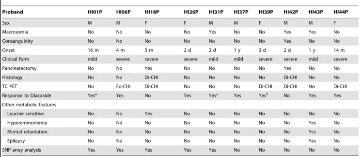
genes implicated in the regulation of insulin secretion (KCNH6, GNAS, ACACB, NOTCH2, RYR3, TRPV3, TRPC5, CAMK2D, PIK3R3, CDKAL1, SCN8A, KCNJ10, PDE4C, NOS2, SLC24A6, CACNA1A, PC) and four patients carrying mutations in four genes suggested by the TDT analysis (SLC37A3,CSMD1,SULF1,TLL1). Overall, we identified: one patient displaying a unique variant in one gene, two patients are displaying mutations in two genes, and the remaining carrying multiple variants in more than two genes. For known genes, we found a compound heterozygous patient carrying two mutations inABCC8: a nonsense mutation pQ953X (CM981878) previously described [19] and a novel c.3989-2A.G splicing variant (patient HI39) abolishing the canonical acceptor splice site. We also identified a homozygous ABCC8 variant (p.A390E) in exon 7 within a ROH on chromosome 11 (proband Table 1.Clinical characteristics of the ten CHI probands.
Proband HI01P HI06P HI18P HI26P HI31P HI37P HI39P HI42P HI43P HI44P
Sex M M F F M M F M M F
Macrosomia No No No No Yes No No Yes Yes No
Consanguinity No No No No No No No Yes No No
Onset 16 m 4 m 5 m 2 d 2 d 1 y 3 d 2 d 1 y 14 m
Clinical form mild severe severe severe mild mild severe severe mild severe
Pancreatectomy No No Yes No No No No Yes No No
Histology No No Di-CHI No No No No Di-CHI No No
TC PET No Fo-CHI Di-CHI No No No Di-CHI Di-CHI No Di-CHI
Response to Diazoxide Yes* Yes No Yes Yes* Yes Yes1
No Yes Yes
Other metabolic features
Leucine sensitive No No Yes No No No No No No No
Hyperammonemia No No No No No No No No Yes No
Mental retardation No No No No No No No No Yes No
Epilepsy No No No No No No No No Yes No
SNP array analysis Yes Yes Yes Yes Yes No No No No No
*Patient in Remission. 1
Medical Treatment with Diazoxide and Octreotide. Abbreviations: m, month; d, day; y, year; Di, Diffuse; Fo Focal. doi:10.1371/journal.pone.0068740.t001
Table 2.Exome coverage and target coverage statistics of ten CHI probands.
CHI patients HI01P HI06P HI18P HI26P HI31P HI37P HI39P HI42P HI43P HI44P
Whole exome sequencing results
Total number of raw reads (10‘6) 81.87 62.75 68.38 52.75 65.58 80.60 84.95 45.53 68.11 88.79
Total number of mapped reads (10‘6) 67.28 49.14 50.77 41.70 52.64 66.24 70.46 36.74 54.61 71.53
Target coverage (106) (%) 88.67 87.38 88.00 86.46 88.40 88.51 88.36 83.55 88.43 88.16
Target coverage (206) (%) 77.84 73.82 74.90 70.14 76.15 77.20 77.48 62.06 76.02 77.08
Mean read depth (x) 50.19 39.26 40.29 33.24 42.06 47.52 49.48 27.04 41.33 49.78
Exonic SNV variants analysis
Total exonic SNP (Nu) 19208 18751 19361 18959 19200 19697 19287 17808 20252 19293
Total exonic Indels (Nu) 281 258 262 240 265 247 257 223 266 282
Missense and Nonsense SNPs (Nu) 9436 9187 9461 9318 9456 9679 9433 8660 9890 9434
Filtering (dbSNP) 722 794 863 830 846 883 905 685 952 765
Filtering (1000g MAF,0.005) 618 705 761 721 737 778 800 608 825 652
Filtering (internal DB) 369 415 480 414 438 423 457 405 495 414
HI42) which has been previously reported in a case report [20]. The GLUD1 mutation (pS498L,) was found at the heterozygous state in patient (HI43) who was affected by a mild CHI-HA (Hyperinsulinism-Hyperammoniemia) form and epilepsy. This mutation is associated to CHI-HA in the Human Gene Mutation Database (CM980942). Finally, we identified a HNF1A gene mutation (proband HI01) that has been previously reported in MODY3 patients [21]. Interestingly, the phenotype of our patient was similar to the one described by Stanescu, who recently reported mutations in theHNF1Agene in two cases of CHI [22]. For novel genes, by literature mining and database queries we have obtained indications about their biological relevance for pancreatic b-cell (Figure S3) and by gene ontology (GO) classification we group them in three main categories of cellular components such as transmembrane, cytosolic and mitochondrial proteins (Table 4).
Transmembrane proteins. The CACNA1A gene encodes the alpha 1A subunit of the voltage-dependent (VDCC), P/Q type calcium channel. The protein is participating in the complex regulation of insulin secretion: the mechanism implicates that the closure of the KATP channels stimulates insulin secretion
depolarizing the plasma membrane; the consequent opening of VDCC results in influx of extracellular Ca2+, which then triggers
insulin exocytosis in pancreatic b-cells [23,24]. CSMD1 gene (belonging to the TDT analysis list) encodes an integral membrane protein with unknown molecular function. The KCNH6 (alias HERG2) gene encodes a voltage-gated K+channel (KV) belonging
to the 6-TM family of potassium channels; interestingly; it has been described that HERG channels have a crucial role in regulating insulin secretion, raising the possibility that HERG mutations might be involved in some hyperinsulinemic diseases of unknown origin [25]. TheKCNJ10gene encodes a member of the inward-rectifier potassium channel family (also known as 2-TM channels) and its mutations have been associated to SeSAME syndromes [26]. TheNOTCH2gene encodes a type 1 transmem-brane receptor that is expressed in the ductal cells during pancreatic organogenesis [27], and is belonging to the Notch family that has been suggested to have a role in diabetes [28]. Recently, aNOTCH2 variant has been reported in one MODY affected patient [16], therefore similarly to other MODY causative genes [29–31], that are resulted also CHI causative, we propose NOTCH2as a good candidate for further investigations. TheRYR3 Table 3.Whole-exome sequencing identification of non-synonymous SNP variants of 10 CHI patients.
Sample ID Gene symbol Chr Position RefSeq ID Amino Acid change PolyPhen/SIFT prediction
HI01 HNF1A chr12 121426790 NM_000545 p.A161T* ++/++
KCNH6 chr17 61615518 NM_030779 p.V532F 2/++
HI06 GNAS chr20 57428464 NM_080425 p.E48D +/++
ACACB chr12 109683531 NM_001093 p.N1760S 2/++
NOTCH2 chr1 120464961 NM_024408 p.R1704H +/++
HI18 SLC37A3 chr7 140035229 NM_032295 p.S389L 2/++
CSMD1 chr8 3889535 NM_033225 p.R168W +/++
RYR3 chr15 33938601 NM_001036 p.T1272M +/++
HI26 TRPV3 chr17 3417258 NM_145068 p.L776F ++/++
TRPC5 chrX 111020098 NM_012471 p.G789R 2/++
CAMK2D chr4 114435066 NM_172115 p.R275C ++/++
HI31 PIK3R3 chr1 46521492 NM_003629 p.R306X Nonsense
CDKAL1 chr6 21231212 NM_017774 p.S561F 2/++
SCN8A chr12 52163721 NM_014191 p.V1148M 2/++
KCNJ10 chr1 160011280 NM_002241 p.R348H 2/++
HI37 PDE4C chr19 18331081 NM_000923 p.R253C ++/++
HI39 ABCC8 chr11 17428964 NM_000352 p.Q953X* Nonsense
ABCC8 chr11 17418595 NM_000352 Splice site
NOS2 chr17 26107857 NM_000625 p.R314C ++/++
HI42 ABCC8 chr11 17474673 NM_000352 p.A390E 2/++
SLC24A6 chr12 113737647 NM_024959 p.Y564H ++/++
SULF1 chr8 70501306 NM_015170 p.A222T 2/++
HI43 GLUD1 chr10 88817449 NM_005271 p.S498L* 2/2
TLL1 chr4 166981332 NM_012464 p.G667S ++/2
HI44 CACNA1A chr19 13325090 NM_001127222 p.R1966Q ++/++
PC chr11 66619274 NM_001040716 p.N657Y ++/++
TLL1 chr4 166986893 NM_012464 p.H689P ++/++
*Mutation reported in Human Genome Mutation Database( HGMD).
PolyPhen/SIFT: (2) benign or tolerated; (+) possibly damaging/DAMAGING Low confidence; (++) probably damaging/DAMAGING.
gene encodes for receptors that are intracellular ion channels expressed in human pancreaticb-cell. Interestingly, the activation of these receptors by ryanodine stimulates the insulin secretion at low glucose concentrations [32]. TheSLC37A3gene (belonging to the TDT analysis list) encodes a transmembrane sugar transport-ers family, SLC37; to date, only theSLC37A4gene has been found mutated in the glycogen storage disease non-1A type [33]. The SCN8A gene encodes a member of the sodium channel alpha subunit gene family (Nav1.6), and it forms the ion pore region of
the gated sodium channel and mediates the voltage-dependent sodium ion permeability of excitable membranes. Nav
1.6 together with Nav1.7 (the otherasubunit of humanb-cells) is
highly expressed in human islets [8,34]. TheTRPV3andTRPC5 genesencode proteins belonging to the transient receptor potential (TRP) cation channels group, they provide the background membrane conductance required for b-cells to depolarize upon KATPchannel closure [9], but their role in human pancreas islets
has not been studied.
Cytosolic proteins. The ACACB gene encodes a complex multifunctional enzyme system that catalyzes the carboxylation of acetyl-CoA to malonyl-CoA (the rate-limiting step in fatty acid synthesis); the malonyl-CoA can act as a coupling factor that regulates insulin secretion [35]. Interestingly,b-cell islets produce high quantity of ACACB enzyme [36]. TheCAMK2D, encodes a protein Ca2+sensing serine/threonine kinases (CaMK2) that it is
involved in the regulation of Ca2+mediated glucose-sensitivity of b-cells; in particular CaMK2 delta play a role as a feedback sensor, linking Ca2+induced exocytosis to the re-synthesis of insulin acting
on transcriptional regulation of genes related to the metabolic control of insulin secretion [37]. In humans, the CaMK2 beta and delta subtypes represent the major isoforms that are highly expressed in b-cells [38]. The CDKAL1 gene, encodes a methylthiotransferase that modifies tRNA to enhance the trans-lational fidelity of proinsulin transcript [39]. Moreover, a study using knockout mice for theCDKAL1 gene revealed its possible role in controlling the first phase of insulin exocytosis inb-cells, through the KATPchannel responsiveness [40]. The GNASgene
encodes the Gsa subunit of the G protein that is ubiquitously expressed. The encoded protein stimulates adenylyl cyclase (AC), thereby generating the second messenger cAMP. It has been shown that mice withb-cell-specific Gsadeficiency develop severe defect in b-cell proliferation and early onset insulin deficient diabetes [41]. The GNAS locus has a highly complex imprinted expression pattern and encodes four main transcripts: Gs-alpha, XLas, NESP55, and the A/B transcript. Specifically, we found that our mutation is referred to the XLas transcript, whose promoter is methylated on the maternal allele and transcription-ally active only on the paternal allele [42]. Therefore, for our proband, we hypothesize that a post-zygotic somatic event (such as methylation) and imprinting defect, resulting in the absence of Table 4.Functional annotation of novel variants.
GO cellular component Relevant pathways ( KEGG) Biological process in pancreas [ref]
Transmembrane proteins
CACNA1A Calcium signaling pathway, Type II diabetes mellitus
insulin exocytosis [23,24]
CSMD1 undetermined unknown
KCNH6 undetermined regulating insulin secretion [25]
KCNJ10 Gastric acid secretion unknown
NOTCH2 Notch signaling pathway role in diabetes, MODY [16,28]
RYR3 Calcium signaling pathway, insulin secretion [32]
SCN8A undetermined ion permeability, insulin secretion [8,34]
SLC37A3 undetermined unknown
TRPC5,TRPV3 undetermined ion permeability [9]
Cytosolic proteins
ACACB Fatty acid biosynthesis, Metabolic pathways, Insulin signaling pathway
insulin secretion [35,36]
CAMK2D Calcium signaling pathway,Wnt signaling pathway
insulin gene expression regulation [37]
CDKAL1 undetermined insulin gene expression, insulin exocytosis [39]
GNAS Calcium signaling pathway b-cell proliferation, diabetes [41]
NOS2 Metabolic pathways, Calcium signaling pathway b-cell proliferation, diabetes [43]
PDE4C Purine metabolism insulin secretion [45]
PIK3R3 Insulin signaling pathway, Type II diabetes mellitus
insulin signaling [47]
SULF1 undetermined unknown
TLL1 undetermined glucose regulation [49]
Mitochondrial proteins
PC Citrate cycle (TCA cycle), Pyruvate metabolism, Metabolic pathways
insulin secretion [10]
SLC24A6 undetermined calcium homeostasis [51]
expression of the maternal Gs-alpha isoform, might lead to the expression of mutated paternal allele in some affected focal area of the pancreas. The NOS2 gene, has been implicated in b-cell damage and death in both type 1 and type 2 diabetes [43]; moreover, it mediates the IRS-2 expression contributing tob-cell failure [44]. ThePDE4Cgene encodes a protein belonging to the cyclic nucleotide phosphodiesterase subfamily PDE4. This protein hydrolyzes the second messenger, cAMP, thus playing a key role in many important physiological processes such as those mediated by hormones and neurotransmitters. It has been demonstrated that PDE1–4 enzymes are expressed in rodent pancreatic islets andb -cells [45] while PDE4C is the major isoform in human islets [46]. Furthermore, it has been shown that family-selective inhibition of PDE (PDE1, PDE3 and to some extent also PDE4) potentiates glucose-stimulated insulin secretion (GSIS); in particular, siRNA-mediated knockdown of PDE4C significantly enhanced GSIS in rat INS-1 (832/13) cells [45]. The PIK3R3encodes a family of lipid kinases that binds to activated (phosphorylated) protein-tyrosine kinases through the SH2 domain, regulates their kinase activity and triggers a plethora of intracellular events involving also the insulin signaling [47]. Notably, we found a mutation in the exon 7 that causes the complete loss of the second SH2 domain that may be deleterious to the protein function (Table 3). The SULF1 gene (belonging to the TDT analysis list) encodes an enzyme with arylsulphatase activity acting on cell-surface heparan sulphate proteoglycans. It has been suggested an important role of this enzyme in pancreatic cancer progression [48]. TheTLL1gene (belonging to the TDT analysis list) encodes an astacin-like zinc-dependent metalloprotease and is a subfamily member of the metzincin family; interestingly, it has been described as a gene responsive to a member of the NR4A subgroup of nuclear receptors that have been implicated in the regulation of glucose and lipid metabolism in insulin-sensitive tissues [49].
Mitochondrial proteins. ThePCgene encodes the enzyme pyruvate carboxylase, which is a mitochondrial enzyme involved in gluconeogenesis, lipogenesis, insulin secretion, and synthesis of the neurotransmitter glutamate. PC seems to play an important role in the ‘‘amplifying signal’’ responsible for insulin secretion trough the KATPindependent pathway [10]. Interestingly, Pineda
et al. [50] reported an infant with what they termed the ‘‘French’’ type of pyruvate carboxylase deficiency, presenting initial neonatal symptoms such as respiratory distress, severe metabolic acidosis, and a tendency to hypoglycaemia. TheSLC24A6gene encodes a mitochondrial Na+/Ca2+ exchanger (NCKX6) that maintain
cellular calcium homeostasis and it plays a critical role in the mitochondrial Ca2+ transport in skeletal muscle, stomach, and
pancreas [51].
Conclusions
In the present investigation, we demonstrated that exome sequencing is useful as a diagnostic tool for CHI because identifies mutations in known genes but also gives the opportunity to discover potential damaging mutations affecting several genes combined in a complex fashion. WES, associated with family-based association analysis, revealed that CHI patients, lacking mutation in known causative genes, carry multiple exonic mutations. Due to the small number of patients of this study, some cautions should be taken into account to interpret the pathological role of identified variants. However, due to difficulties to collect large samples for such a rare disease, our results may be useful for future meta-analyses that will combine genetic data provided by other individual studies. The present study should be regarded as a starting point to design further investigations aimed from one side to the molecular characterization of the identified
mutations, and from the other to investigate possible modifier genes, which might be relevant for the CHI pathogenesis. Increasing the number of well-characterized clinical CHI patients will allow the evaluation of the mutation frequency of novel gene variants and the identification of a clearer genotype-phenotype correlation.
Finally, we indicate that exome sequencing should be consid-ered a valuable, time- and cost-effective diagnostic tool providing a fine molecular classification of CHI patient in alternative to Sanger sequencing, offering also the opportunity to better understand the genetic basis of CHI.
Materials and Methods
Study Sample and Strategy
The study was approved by the Ethical Committee of the San Raffaele Scientific Institute and performed according to the principles of the Declaration of Helsinki. Written informed consent was obtained from the next of kin on the behalf of the minors/children participants involved in this study. A detailed description of the study design is available through the Italian Registry of Congenital Hyperinsulinism (http://www.progettorici. it).
The diagnosis of CHI was carried out according to the following criteria: a fasting and post-prandial hypoglycemia with unsup-pressed insulin secretion, a positive response to the administration of glucagon, negative ketone bodies in urine and plasma, and a prolonged dependence to treatment to prevent hypoglycemia. From the study we excluded infants from diabetic mothers, children with transient neonatal hyperinsulinism, with insulinoma, or Beckwith-Wiedemann Syndrome. Patients were considered to be diazoxide responsive if satisfactory glycemic control was achieved with doses of oral diazoxide not exceeding 20 mg/kg per day and were considered to be in remission if good glycemic control was maintain without further medical or surgical therapy. Patients were considered to be unresponsive to medical treatment if recurrent hypoglycemia episodes (,3 mmol/L) occurred during diet treatment (milk enriched with malto-dextrine or continuous enteral feeding) and treatment with diazoxide and octreotide at maximum dosage.
The study design is shown in Figure S1. We enrolled a total of 38 probands and their families; 33 probands were subjected to mutational screening of two ‘‘major’’ CHI genes (ABCC8,KCNJ11) by Sanger sequencing, following the procedure previously described [52]. After the mutational screening, 17 non consan-guineous probands remained without a genetic diagnosis: together with their parents, they were hence processed for SNP genotyping and family-based TDT association analysis. Finally, we used WES analysis in 10 probands (for five of them, no mutational pre-screening was performed), either to find novel causative gene mutations or to evaluate WES as an alternative approach to classical Sanger sequencing. The clinical features of these patients are reported in Table 1.
homozygosity mapping was carried out using CNAG 3.3, as previously described [53]. Allele and genotype frequencies were calculated for eachlocus and tested for Hardy-Weinberg equilib-rium (HWE) considering only parents. For SNP quality control (QC), we employed predetermined QC inclusion criteria [minor allele frequency (MAF).1%, SNP call rate .90%, HWE P.0.01]. The QC procedure also included a check for Mendelian errors (predetermined threshold: 1%), as well as sex and pedigree check. All procedures were performed by using the PLINK software [54], obtaining a set of 221,605 high-performing SNP markers for further analysis. A subset of SNPs that are in approximate linkage equilibrium with each other were hence selected by linkage disequilibrium (LD)-based pruning: 112,740 SNPs remained for family-based TDT association analysis.
We proceeded further performing the TDT association analysis on the 17 trios to search for association signals highlighting novel CHI candidate/modifier genes. Adjusted P-values were assessed by gene-dropping permutation, flipping the allele transmitted from parent to affected offspring, and using the default mode with the adaptative permutation approach. The maximum number of permutations per SNPs was 1,000,000. Considering the number of tests conducted, any empirical P-value at 4.461027 would have
been considered significant [55]; however, none of the SNPs reached the genome-wide significance. Hence, considering that: 1) the study cohort is obviously underpowered by its size; 2) ‘‘suggestive’’ P-values still may reflect evidence for association; and that 3) it is possible to make use of these suggestive P-values through the incorporation of prior biological knowledge; we decided to adopt P = 4.961025as significance threshold to build a
SNP list to be used in all the subsequent analyses. All P-values are hence presented as not corrected.
Finally, we refined the association analysis on theTLL1 gene performing a haplotype analysis. It was performed using all SNPs mapping in the TLL1 genomic region and the sliding window specification option of the PLINK program (width of the window: 5 SNPs; shifting: 1 SNP at a time).
WES Analysis
Three micrograms of genomic DNA was fragmented using the Covaris shearing system (Covaris inc., MA, USA). Exome capture was performed with the SureSelect Human All Exon Kit (v3, target size 50 Mb) according to the manufacturer’s instructions (Agilent technologies, Santa Clara, CA, USA). The platform design covers 1.22% of human genomic regions corresponding to the NCBI Consensus CDS Database (CCDS), including more than 700 human miRNAs from the Sanger v13 database and more than 300 additional human non-coding RNAs in a single tube. The selected coding regions were then massively sequenced by the Illumina GAIIx technology (Illumina, San Diego, CA, USA); following proprietary reversible terminator-based method and producing 2676 bp read lengths. The paired-end sequence reads
were mapped to the reference genome (UCSC NCBI37/hg19) using the Burrows-Wheeler Aligner (BWA) software [56] and the variant calling was performed using SAMtools [57]. The entire dataset of mapped reads (.bam files) is available on Sequence Read Archive, accession number ERP002635. The detected variations in each sample were filtered out using the minimum Phred SNP quality score of 100, the minimum depth of 10 reads, and the minor allele with an allelic fraction.0.25. The variants were then annotated by the ANNOVAR software (http://www. openbioinformatics.org/annovar/annovar_db.html), for filtering common variants reported in dbSNP132 (http://www.ncbi.nlm. nih.gov), in 1000 Genomes Project (http://www.1000genomes. org/), andin-housedatabase.
Selection of Gene of Interest and Validation
For a focused evaluation of the whole-exome data, a list of 272 genes arising from three functional criteria of inclusion was used: ten genes known to be causative for CHI (ABCC8, KCNJ11, GLUD1, GCK, HADH, HFN4A, SLC16A1, UCP2, INSR and HNF1A); 51 genes suggested by TDT association analysis (Table S1); 145 genes associated to regulation of insulin secretion reported in the Rat Genome Database [58] and 66 genes indicated by reviewing the literature concerning theb-cell function and its metabolic regulation (Table S3).
To predict the effect of non-synonymous mutations on the encoded proteins of the selected gene list, we used two different web tools: SIFT [59] and PolyPhen v2.0 [60]. We reported non-synonymous mutations that were predicted deleterious at least by one predictor tool with a high score. To investigate the presence of functional interactions among genes, including direct (physical) and indirect (functional) associations, we used STRING, a database of known and predicted protein interactions [61]. Computer-assisted analysis for splice-site prediction was accom-plished using the Neural Network Promoter Prediction Tool (NNPPT) program (http://www.fruitfly.org/seq_tools/splice. html) and the NetGene2 (release 2.4) program (http://www.cbs. dtu.dk/services/NetGene2).
All variants were validated by PCR amplification of exons carrying the identified mutation (primers sequences and PCR conditions are available on request)using genomic DNA from CHI probands. The amplified products were purified using montage the Micro PCR96 plates (Merck Millipore, Billerica, MA, USA) and then directly sequenced with the forward and reverse primers previously used for the amplification. Sequenced products were purified using the Montage SEQ 96 Sequencing Reaction Cleanup kit (Merck Millipore). Sequencing analysis was carried out using the Big Dye Terminator Cycle Sequencing Ready Reaction Kit v3.1 and an automated DNA sequencer (ABI-3100 XL Genetic Analyser; Applied Biosystems, Foster City, CA, USA). Factura and Sequence Navigator software Packages (Applied Biosystems) were used for mutation detection.
Supporting Information
Figure S1 Study design.The initial study sample consisted of a total of 65 subjects, belonging to 32 CHI families. Overall 33 probands of these families were pre-screened for mutations in the two main CHI genes (ABCC8,KCNJ11). The 17 probands non consanguineous (lacking causal mutations in ABCC8/KCNJ11) together with their relatives were taken forward to whole-genome scan by the Affymetrix GeneChip Human Mapping 250 K Array. We applied TDT and haplotype analyses to highlight possible susceptibility/modifier CHI genes. Finally, 5 patients from the TDT study and 5 newly-enrolled CHI patients (not pre-screened forABCC8/KCNJ11 were analyzed by whole-exome sequencing (WES).
(TIF)
Figure S2 Manhattan plot. Results of single-locus test of association between each of the 112,740 performing SNPs and CHI using the TDT association analysis. 112,740 single-marker permuted results (–log10 p-values) from the TDT test are plotted on each chromosome; red line represents P#1023
threshold, blue line represents P#561023threshold.
(TIF)
(GLUT2) stimulates insulin secretion by metabolic amplifying pathways producing ATP. The increase of ATP/ADP ratio leads to the closure of KATP channels, to the depolarization of the
plasma membrane and to the subsequent activation of VDCC promoting influx of calcium into the cell. The overall modulation of the cytosolic free concentration [Ca2+] is essential for the
triggering pathways of the insulin secretion. The binding of secreted insulin to its receptors (INSR), might activates the PI3K/ Akt pathway and some transcription factors controlling insulin gene expression. Insulin exocytosis can also be influenced by neurotransmitters and hormones. Indeed, GLP1 actives AC leading the elevation of cAMP and the consequent PKA activation which finally mediates insulin exocytosis; alternatively the Ach mobilizes intracellular Ca2+activating of IP3 receptor; then [Ca2+]
binds to CaM activating CaMK and inducing the secretory process of insulin. Moreover, CDKAL1 is implicated in the control of the first phase of insulin exocytosis via KATPresponsiveness.
Other transmembrane ion channels might modulate electrical activity of the cellular membrane regulating the insulin secretion (KCN, TRP, SCN). Abbreviations: VDCC, voltage dependent calcium channel; TRP, transient receptor potential channels; KCN, potassium voltage-gated channel; SCN, sodium channel voltage-gated; ER, endoplasmic reticulum; SERCA, sarco/ endoplasmic reticulum Ca2+ATPase; GIP, glucose-dependent
insulinotropic peptide; AC, adenyl cyclase; GLP1, glucagon like peptide 1; INS, insulin; IRS1/2, Insulin receptor substrate 1/2; PLC, phospholipase C; IP3, Inositol trisphosphate; PKC, protein kinase C; DAG, diacylglycerol; Gs,Gi,Gq, G proteins; PKA, Protein kinase A; PI3K, phosphatidylinositol; CaM, calmodulin;
Ach, acetylcholine; FA, fatty acid; FFA, free fatty acid. Yellow box indicate the CHI causative genes.
(TIF)
Table S1 TDT data and associated alleles and genes.
144 SNP resulted from TDT analysis at P#0.005. OR, TDT odd ratio; CI L95 and CI U95, lower and upper 95% confidence interval for TDT odds ratio; Adjusted P-value, empirical p-value by adaptive procedure; A1/A2, A1: minor allele, A2: major allele; NP: number of permutations.
(XLS)
Table S2 Haplotype analysis of TLL1 locus.
(XLS)
Table S3 Refined gene list.The table is including 221 gene symbols and criteria of inclusion. PMID, pubmed identification number; RGD, rat genome database (http://rgd.mcw.edu/). (XLS)
Acknowledgments
We thank all patients with CHI disease and their families for participating in this study. We also thank Maria Vurchio (CNR-ITB) for technical and administrative support.
Author Contributions
Conceived and designed the experiments: PSV SDC SM AS MCP CB GDB. Performed the experiments: AG MCP RB IZ CD. Analyzed the data: EM RS AG MCP RA GDB CB. Contributed reagents/materials/ analysis tools: AS MCN SDC PSV SM. Wrote the paper: CB MCP GDB. Clinical and ethical issues of the study: AS MCN PSV SDC SM.
References
1. Hussain K (2008) Diagnosis and management of hyperinsulinaemic hypogly-caemia of infancy. Hormone research 69: 2–13. Available: http://www.ncbi. nlm.nih.gov/pubmed/18059080. Accessed 16 September 2012.
2. Arnoux J-B, Verkarre V, Saint-Martin C, Montravers F, Brassier A, et al. (2011) Congenital hyperinsulinism: current trends in diagnosis and therapy. Orphanet journal of rare diseases 6: 63. Available: http://www.pubmedcentral.nih.gov/ articlerender.fcgi?artid = 3199232&tool = pmcentrez&rendertype = abstract. 3. Senniappan S, Shanti B, James C, Hussain K (2012) Hyperinsulinaemic
hypoglycaemia: genetic mechanisms, diagnosis and management. Journal of inherited metabolic disease 35: 589–601. Available: http://www.ncbi.nlm.nih. gov/pubmed/22231386. Accessed 26 September 2012.
4. Flanagan SE, Kapoor RR, Hussain K (2011) Genetics of congenital hyperinsulinemic hypoglycemia. Seminars in pediatric surgery 20: 13–17. Available: http://www.ncbi.nlm.nih.gov/pubmed/21185998. Accessed 9 Au-gust 2012.
5. Hojlund K, Hansen T, Lajer M, Henriksen JE, Levin K, et al. (2004) A Novel Syndrome of Autosomal-Dominant Hyperinsulinemic Hypoglycemia Linked to a Mutation in the Human Insulin Receptor Gene. Diabetes 53: 1592–1598. Available: http://diabetes.diabetesjournals.org/content/53/6/1592.short. Ac-cessed 13 March 2013.
6. Stanescu DE, Hughes N, Kaplan B, Stanley CA, De Leo´n DD (2012) Novel Presentations of Congenital Hyperinsulinism due to Mutations in the MODY genes: HNF1A and HNF4A. The Journal of clinical endocrinology and metabolism 97: 1–5. Available: http://www.ncbi.nlm.nih.gov/pubmed/ 22802087. Accessed 3 August 2012.
7. Snider KE, Becker S, Boyajian L, Shyng S-L, Macmullen C, et al. (2013) Genotype and phenotype correlations in 417 children with congenital hyperinsulinism. The Journal of clinical endocrinology and metabolism 98: E355–63. Available: http://www.ncbi.nlm.nih.gov/pubmed/23275527. Ac-cessed 16 February 2013.
8. Rorsman P, Braun M (2012) Regulation of Insulin Secretion in Human Pancreatic Islets. Annual review of physiology: 1–25. Available: http://www. ncbi.nlm.nih.gov/pubmed/22974438. Accessed 31 October 2012.
9. Colsoul B, Vennekens R, Nilius B (2011) Transient Receptor Potential cation channels in PancreaticbCells. Rev Physiol Biochem Pharmacol 161: 87–111. doi:10.1007/112.
10. Jitrapakdee S, Wutthisathapornchai A, Wallace JC, MacDonald MJ (2010) Regulation of insulin secretion: role of mitochondrial signalling. Diabetologia 53: 1019–1032. doi:10.1007/s00125-010-1685-0.Regulation.
11. Dipple KM, McCabe ER (2000) Phenotypes of patients with ‘‘simple’’ Mendelian disorders are complex traits: thresholds, modifiers, and systems dynamics. American journal of human genetics 66: 1729–1735. Available:
h t t p : / / w w w . p u b m e d c e n t r a l . n i h . g o v / a r t i c l e r e n d e r . fcgi?artid = 1378056&tool = pmcentrez&rendertype = abstract.
12. Ott J, Kamatani Y, Lathrop M (2011) Family-based designs for genome-wide association studies. Nature reviews Genetics 12: 465–474. Available: http:// www.ncbi.nlm.nih.gov/pubmed/21629274. Accessed 16 October 2012. 13. Bailey-Wilson JE, Wilson AF (2011) Linkage analysis in the next-generation
sequencing era. Human heredity 72: 228–236. Available: http://www.ncbi.nlm. nih.gov/pubmed/22189465. Accessed 31 October 2012.
14. Ng SB, Buckingham KJ, Lee C, Bigham AW, Tabor HK, et al. (2010) Exome sequencing identifies the cause of a Mendelian disorder. Nature genetics 42: 30– 35. doi:10.1038/ng.499.Exome.
15. Bamshad MJ, Ng SB, Bigham AW, Tabor HK, Emond MJ, et al. (2011) Exome sequencing as a tool for Mendelian disease gene discovery. Nature reviews Genetics 12: 745–755. Available: http://www.ncbi.nlm.nih.gov/pubmed/ 21946919. Accessed 13 July 2012.
16. Johansson S, Irgens H, Chudasama KK, Molnes J, Aerts J, et al. (2012) Exome sequencing and genetic testing for MODY. PloS one 7: e38050. Available: h t t p : / / w w w . p u b m e d c e n t r a l . n i h . g o v / a r t i c l e r e n d e r . fcgi?artid = 3360646&tool = pmcentrez&rendertype = abstract. Accessed 31 July 2012.
17. Bonnefond A, Philippe J, Durand E, Dechaume A, Huyvaert M, et al. (2012) Whole-Exome Sequencing and High Throughput Genotyping Identified KCNJ11 as the Thirteenth MODY Gene. PloS one 7: e37423. Available: h t t p : / / w w w . p u b m e d c e n t r a l . n i h . g o v / a r t i c l e r e n d e r . fcgi?artid = 3372463&tool = pmcentrez&rendertype = abstract. Accessed 3 Au-gust 2012.
18. Lehne B, Lewis CM, Schlitt T (2011) From SNPs to genes: disease association at the gene level. PloS one 6: e20133. Available: http://dx.plos.org/10.1371/ journal.pone.0020133. Accessed 13 March 2013.
19. Nestorowicz A, Glaser B, Wilson BA, Shyng SL, Nichols CG, et al. (1998) Genetic heterogeneity in familial hyperinsulinism. Human molecular genetics 7: 1119–1128. Available: http://www.ncbi.nlm.nih.gov/pubmed/9618169. 20. Parappil HA, Rahman SA, Soliman AB, Ismail AC, AL-Bozom I, et al. (2009)
Congenital Hyperinsulinism in a Neonate Due to a Novel Homozygous Mutation (ABCC8): A case report. Int J Endocrinol Metab 2: 95–100. 21. Che`vre JC, Hani EH, Boutin P, Vaxillaire M, Blanche´ H, et al. (1998) Mutation
screening in 18 Caucasian families suggest the existence of other MODY genes. Diabetologia 41: 1017–1023. Available: http://www.ncbi.nlm.nih.gov/ pubmed/9754819.
metabolism 97: 1–5. Available: http://www.ncbi.nlm.nih.gov/pubmed/ 22802087. Accessed 3 August 2012.
23. Rorsman P, Braun M, Zhang Q (2011) Regulation of calcium in pancreatica -andb-cells in health and disease. Cell calcium 51: 300–308. Available: http:// w w w . p u b m e d c e n t r a l . n i h . g o v / a r t i c l e r e n d e r . fcgi?artid = 3334273&tool = pmcentrez&rendertype = abstract. Accessed 7 Sep-tember 2012.
24. Dunne MJ, Shepherd RM, Cosgrove KE, Macfarlane WM, Lindley KJ, et al. (2000) Persistent hyperinsulinaemic hyperglycaemia of infancy-derived cells; implications for beta-cells that replicate in vitro. Journal of molecular endocrinology 24: 313–320. Available: http://www.ncbi.nlm.nih.gov/ pubmed/10828824.
25. Rosati B, Marchetti P, Crociani O, Lecchi M, Lupi R, et al. (2000) Glucose- and arginine-induced insulin secretion by human pancreaticb-cells: the role of HERG K+channels in firing and relase. FASEB journal 14: 2601–2610. 26. Scholl UI, Choi M, Liu T, Ramaekers VT, Ha¨usler MG, et al. (2009) Seizures,
sensorineural deafness, ataxia, mental retardation, and electrolyte imbalance (SeSAME syndrome) caused by mutations in KCNJ10. Proceedings of the National Academy of Sciences of the United States of America 106: 5842–5847. A v a i l a b l e : h t t p : / / w w w . p u b m e d c e n t r a l . n i h . g o v / a r t i c l e r e n d e r . fcgi?artid = 2656559&tool = pmcentrez&rendertype = abstract. Accessed 13 March 2013.
27. Lammert E, Brown J, Melton DA (2000) Notch gene expression during pancreatic organogenesis. Mechanisms of development 94: 199–203. Available: http://www.ncbi.nlm.nih.gov/pubmed/10842072. Accessed 13 March 2013. 28. Kim W, Shin Y-K, Kim B-J, Egan JM (2010) Notch signaling in pancreatic
endocrine cell and diabetes. Biochemical and biophysical research communi-cations 392: 247–251. Available: http://www.ncbi.nlm.nih.gov/pubmed/ 20035712. Accessed 31 October 2012.
29. Vionnet N, Stoffel M, Takeda J, Yasuda K, Bell GI, et al. (1992) Nonsense mutation in the glucokinase gene causes early-onset non-insulin-dependent diabetes mellitus. Nature 356: 721–722. Available: http://www.ncbi.nlm.nih. gov/pubmed/1570017. Accessed 13 March 2013.
30. Yamagata K, Furuta H, Oda N, Kaisaki PJ, Menzel S, et al. (1996) Mutations in the hepatocyte nuclear factor-4alpha gene in maturity-onset diabetes of the young (MODY1). Nature 384: 458–460. Available: http://www.ncbi.nlm.nih. gov/pubmed/8945471. Accessed 13 March 2013.
31. Yamagata K, Oda N, Kaisaki PJ, Menzel S, Furuta H, et al. (1996) Mutations in the hepatocyte nuclear factor-1alpha gene in maturity-onset diabetes of the young (MODY3). Nature 384: 455–458. Available: http://www.ncbi.nlm.nih. gov/pubmed/8945470. Accessed 13 March 2013.
32. Johnson JD, Kuang S, Misler S, Polonsky KS (2004) Ryanodine receptors in human pancreatic beta cells: localization and effects on insulin secretion. FASEB journal 18: 878–880. Available: http://www.ncbi.nlm.nih.gov/pubmed/ 15033925.
33. Bartoloni L, Antonarakis SE (2004) The human sugar-phosphate/phosphate exchanger family SLC37. European journal of physiology 447: 780–783. Available: http://www.ncbi.nlm.nih.gov/pubmed/12811562. Accessed 26 Sep-tember 2012.
34. Braun M, Ramracheya R, Bengtsson M, Zhang Q, Karanauskaite J, et al. (2008) Voltage-Gated Ion Channels in Human Pancreaticb-Cells: Electrophysiological Characterization and Role in Insulin Secretion. Diabetes 57: 1618–1628. doi:10.2337/db07-0991.M.B.
35. Prentki M, Vischer S, Glennon MC, Regazzi R, Deeney JT, et al. (1992) Malonyl-CoA and long chain acyl-CoA esters as metabolic coupling factors in nutrient-induced insulin secretion. The Journal of biological chemistry 267: 5802–5810. Available: http://www.ncbi.nlm.nih.gov/pubmed/1556096. 36. Zhang S, Kim K-H (1995) Glucose activation of acetyl-CoA carboxylase in
association with insulin secretion in a pancreatic b-cell line. Journal of Endocrinology 147: 33–41. Available: http://joe.endocrinology-journals.org/ c o n t e n t / 1 4 7 / 1 / 3 3 .
abstract?ijkey = 8a76ed9c70df96aeae6c97c33f117d4df096fb6e&keytype2 = tf_ ipsecsha. Accessed 6 November 2012.
37. Osterhoff M, Mo¨hlig M, Schwanstecher M, Seufert J, Ortmann J, et al. (2003) Ca2+/calmodulin-dependent protein kinase IId2 regulates gene expression of insulin in INS-1 rat insulinoma cells. Cell Calcium 33: 175–184. Available: http://linkinghub.elsevier.com/retrieve/pii/S0143416002002270. Accessed 31 October 2012.
38. Rochlitz H, Voigt A, Lankat-Buttgereit B, Go¨ke B, Heimberg H, et al. (2000) Cloning and quantitative determination of the human Ca2+/calmodulin-dependent protein kinase II (CaMK II) isoforms in human beta cells. Diabetologia 43: 465–473. Available: http://www.ncbi.nlm.nih.gov/pubmed/ 10819240.
39. Brambillasca S, Altkrueger A, Colombo SF, Friederich A, Eickelmann P, et al. (2012) CDK5 regulatory subunit-associated protein 1-like 1 (CDKAL1) is a tail-anchored protein in the endoplasmic reticulum (ER) of insulinoma cells. The Journal of biological chemistry 287: 41808–41819. Available: http://www.ncbi. nlm.nih.gov/pubmed/23048041. Accessed 13 March 2013.
40. Ohara-Imaizumi M, Yoshida M, Aoyagi K, Saito T, Okamura T, et al. (2010) Deletion of CDKAL1 affects mitochondrial ATP generation and first-phase insulin exocytosis. PloS one 5: e15553. Available: http://www.pubmedcentral. n i h . g o v / a r t i c l e r e n d e r .
fcgi?artid = 3000340&tool = pmcentrez&rendertype = abstract. Accessed 31 Oc-tober 2012.
41. Xie T, Chen M, Zhang Q-H, Ma Z, Weinstein LS (2007) Beta cell-specific deficiency of the stimulatory G protein alpha-subunit Gsalpha leads to reduced beta cell mass and insulin-deficient diabetes. Proceedings of the National Academy of Sciences of the United States of America 104: 19601–19606. A v a i l a b l e : h t t p : / / w w w . p u b m e d c e n t r a l . n i h . g o v / a r t i c l e r e n d e r . fcgi?artid = 2148335&tool = pmcentrez&rendertype = abstract.
42. Bastepe M (2007) The GNAS Locus: Quintessential Complex Gene Encoding Gsalpha, XLalphas, and other Imprinted Transcripts. Current genomics 8: 398– 414. Available: http://www.pubmedcentral.nih.gov/articlerender. fcgi?artid = 2671723&tool = pmcentrez&rendertype = abstract.
43. Corbett BJA, Mcdaniel ML (1995) Intraislet Release of Interleukin 1 Inhibits 3 Cell Function by Inducing 3 Cell Expression of Inducible Nitric Oxide Synthase. J Exp Med 181.
44. Tanioka T, Tamura Y, Fukaya M, Shinozaki S, Mao J, et al. (2011) Inducible nitric-oxide synthase and nitric oxide donor decrease insulin receptor substrate-2 protein expression by promoting proteasome-dependent degradation in pancreatic beta-cells: involvement of glycogen synthase kinase-3beta. The Journal of biological chemistry 286: 29388–29396. Available: http://www. p u b m e d c e n t r a l . n i h . g o v / a r t i c l e r e n d e r . fcgi?artid = 3190744&tool = pmcentrez&rendertype = abstract. Accessed 31 Oc-tober 2012.
45. Waddleton D, Wu W, Feng Y, Thompson C, Wu M, et al. (2008) Phosphodiesterase 3 and 4 comprise the major cAMP metabolizing enzymes responsible for insulin secretion in INS-1 (832/13) cells and rat islets. Biochemical pharmacology 76: 884–893. Available: http://www.ncbi.nlm.nih. gov/pubmed/18706893. Accessed 9 September 2012.
46. Heimann E, Jones HA, Resjo¨ S, Manganiello VC, Stenson L, et al. (2010) Expression and regulation of cyclic nucleotide phosphodiesterases in human and rat pancreatic islets. PloS one 5: e14191. Available: http://www.pubmedcentral. n i h . g o v / a r t i c l e r e n d e r .
fcgi?artid = 2995729&tool = pmcentrez&rendertype = abstract. Accessed 26 Sep-tember 2012.
47. Hirsch E, Costa C, Ciraolo E (2007) Phosphoinositide 3-kinases as a common platform for multi-hormone signaling. The Journal of endocrinology 194: 243– 256. Available: http://www.ncbi.nlm.nih.gov/pubmed/17641274. Accessed 7 November 2012.
48. Li J, Kleeff J, Abiatari I, Kayed H, Giese NA, et al. (2005) Enhanced levels of Hsulf-1 interfere with heparin-binding growth factor signaling in pancreatic cancer. Molecular cancer 4: 14. Available: http://www.pubmedcentral.nih.gov/ articlerender.fcgi?artid = 1087876&tool = pmcentrez&rendertype = abstract. Ac-cessed 5 November 2012.
49. Chao LC, Bensinger SJ, Villanueva CJ, Wroblewski K, Tontonoz P (2008) Inhibition of adipocyte differentiation by Nur77, Nurr1, and Nor1. Molecular endocrinology (Baltimore, Md) 22: 2596–2608. Available: http://www. p u b m e d c e n t r a l . n i h . g o v / a r t i c l e r e n d e r . fcgi?artid = 2610364&tool = pmcentrez&rendertype = abstract. Accessed 5 No-vember 2012.
50. Pineda M, Campistol J, Vilaseca MA, Briones P, Ribes A, et al. (1995) An atypical French form of pyruvate carboxylase deficiency. Brain & development 17: 276–279. Available: http://www.ncbi.nlm.nih.gov/pubmed/7503391. Ac-cessed 5 November 2012.
51. Palty R, Ohana E, Hershfinkel M, Volokita M, Elgazar V, et al. (2004) Lithium-calcium exchange is mediated by a distinct potassium-independent sodium-calcium exchanger. The Journal of biological chemistry 279: 25234–25240. Available: http://www.ncbi.nlm.nih.gov/pubmed/15060069. Accessed 8 March 2013.
52. Biagiotti L, Proverbio MC, Bosio L, Gervasi F, Rovida E, et al. (2007) Identification of two novel frameshift mutations in the KCNJ11 gene in two Italian patients affected by Congenital Hyperinsulinism of Infancy. Experimental and molecular pathology 83: 59–64. Available: http://www.ncbi.nlm.nih.gov/ pubmed/17316607. Accessed 31 October 2012.
53. Cazzaniga G, Lo Nigro L, Cifola I, Milone G, Schnittger S, et al. (2009) Simultaneous occurrence of acute myeloid leukaemia with mutated nucleo-phosmin (NPM1) in the same family. Leukemia 23: 199–203. Available: http:// www.ncbi.nlm.nih.gov/pubmed/18596743. Accessed 31 October 2012. 54. Purcell S, Neale B, Todd-Brown K, Thomas L, Ferreira MAR, et al. (2007)
PLINK: a tool set for whole-genome association and population-based linkage analyses. American journal of human genetics 81: 559–575. Available: http:// w w w . p u b m e d c e n t r a l . n i h . g o v / a r t i c l e r e n d e r . fcgi?artid = 1950838&tool = pmcentrez&rendertype = abstract. Accessed 12 July 2012.
55. Dudbridge F, Gusnanto A (2008) Estimation of significance thresholds for genomewide association scans. Genetic epidemiology 32: 227–234. Available: h t t p : / / w w w . p u b m e d c e n t r a l . n i h . g o v / a r t i c l e r e n d e r . fcgi?artid = 2573032&tool = pmcentrez&rendertype = abstract. Accessed 28 February 2013.
56. Li H, Durbin R (2009) Fast and accurate short read alignment with Burrows-Wheeler transform. Bioinformatics (Oxford, England) 25: 1754–1760. Available: h t t p : / / w w w . p u b m e d c e n t r a l . n i h . g o v / a r t i c l e r e n d e r . fcgi?artid = 2705234&tool = pmcentrez&rendertype = abstract. Accessed 4 Oc-tober 2012.
fcgi?artid = 2723002&tool = pmcentrez&rendertype = abstract. Accessed 13 July 2012.
58. Twigger SN, Shimoyama M, Bromberg S, Kwitek AE, Jacob HJ (2007) The Rat Genome Database, update 2007–easing the path from disease to data and back again. Nucleic acids research 35: D658–62. Available: http://www. p u b m e d c e n t r a l . n i h . g o v / a r t i c l e r e n d e r . fcgi?artid = 1761441&tool = pmcentrez&rendertype = abstract. Accessed 31 Oc-tober 2012.
59. Kumar P, Henikoff S, Ng PC (2009) Predicting the effects of coding non-synonymous variants on protein function using the SIFT algorithm. Nature protocols 4: 1073–1081. Available: http://www.ncbi.nlm.nih.gov/pubmed/ 19561590. Accessed 16 July 2012.
60. Adzhubei IA, Schmidt S, Peshkin L, Ramensky VE, Gerasimova A, et al. (2010) A method and server for predicting damaging missense mutations. Nature methods 7: 248–249. Available: http://www.pubmedcentral.nih.gov/ articlerender.fcgi?artid = 2855889&tool = pmcentrez&rendertype = abstract. Ac-cessed 8 October 2012.