Received on 15 April 2008; revised 20 September 2008.
Address for correspondence: Dr. Kleber Dias do Prado. Emilio Ribas Infectious Diseases Institute, Scientific Department. Av. Dr. Arnaldo 165, São Paulo, SP, Brazil, Zip code: 01246-900. E-mail: [email protected] - Phone/Fax: (55 11) 3896-1241.
The Brazilian Journal of Infectious Diseases 2008;12(5):362-367. © 2008 by The Brazilian Journal of Infectious Diseases and Contexto Publishing. All rights reserved.
Histological Response Study of Chronic Viral Hepatitis C Patients Treated With Interferon Alone or Combined With Ribavirin
Kleber Prado1, Rosely Patzina2, Denise Bergamaschi3 and Roberto Focaccia1
1Viral Hepatitis Clinic - Emilio Ribas Infectious Diseases Institute; 2Department of Pathology – Emilio Ribas Infectious Diseases Institute; 3Department of Statistics - Public Health College – University of São Paulo; São Paulo, SP, Brazil
Chronic hepatitis C is often a progressive, fibrotic disease that can lead to cirrhosis and other complications. The recommended therapy is a combination of interferon and ribavirin. Besides its antiviral action, interferon is considered to have antifibrotic activity. We examined the outcome of hepatic fibrosis and inflammation in chronic hepatitis C patients who were non-responders to interferon. We made a case series, retrospective study, based on revision of medical records and reassessment of liver biopsies. For inclusion, patients should have been treated with interferon alone or combined with ribavirin, with no virological response (non responders and relapsers) and had a liver biopsy before and after treatment. Histological evaluation included: i-outcome of fibrosis and necroinflammation; ii-annual fibrosis progression rate evaluation, before and after treatment. Seventy-five patients were included. Fifty-seven patients (76%) did not show progression of fibrosis after treatment, compared to six (8%) before treatment (p < 0.001). The mean annual fibrosis progression rate was significantly reduced after treatment (p = 0.036). Inflammatory activity improved in 19 patients (25.3%). The results support the hypothesis of an antifibrotic effect of interferon-based therapy, in non-responder patients. There was evidence of anti-inflammatory effects of treatment in some patients.
Key Words: Chronic hepatitis C, fibrosis, interferon-alfa, ribavirin, treatment result.
Hepatitis C virus (HCV) infection is a major worldwide public health issue. It is estimated that between 170 and 250 million people are chronic carriers [1]. There is no available information about the prevalence rate of chronic hepatitis C in Brazil, although Focaccia et al. found an estimated prevalence of 1.42% in São Paulo City, rising to over 3% within the 30 and older age group. We examined the medical records of patients from 2 to 80 years old in a serological survey of a stratified, randomized and residence-based population [2].
HCV infection becomes a chronic disease in a large proportion of patients, ranging from 55% to 90%; it has a progressive fibrotic nature. Up to 20% of the cases may culminate in cirrhosis; which may cause complications, such as hepatocellular carcinoma and end-stage liver disease [3-5]. The combination of pegylated interferon (PEG) and ribavirin, which is currently the best available treatment, given during 48 weeks, only reaches a sustained virological response (SVR) in 54%-56% of naive patients [6,7]. Non-responder patients to interferon alone or in combination with ribavirin have an even smaller frequency of virological response [8]. Currently, for non-responders to a combination of PEG and ribavirin, there is no consensus therapeutic option that considers the histological response among treatment efficacy end-points [3,9-11].
Several studies have demonstrated the inherent antifibrinogenic action of interferon, which is not related to
its antiviral and anti-inflammatory effects [12-16]. Furthermore, it has also been proven through several clinical trials that interferon-based treatments provoke histological improvements, even in virological non-responders [17-22]. We evaluated hepatic fibrosis and necro-inflammatory activity progression in chronic hepatitis C patients who were non-responders to interferon-based treatments or relapsers.
Material and Methods
A retrospective study was made of chronic hepatitis C patients who had been treated at the Viral Hepatitis Clinic of the Emilio Ribas Infectious Diseases Institute (ERIDI), in São Paulo, Brazil, from January 1997 to December 2002. We reviewed patient medical records to collect demographic, anthropometric, epidemiological and clinical information, as information on liver biopsies. The study protocol was approved by ERIDI’s Ethics Committee.
We identified 8,192 medical records of hepatitis patients within this period. A preliminary analysis ruled out acute hepatitis cases, HIV or HVB coinfections, HCV spontaneous clearance, patients who had never been under treatment or had not undergone liver biopsy before and after treatment. The remaining 615 medical charts were reviewed for inclusion. The inclusion criteria were: 1 – previous treatment for at least six months with interferon-alfa, with or without ribavirin, using standardized doses and routes of administration [6,7,23,24]; 2 – a liver biopsy performed or reviewed by an ERIDI pathologist, performed within six months prior to treatment and at least six months after treatment; 3 – non-response to treatment or relapse. The exclusion criteria were: 1 – chronic liver diseases not related to HCV; 2 – current alcohol or illegal drug abuse; 3 - use of hepatotoxic or immunosuppressive drugs; 4 - immunosuppressive illness and HIV coinfection.
sensitivity of 50 IU/mL) at the end of treatment and relapse based on undetectable serum HCV-RNA using the same technique at the end of treatment, which became detectable six months later. To determine the HCV genotype, the INNO-LIPA HCV II technique was used, with the Versant kit (Lipa)®. Liver biopsies were read blind, always by the same pathologist from the Department of Pathology of ERIDI, applying the METAVIR score. Fibrosis qualitative evolution was evaluated by making a fibrosis stage comparison between the two biopsy tests (before and after treatment), with three possible outcomes: 1 – progression (after-treatment fibrosis stage more advanced than before-treatment stage); 2 – stabilization (after-treatment fibrosis stage not changed compared to before-treatment stage); and 3 – regression (after-treatment fibrosis stage lower than before-(after-treatment stage). Patients classified as stage four (cirrhosis) at the first liver biopsy were classified in the progression category whenever decompensated cirrhosis was identified. In such cases, a new biopsy was not required. Otherwise, patients were designated in the non-progression category if the new biopsy did not demonstrate regression.
The annual fibrosis progression rate, both pre and post-treatment, was calculated based on the following definitions: 1- pre-treatmentannual fibrosisprogression rate =the ratio between fibrosis stage measured in METAVIR units at the first biopsy (pre-treatment) and the estimated time of infection in years; 2 – post-treatmentannual fibrosisprogression rate = the ratio between the difference in fibrosis stage measured in METAVIR units of the pre and post-treatment biopsies and the delay between the two biopsies in months multiplied by 12 [4,18].
The calculation of the probable infection date, and consequently, the time of infection, was based on: a – for transfusional infections, the date of the first blood and/or plasma derived products transfusion was considered, if the patient had gone through more than one transfusion session; b – for infection caused by injectable drugs, the first year of use of such drugs [12,25,26]. For any other cause of HCV infection, only infection dates based on unequivocal evidence of a temporal relation between exposure and HCV infection were used.
The liver biopsy samples were at least 10mm long or had six portal tracts [27]. The liver tissue fragments were colored by hematoxilin-eosine, Masson’s trichrome, Perls and reticuline.
Statistical Analysis
For comparison of proportions (pre and post-treatment situations), we used the McNemar test for dependent samples and the Pearson’s chi-square for independent samples. For mean values comparison, we used the “Student” t test (on independent samples) and a paired student t (on dependent samples), when there were only two means involved in the comparison. When the variables did not conform to a normal distribution, we used the Wilcoxon signed-rank test [28], for
population comparisons. The statistical computer program Stata was used for this analysis [29]. Statistical decisions were made considering the descriptive value of the tests (p value).
Results
Seventy-five patients met the inclusion criteria. Table 1 shows the main characteristics of the 75 patients included in this study.
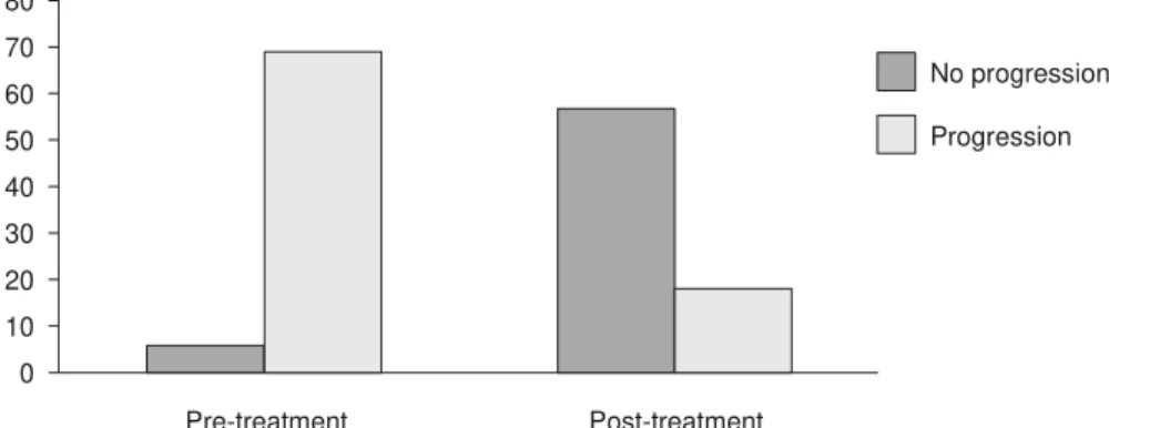
Seventy-nine percent of the patients had undergone a liver biopsy one year or more after the end of treatment (median time: 20 months; range: 1 - 67 months). Seventy-six percent of patients showed stabilization or regression of fibrosis (no progression) at the second liver biopsy (Table 2 and Figure 1). Before treatment, the vast majority of patients had evidence of fibrosis progression (fibrosis stage > 0). In contrast, after treatment, most patients displayed a tendency towards no progression (p< 0.001), at least over the period the patients were observed (Figure 2).
A significant association was found between fibrosis qualitative evolution and pre-treatment fibrosis stages (p=0.005), with higher rates of progression among patients at stages 0, 1 and 2, and regression among patients at stages 3 and 4 (Table 3).
The pre-treatment annual fibrosis progression rate was defined for 42 patients. There was a mean rate of 0.096 FMU/ year (fibrosis METAVIR units per year) (CI 95%: 0.075 – 0.117 FMU/year), a median value of 0.096 FMU/year, with minimum and maximum values of 0.000 and 0.272 FMU/year, respectively. The post-treatment fibrosis annual progression rate was calculated for 75 patients. There was a mean rate of 0.007 FMU/year, (CI 95%: -0.066 – 0.081 FMU/year), a median value of 0.000 FMU/year, with minimum and maximum values of -1.500 and 0.827 FMU/year, respectively. The paired pre and post –treatment rate analyses, restricted to the 42 patients with known pre-treatment rates, showed a significant decrease of the fibrosis annual progression rate. The post-treatment mean rate in these patients was 0.005 FMU/year (CI 95%: -0.088 – 0.078 FMU/year); there was a difference of 0.101 FMU/ year (CI 95%: 0.007 – 0.194FMU/year, p=0.036) between the mean rates (paired Student-t test).
The post-treatment fibrosis annual progression rate in comparison with the fibrosis stage on the pre-treatment biopsy showed a significantly higher decrease of this progression among the patients that were in an advanced stage of fibrosis (F3/F4) at the time of the pre-treatment biopsy when compared to those who had had mild-to-moderate fibrosis (F0/F1/F2) (p=0.0143). Among the 31 patients that had showed mild-to-moderate fibrosis at pre-treatment, the mean decrease in fibrosis progression after treatment was 0.034 FMU/year, while in the 11 patients who had shown advanced fibrosis pre-treatment, the mean decrease was 0.287 FMU/year.
patients who had shown fibrosis regression and progression (p=0.024), with a higher regression rate and a lower progression rate of liver inflammation in the former compared to the latter. There was information available about previous alcohol consumption in the medical records of the 44 patients: 14 (31.8%) reported no alcohol use, 15 (34.1%) reported occasional use and 15 (34.1%) admitted daily use of alcoholic beverages. Statistical differences in alcohol consumption between mild-to-moderate (F0/F1/F2) and advanced (F3/F4) fibrosis patients were not found (p=0.660). Among the 15 patients who admitted daily use of alcohol, 13 (86.7%) showed mild-to-moderate liver fibrosis before treatment.
Discussion
The main obstacle against including patients was the unavailability, for various reasons, of liver biopsies for revision by the pathologist. This occurred in 355 cases. Some other reasons for not including patients were: patients that had been treated by other health facilities outside of the ERIDI; patients who had been under irregular treatment, with discontinuity or reduced doses of the treatment drugs; patients whose time interval between the first biopsy and treatment exceeded one year; and patients who had made the liver biopsy less than six months after the end of treatment.
As a retrospective case series on a medical records revision basis, this study has some serious limitations. The most important of them comes from the lack of standardization of the medical record information. Another weakness originates from the adoption of a consensual and universal treatment that has prevented the use of a control group of patients without treatment for comparison. Lastly, the period of time during which the patients were under observation after treatment was significantly shorter than the pre-treatment period. This may overestimate the therapeutic benefits in cases of slowly progressing diseases.
Statistical associations were found among treatment, hepatic fibrosis non-progression and decreases in the annual
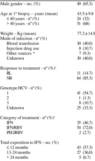
Table 1. Characteristics of the patients.
Male gender – no. (%) 49 (65.3)
Age at 1st biopsy – years (mean) 43.5 ± 9.8
≤ 40 years - nº (%) 24 (32)
> 40 years - nº (%) 51 (68)
Weight – Kg (mean) 77.2 ± 14.8
Mode of infection - nº (%)
Blood transfusion 30 (40.0)
Injection drug use 8 (10.7)
Other sources * 7 (9.3)
Unknown 30 (40.0)
Response to treatment - nº (%)†
RL 11 (14.7)
NR 64 (85.3)
Genotype HCV - nº (%)
1 41 (54.7)
2 1 (1.3)
3 8 (10.7)
Unknown 25 (33.3)
Category of treatment - nº (%)‡
IFN 35 (46.7)
IFN/RBV 54 (72.0)
PEG/RBV 2 (2.7)
Total exposition to IFN – no. (%)
≤ 12 months 43 (57.3)
13-24 months 27 (36.0)
> 24 months 5 (6.7)
*Occupational exposure to blood (needles, contaminated syringes) or sexual relationship with an infected individual. † RL
– relapser; NR – non-responder. ‡ IFN – interferon; RBV –
ribavirin; PEG – pegylated interferon.
Table 2. Post-treatment fibrosis qualitative evolution.
Evolution N %
N* 43 57.3
P 18 24.0
R 14 18.7
Total 75 100.0
*N – non-progression (stabilization) P – progression; R – regression.
Table 3. Post-treatment fibrosis qualitative evolution according to pre-treatment fibrosis stages.
Table 4. Qualitative evolution of inflammatory activity.
Evolution N %
Non-regression* 56 74.7
Regression 19 25.3
Total 75 100.0
*Non-regression: patients with impairment or stabilization of inflammatory activity.
Pre-treatment fibrosis stages Fibrosis evolution n (%) P value
Non progression Progression Regression
Stages 0, 1 and 2 32 (58.2) 17 (30.9) 6 (10.9) 0.005*
Stages 3 and 4 11 (55.0) 1 (5.0) 8 (40.0)
Total 43 (57.3) 18 (24.0) 14 (18.7)
Figure 1. Pre and post-treatment fibrosis stage. The shadow zone indicates stabilization of fibrosis. Above and to the right of the shadow zone indicates fibrosis progression. Below and to the left indicates fibrosis regression.
Figure 2. Fibrosis progression before and after treatment.
Table 5. Inflammatory activity evolution according to fibrosis evolution.
Fibrosis evolution Inflammatory activity evolution - N (%)
Regression Stabilization Progression Total
Regression 7 (50)* 7 (50) 0 14 (100)
Stabilization 10 (23.3) 25 (58.1) 8 (18.6) 43 (100)
Progression 2 (11.2) 8 (44.4) 8 (44.4)* 18 (100)
Total 19 40 16 75
*p = 0.024 (Pearson’s chi-square).
Post - treatment fibrosis – n (%)
Pre -treatment
fibrosis
0 1 2 3 4
0 3
(4.0)
2
(2.7)
1
(1.3)
1 3
(4.0)
14
(18.7)
4
(5.3)
1
(1.3)
1
(1.3)
2 3
(4.0)
15
(20.0)
6
(8.0)
2
(2.7)
3 1
(1.3)
3
(4.0)
8
(10.7)
1
(1.3)
4 4
(5.3)
3
(4.0)
0 10 20 30 40 50 60 70 80
Pre-treatment Post-treatment
No progression
fibrosis progression rate, in comparison with the chronic hepatitis C natural history. Similar results have been found in other studies [8,18,20,30].
The observation of hepatic fibrosis regression in 14 patients is a very important piece of information, since spontaneous regression of hepatic fibrosis is not usually seen in patients without treatment. We speculate that the regression in fibrosis is due to an antifibrogenic effect of interferon, regardless of its antiviral action. Poynard et al. demonstrated that hepatitis C is indeed a progressive fibrotic disease, which is the reason for its morbidity and mortality [4]. Therefore, a therapeutic intervention to decrease or revert hepatic fibrosis progression will probably have a relevant clinical impact. In our study, seven out of 14 patients with fibrosis regression also showed post-treatment inflammatory activity regression, while the other seven patients remained at the same activity level. It seems reasonable to attribute fibrosis regression in the latter group to an intrinsic antifibrogenic effect.
On the other hand, the relevant fibrosis stabilization rates in patients with mild-to-moderate, as well as advanced fibrosis (58.2% and 55.0%, respectively), are probably due to the anti-inflammatory action of ribavirin and interferon. As a matter of fact, among the 75 patients, only 21 (28%) had made use of standard interferon (IFN) alone, while the other 54 (72%), had made use of ribavirin along with IFN or PEG, with the possibility that the ribavirin indirectly facilitated the interferon antifibrogenic effect through its anti-inflammatory action.
It has been noticed among patients with advanced fibrosis before treatment, that there is a higher proportion of patients with fibrosis regression and a larger reduction in the annual fibrosis progression rate, after treatment in comparison with patients who had mild-to-moderate fibrosis. Again, there was a good correlation between the qualitative evolution and the annual fibrosis progression rate. Poynard et al. found similar results among 1,509 French patients, though they used a different methodology for defining mild-to-moderate and advanced fibrosis [20]. However, in our study, the small number of patients with advanced fibrosis did not make it possible to make conclusions from the results.
Treatment effects on inflammatory activity evolution were less evident. About 75% of the patients worsened or did not present any variation in hepatic inflammatory activity, in disagreement with several other studies that reported on the anti-inflammatory effect of chronic hepatitis C treatment [8,17,31-35]. Even so, 66% of the patients showed null or mild degrees of inflammatory activity after treatment, foreseeing an absence of fibrosis progression on the short and probably the mid-term. Studies of Poynard et al. about the natural history of hepatitis C show that the degree of inflammatory activity is less associated with the time of disease than fibrosis, which reflects the oscillating inflammatory activity due to infection [4]. For a better measurement of the treatment’s impact upon inflammatory
liver activity, a post-treatment hepatic biopsy should have been made, preferably right away after the end of the treatment, or within the first six months, as has been done in other studies [17,32-34].
Finally, we did not find histological benefits for longer exposure to interferon in terms of fibrosis or inflammatory activity improvements. A possible reason was the small number of patients exposed to interferon over 24 months. In conclusion, the significant proportion of patients who have not shown fibrosis progression, associated with the significant fibrosis progression rate reduction after an interferon-based treatment, support the hypothesis of an antifibrogenic effect of interferon, associated or not with ribavirin, in patients that still carry the virus after treatment. We conclude that long-term reduced dosages of interferon for patients with advanced fibrosis and who have no virological response to classic treatment would help prevent liver damage.
Acknowledgements
The authors thank the Department of Pathology for logistical support and the Documentation and Medical Records Service of Emilio Ribas Infectious Diseases Institute for allowing the revision of liver biopsies and medical charts of the patients.
References
1. WHO Consultation. Global surveillance and control of hepatitis C: report of a WHO consultation organized in collaboration with the Viral Hepatitis Prevention Board, Antwerp, Belgium. J Viral Hepat 1999;6:35-47.
2. Focaccia R., Conceição O.J., Sette Jr. H., et al. Estimated prevalence of viral hepatitis in the general population of the municipality of São Paulo, measured by a serologic survey of a stratified, randomized and residence-based population. Braz J Infect Dis 1998;2(6):269-84.
3. Centers for Disease Control and Prevention. Recommendations for prevention and control of hepatitis C virus (HCV) infection and HCV-related chronic disease. MMWR 1998;47(RR-19):1-40.
4. Poynard T., Bedossa P., Opolon P. for the OBSVIRC, and METAVIR, CLINIVIR, and DOSVIRC groups. Natural history of liver fibrosis progression in patients with chronic hepatitis C. Lancet 1997;349:825-32.
5. Seeff L.B. Natural history of chronic hepatitis C. Hepatology 2002;36(Suppl.1):S35-46.
6. Manns M.P., McHutchison J.G., Gordon S.C., et al. Peginterferon alfa-2b plus ribavirin compared with interferon alfa-2b plus ribavirin for initial treatment of chronic hepatitis C: a randomized trial. Lancet 2001;358:958-65.
7. Fried M.W., Shiffman M.L., Reddy R., et al. Peginterferon alfa-2a plus ribavirin for chronic hepatitis C virus infection. N Engl J Med 2002;347:975-82.
8. Poynard T., McHutchison J., Manns M., et al. Impact of pegylated interferon alfa-2b and ribavirin on liver fibrosis in patients with chronic hepatitis C. Gastroenterology 2002;122:1303-13.
9. EASL. Consensus Statement. EASL International Consensus Conference on Hepatitis C. J Hepatol 1999;30:956-61. 10. National Institutes of Health Consensus Development Conference
11. Wejstal R., Alaeus A., Fischler B., et al. Chronic hepatitis C: updated Swedish consensus. Scand J Infect Dis 2003;35(8):445-51. 12. Li D., Friedman S.L. Liver fibrogenesis and the role of hepatic
stellate cells: new insights and prospects for therapy. J Gastroenterol Hepatol 1999;14(7):618-33.
13. Sakaida I., Nagatomi A., Hironaka K., et al. Quantitative analysis of liver fibrosis and stellate cell changes in patients with chronic hepatitis C after interferon therapy. Am J Gastroenterol 1999;94(2):489-96.
14. Duchatelle V., Marcellin P., Giostra E., et al. Changes in liver fibrosis at the end of alpha interferon therapy and 6 to 18 months later in patients with chronic hepatitis C: quantitative assessment by a morphometric method. J Hepatol 1998;29:20-8.
15. Abe S., Tabaru A., Ono M., et al. High-dose interferon-a therapy lowers the levels of serum fibrogenesis markers over 5 years in chronic hepatitis C. Hepatology Research 2003;25:22-31. 16. Caballero T., Pérez-Milena A., Masseroli M., et al. Liver fibrosis
assessment with semiquantitative indexes and image analysis quantification in sustained-responder and non-responder interferon-treated patients with chronic hepatitis C. J Hepatol 2001;34:740-7.
17. Shiffman M.L., Hofmann C.M., Contos M.J., et al. A randomized, controlled trial of maintenance interferon therapy for patients with chronic hepatitis C virus and persistent viremia. Gastroenterology 1999;117:1164-72.
18. Sobesky R., Mathurin P., Charlotte F., et al. Modeling the impact of interferon alfa treatment on liver fibrosis progression in chronic hepatitis C: a dynamic view. Gastroenterology 1999;116:378-86.
19. Guerret S., Desmoulière A., Chossegros P., et al. Long-term administration of interferon-[alpha] in non-responder patients with chronic hepatitis C: follow-up of liver fibrosis over 5 years. J Viral Hepat 1999;6(2):125-33.
20. Poynard T., McHutchison J.G., Davis G.L., et al. Impact of interferon alfa-2b and ribavirin on progression of liver fibrosis in patients with chronic hepatitis C. Hepatology 2000;32:1131-7.
21. Kojima H., Hongo Y., Harada H., et al. Long-term histological prognosis and serum fibrosis markers in chronic hepatitis C patients treated with interferon. J Gastroenterol Hepatol 2001;16:1015-21.
22. Pol S., Carnot F., Nalpas B., et al. Reversibility of hepatitis C virus-related cirrhosis. Human Pathol 2004 ;35(1):107-1 2 .
23. Poynard T., Marcellin P., Lee S.S., et al for the International Hepatitis Interventional Therapy Group (IHIT). Randomised trial of interferon alfa-2b plus ribavirin for 48 weeks or for 24 weeks versus interferon alfa-2b plus placebo for 48 weeks for treatment of chronic infection with hepatitis C virus. Lancet 1998;352:1426-32.
24. McHutchison J.G., Gordon S.C., Schiff E.R., et al. For the Hepatitis Interventional Therapy Group. Interferon alfa-2b alone or in combination with ribavirin as initial treatment for chronic hepatitis C. N Engl J Med 1998;339:1485-92.
25. Alter M.J., Kruszon-Moran D., Nainan O.V., et al. The prevalence of hepatitis C virus infection in the United States, 1988 through 1994. N Engl J Med 1999;341:556-62.
26. Ferrell G.C., Bacon B.R., Goldin R.D. Lymphoblastoid interferon alfa-n1 improves the long-term response to a 6-month course of treatment in chronic hepatitis C compared with recombinant interferon alfa-2b: results of an international randomized controlled trial. Hepatology 1998;27:1121-7.
27. Gayotto LCC e Comitê SBP/SBH. Visão histórica e consenso nacional sobre a classificação das hepatites crônicas. GED. 2000;19(3):137-40.
28. Snedecor GW & Cochran WG. Statistical Methods. Sixth Edition, 1967. The Iowa State University Press.
29. StataCorp. Stata statistical Software. Release 7.0. College station, TX: Stata Corporation, 2001.
30. Shiratori Y., Imazeki F., Moriyama M., et al. Histologic improvement of fibrosis in patients with hepatitis C who have sustained response to interferon therapy. Ann Intern Med 2000;132:517-24. 31. The French METAVIR Cooperative Study Group. Intraobserver
and interobserver variations in liver biopsy interpretation in patients with chronic hepatitis C. Hepatology 1994;20:15-20. 32. Bedossa P., Poynard T. An algorithm for the grading of activity in chronic hepatitis C. The METAVIR Cooperative Study Group. Hepatology 1996;24:289-93.
33. Poynard T., Bedossa P., Chevallier M., et al. A comparison of three interferonalfa-2b regimens for the long-term treatment of chronic non-A, non-B hepatitis. N Engl J Med 1995;332:1457-62.
34. Shiffman M.L., Hofmann C.M., Thompson E.B., et al. Relationship between biochemical, virological, and histological response during interferon treatment of chronic hepatitis C. Hepatology 1997;26:780-5.