BGD
12, 9443–9463, 2015Technical Note: Towards resolving in situ, centimeter-scale
location
J. M. Bernhard et al.
Title Page
Abstract Introduction
Conclusions References
Tables Figures
◭ ◮
◭ ◮
Back Close
Full Screen / Esc
Printer-friendly Version
Interactive Discussion
Discussion
P
a
per
|
Discussion
P
a
per
|
Discussion
P
a
per
|
Discussion
P
a
per
Biogeosciences Discuss., 12, 9443–9463, 2015 www.biogeosciences-discuss.net/12/9443/2015/ doi:10.5194/bgd-12-9443-2015
© Author(s) 2015. CC Attribution 3.0 License.
This discussion paper is/has been under review for the journal Biogeosciences (BG). Please refer to the corresponding final paper in BG if available.
Technical Note: Towards resolving in situ,
centimeter-scale location and timing of
biomineralization in calcareous
meiobenthos – the Calcein-Osmotic pump
method
J. M. Bernhard1, W. G. Phalen2, A. McIntyre-Wressnig1, F. Mezzo3,a, J. C. Wit1, M. Jeglinski1, and H. L. Filipsson4
1
Geology and Geophysics Department, Woods Hole Oceanographic Institution, Woods Hole, MA 02543, USA
2
University of Georgia, Department of Geology, 210 Field Street, Athens, GA 30602, USA
3
University of Bologna, Department of Biological, Geological and Environmental Sciences – BiGeA, Via Selmi 3, 40126, Bologna, Italy
4
Department of Geology, Lund University, Sölvegatan 12, 223 62 Lund, Sweden
a
BGD
12, 9443–9463, 2015Technical Note: Towards resolving in situ, centimeter-scale
location
J. M. Bernhard et al.
Title Page
Abstract Introduction
Conclusions References
Tables Figures
◭ ◮
◭ ◮
Back Close
Full Screen / Esc
Printer-friendly Version
Interactive Discussion
Discussion
P
a
per
|
Discussion
P
a
per
|
Discussion
P
a
per
|
Discussion
P
a
per
|
Received: 12 May 2015 – Accepted: 04 June 2015 – Published: 25 June 2015
Correspondence to: J. M. Bernhard ([email protected])
BGD
12, 9443–9463, 2015Technical Note: Towards resolving in situ, centimeter-scale
location
J. M. Bernhard et al.
Title Page
Abstract Introduction
Conclusions References
Tables Figures
◭ ◮
◭ ◮
Back Close
Full Screen / Esc
Printer-friendly Version
Interactive Discussion
Discussion
P
a
per
|
Discussion
P
a
per
|
Discussion
P
a
per
|
Discussion
P
a
per
Abstract
Insights into oceanographic environmental conditions such as paleoproductivity, sea-surface temperatures, deep-water temperatures, salinity, ice volumes, circulation pat-terns, and nutrient cycling have all been obtained from geochemical analyses of biomineralized carbonate of marine organisms. However, we cannot fully
under-5
stand geochemical proxy incorporation and the fidelity of such in species until we better understand fundamental aspects of their ecology such as where and when these (micro)organisms calcify. Here, we present an innovative method using osmotic pumps and the fluorescent marker calcein to help identify where and when calcare-ous meiofauna calcify in situ. Method development initially involved juvenile quahogs
10
(Mercenaria mercenaria); subsequent method refinement involved a neritic benthic foraminiferal community. Future applications of this method will allow determinations of in situ growth rate in calcareous organisms and provide insights about microhabitats where paleoceanographically relevant benthic foraminifera actually calcify.
1 Introduction
15
Biomineralized carbonate of marine organisms such as foraminifera, coccolithophores, and ostracods has provided an abundance of geochemical data critical to our under-standing of modern-day oceanographic conditions and processes as well as critical to reconstructions of paleoceanographic conditions and processes. While geochem-ical proxies of planktic and benthic foraminiferal tests (shells) have yielded copious
20
insights to past sea-surface temperatures, salinity, ice volumes, deep-water temper-atures, oceanic circulation patterns, nutrient cycling and paleoproductivity (e.g., Katz et al., 2010; Allen and Honisch, 2012), in the vast majority of cases, initial proxy cali-bration was developed from core-top sampling and field calicali-brations. Culturing studies have also contributed greatly to our understanding of the mechanisms controlling these
25
knowl-BGD
12, 9443–9463, 2015Technical Note: Towards resolving in situ, centimeter-scale
location
J. M. Bernhard et al.
Title Page
Abstract Introduction
Conclusions References
Tables Figures
◭ ◮
◭ ◮
Back Close
Full Screen / Esc
Printer-friendly Version
Interactive Discussion
Discussion
P
a
per
|
Discussion
P
a
per
|
Discussion
P
a
per
|
Discussion
P
a
per
|
edge on these topics (reviewed by Katz et al., 2010), there remain some significant issues regarding fundamental and emerging proxies. In brief, a variety of factors com-plicate proxy interpretations; the most common ones in this context include “microhab-itat preferences”, “vital effects”, and rapid changes in carbonate chemistry occurring in the uppermost sediment column. Microhabitats refer to the micron- or millimeter-scale
5
distribution of foraminifera with respect to the sediment-water interface, some other physical structure (e.g., worm tube), or chemocline. Vital effects, which can include on-togenetic differences (McCorkle et al., 2008; Filipsson et al., 2010), are physiological processes that impact test geochemistry, although some researchers include environ-mental processes in the definition of “vital effects” (de Nooijer et al., 2014).
10
In particular, changes in environmental parameters occurring in the uppermost part of the sediment might affect proxy reconstruction and it is crucial to obtain an increased understanding of where in the sediment biomineralization occurs. For example, one of the most often used temperature proxies, foraminiferal Mg/Ca, relies on temperature, but is also influenced by carbonate ion concentration (Elderfield et al., 2006;
Rosen-15
thal et al., 2006; Healey et al., 2008; Raitzsch et al., 2008) and pH (e.g., Russell et al., 2004), which vary significantly with sediment depth. Stable isotopes of oxygen and car-bon (δ18O andδ13C) also are impacted by the carbonate ion effect (Spero et al., 1997; Bijma et al., 1999; Lea et al., 1999; McCorkle et al., 2008). With increasing sediment depth, pore water becomes increasingly depleted inδ13C due to a steep gradient in
20
pore-water dissolved inorganic carbon (DIC) because of organic carbon remineraliza-tion. This same process is simultaneously lowering the carbonate ion concentraremineraliza-tion. Thus, for benthic foraminifera, determining where within (or on) the sediments they cal-cify is very important for determining the fidelity of their test chemistry and the resulting proxy relationships, as well for improving the precision of proxy reconstructions.
25
BGD
12, 9443–9463, 2015Technical Note: Towards resolving in situ, centimeter-scale
location
J. M. Bernhard et al.
Title Page
Abstract Introduction
Conclusions References
Tables Figures
◭ ◮
◭ ◮
Back Close
Full Screen / Esc
Printer-friendly Version
Interactive Discussion
Discussion
P
a
per
|
Discussion
P
a
per
|
Discussion
P
a
per
|
Discussion
P
a
per
by McCorkle et al. (1990), abundance peaks of Rose Bengal stained foraminifera are typically several centimeters thick yet these authors showed species’δ13C ranges of 1–2 ‰, which may suggest calcification in a narrower depth horizon. While it is pos-sible that foraminiferal species calcify at the sediment-water interface or a particular sediment horizon, thereby incorporating only the DIC from bottom waters or the
hori-5
zon’s pore waters, subsequent migration into more oxygen-depleted zones character-ized by extremely lowδ13C values hypothetically results in an apparent disequilibrium between ambient conditions and foraminiferal calcitic tests (McCorkle et al., 1990; Stott et al., 2002). Such activity would explain at least in part the disequilibrium observed in down core studies (e.g., McCorkle et al., 1990). In reality, while benthic foraminiferal
10
calcification horizons are inferred from distribution studies (e.g., Stott et al., 2002), the actual depth of calcification or the related geochemistry is not known, especially for pa-leoceanographically relevant species, such as, for example,Cibicides spp.,Uvigerina
spp., andOridorsalis umbonatus. Furthermore, distribution patterns may not be reliable given that the classically employed method to distinguish live from dead foraminifera,
15
Rose Bengal stain, has been shown to undoubtedly also stain foraminiferal carcasses (i.e., dead foraminifera, Bernhard et al., 2010). The requirements for monospecific (sin-gle species’) analyses as well analyzing specimens within a well-defined size range to avoid biases caused by vital effects or microhabitat effects can minimize geochemical proxy uncertainty (Ravelo and Hillaire-Marcel, 2007; Katz et al., 2010), but at this time
20
it is not established that all conspecifics calcify in the same microhabitats and/or depth horizons or that vital and ontogenetic effects are consistent among an entire population of a given species
To resolve some of these unknowns, we developed a method that will assist in doc-umenting the timing and location of calcification in sediments for calcareous benthic
25
incu-BGD
12, 9443–9463, 2015Technical Note: Towards resolving in situ, centimeter-scale
location
J. M. Bernhard et al.
Title Page
Abstract Introduction
Conclusions References
Tables Figures
◭ ◮
◭ ◮
Back Close
Full Screen / Esc
Printer-friendly Version
Interactive Discussion
Discussion
P
a
per
|
Discussion
P
a
per
|
Discussion
P
a
per
|
Discussion
P
a
per
|
bations, calcein has been used to mark bivalves (e.g., Kaehler and McQuaid, 1999; Moran and Marko, 2005; van der Geest et al., 2011) and also has been used in lab-oratory studies regarding foraminiferal calcification (Bernhard et al., 2004; Dissard et al., 2009; Filipsson et al., 2010; Denoyelle et al., 2012; Nardelli et al., 2014; Kurtarkar et al., 2015). In this contribution, we describe a novel point-source calcein
dispensa-5
tion method and show proof-of-concept for quahog (hard clam) bivalves and benthic foraminifera.
2 Materials and methods
2.1 Osmotic pumps and calcein
The means used to dispense the calcein are ALZET™ osmotic pumps (Fig. 1a;
10
DURECT Corporation, Cupertino, CA, USA). Osmotic pumps are devices designed to deliver pharmaceuticals to animals; as originally intended, they are installed under the skin of an animal. Different osmotic pump models allow different delivery rates and durations. We used model 2ML2 or 2ML4, each with a reservoir of 2 mL. The 2ML2 was designed to dispense (in mammals) at a rate of 5 µL h−1 for 14 days; the 15
2ML4 at a rate of 2.5 µL h−1 for 28 days. Dispensation rate depends on the model, as
noted, but also on osmolality and temperature of the environment. A calculator to de-termine flow rate under specific relevant conditions conveniently exists on the ALZET web page: (http://www.alzet.com/products/guide_to_use/pump_selection.html). In our quahog incubations, we expected each 2ML2 to dispense for about 2 months and, in
20
the foraminifera incubations, for about 4 months. Because incubations were performed in seawater, to avoid corrosion, we replaced the stainless steel tubing that is standard in the ALZET osmotic pumps with PEEK (PolyEtherEtherKetone) tubing.
The osmotic pumps were filled with a concentrated solution of calcein (100 mg L−1;
Fig. 1a). A thin wooden rod was secured to each osmotic pump via elastic bands to
25
BGD
12, 9443–9463, 2015Technical Note: Towards resolving in situ, centimeter-scale
location
J. M. Bernhard et al.
Title Page
Abstract Introduction
Conclusions References
Tables Figures
◭ ◮
◭ ◮
Back Close
Full Screen / Esc
Printer-friendly Version
Interactive Discussion
Discussion
P
a
per
|
Discussion
P
a
per
|
Discussion
P
a
per
|
Discussion
P
a
per
heretofore referred to as the “port”, could be placed facing downward or upward, de-pending on the research objective. To avoid clogging of the port, for downward point-ing osmotic pumps, a thin plastic film (conventional kitchen plastic wrap) was loosely wrapped over the dispensation end during pump emplacement into sediments. After the pump was located within sediments as desired, the thin film was removed by gently
5
pulling one edge vertically so as to minimize disturbance to the sediments. Upward facing osmotic pumps did not require such protection during emplacement.
2.2 Bivalve incubations
Our initial incubations employed juvenile bivalves (quahogs and surf clams; initially ∼5 mm and 1 cm in length, respectively), starting in December 2012. Intertidal
sedi-10
ments that were collected from a local salt pond were divided into four containers so that each had a sediment layer of∼10 cm of sediment. Bivalves were seeded into the sediments at a density of about 1 bivalve per square centimeter. Both species ( Merce-naria merceMerce-naria;Spisula solidissima similus) are surface dwelling or shallow-infaunal taxa. One calcein-filled osmotic pump was placed into each container so that the port
15
was located in the container center, just below the sediment-water interface so that the calcein would emanate near the sediment-water interface.
These containers were initially maintained at 7◦C. During that time, containers were
installed in a recirculating seawater system containing∼10 L. Two containers were in-stalled in each circuit. Salinity was monitored weekly with a refractometer and adjusted
20
to 35 as needed. After the first∼3 weeks, in order to increase bivalve calcification rate, the containers were thereafter maintained at room temperature (21◦C). Due to logistic
reasons, circulating the containers at 21◦C was not possible so each container was
aerated with an aquarium bubbler. During this time, salinity was monitored and new seawater was added approximately every 2 weeks.
25
BGD
12, 9443–9463, 2015Technical Note: Towards resolving in situ, centimeter-scale
location
J. M. Bernhard et al.
Title Page
Abstract Introduction
Conclusions References
Tables Figures
◭ ◮
◭ ◮
Back Close
Full Screen / Esc
Printer-friendly Version
Interactive Discussion
Discussion
P
a
per
|
Discussion
P
a
per
|
Discussion
P
a
per
|
Discussion
P
a
per
|
concentrated via gentle centrifugation and∼40–50 mL was introduced into each con-tainer, so as not to disturb the sediment-water interface, each week.
Every 2–3 weeks whole specimens (live) were removed from containers noting their location with respect to the osmotic pump. Specimens were typically burrowed into the top cm. Each bivalve was examined with epifluorescence microscopy (see below) to
5
determine if they had incorporated the fluorescent marker calcein. After examination, each individual was placed back in the sediment near its original location. After ∼2 months (i.e., the approximate end of calcein dispensation), the osmotic pumps were removed from the sediments and the quahogs allowed to grow for another∼2 weeks. Then, the quahogs were removed from the sediments and preserved in 70 % ethanol.
10
2.3 Foraminiferan incubations
Sediment cores containing benthic foraminifera were collected in May 2013 on a 3-day RV Endeavor cruise. Material was collected from a site south of Martha’s Vineyard. This site, the Mud Patch, is on the broad continental shelf (40◦30′N, 70◦45′W), at a
depth of approximately 75 m (Bothner et al., 1981). The site is well known as being a
15
sediment-focus area, causing sediments to be muddy. Sediments were collected with an Ocean Instruments MC800 multicorer. The top 14–18 cm of designated multicores was extruded into 40 cm long core liners of identical internal diameter (i.e., 10 cm). Bottom waters from the collection site, which were collected in Niskin bottles attached to a CTD-rosette sampler, were carefully introduced into each core liner to produce a
20
seawater header of∼20 cm. These introductions did not visibly disturb the sediment-water interface of any core. Cores were maintained near in situ temperature (8–10◦C)
and brought back to our shore-based cold room (7◦C).
As with the bivalve incubations, one calcein-filled osmotic pump was placed into each core so that the dispensation port was located in the core center. In most cases, the
25
BGD
12, 9443–9463, 2015Technical Note: Towards resolving in situ, centimeter-scale
location
J. M. Bernhard et al.
Title Page
Abstract Introduction
Conclusions References
Tables Figures
◭ ◮
◭ ◮
Back Close
Full Screen / Esc
Printer-friendly Version
Interactive Discussion
Discussion
P
a
per
|
Discussion
P
a
per
|
Discussion
P
a
per
|
Discussion
P
a
per
so each core was aerated with an aquarium bubbler. Salinity was monitored with a refractometer weekly and adjusted to 35 as required. Throughout the foraminiferan incubations, living algal food (Dunaliella tertiolecta;Isochrysis galbana) was provided to each core every week, as noted above for bivalves.
After∼4 months, each osmotic pump was gently removed and each core was
sub-5
sampled as follows. All overlying water was carefully removed. A plastic ring identical in diameter to the core barrel was placed atop the barrel and the core barrel was gently lowered 1 cm so that 1 cm of the core extended into the ring. A thin stainless steel plate was then passed between the core barrel and ring to isolate the surface cm. Our goal was to obtain samples at horizontally and vertically discreet distances from the osmotic
10
pump port. Because surface sediments had high water content, the first core sectioned was subsampled by taking 8-mm-diameter syringe cores along four radii in the surface centimeter (Fig. 1b). The remaining 0–1 cm sediments were retained separately.
All subsequent subsampling of 1 cm intervals was configured in concentric rings (Fig. 1b, c). Thus, the next 1 cm interval was extruded into the large diameter ring,
15
three thin-walled plastic rings were concentrically placed into the core∼1 cm, the thin stainless steel plate was used to slice the core horizontally while the concentric rings were held in place, and the sediments delimited between concentric rings were placed into plastic bottles, and properly labeled (as center, inner, outer or rim) along with depth interval below the sediment-water interface. The 1–2, 2–3, 3–4, and 4–5 cm intervals
20
of each core were subsampled using this concentric ring approach. All sediment sam-ples were preserved in 70 % ethanol. Each sediment sample was sieved with artificial seawater over a 63 µm screen and>63 µm fraction microscopically examined.
2.4 Microscopy
Epifluorescence microscopy (480 nm excitation; long pass 518 nm emission) was used
25
BGD
12, 9443–9463, 2015Technical Note: Towards resolving in situ, centimeter-scale
location
J. M. Bernhard et al.
Title Page
Abstract Introduction
Conclusions References
Tables Figures
◭ ◮
◭ ◮
Back Close
Full Screen / Esc
Printer-friendly Version
Interactive Discussion
Discussion
P
a
per
|
Discussion
P
a
per
|
Discussion
P
a
per
|
Discussion
P
a
per
|
digital camera. Whole quahogs were examined. Whole foraminifera, obtained from the
>63 µm fraction of sieved sediment aliquots, were also examined.
Once imaged at low magnification, select quahog shells were cut with an Isomet slow-speed rock saw (0.4-mm-thick blade) to obtain valve cross sections. These valve cross sections had to be polished with fine grit wet-dry sandpaper to obtain a
5
smooth surface. To remove organics, shells were exposed to 3 % sodium hypochlo-rite for 20 min. After rinsing and drying, these were examined and imaged with the epifluorescence-equipped Leica FLIII stereo-dissecting microscope and DMP digi-tal camera and/or with an Olympus Fluoview Confocal Laser Scanning Microscope (CLSM).
10
3 Results and discussion 3.1 Proof of concept
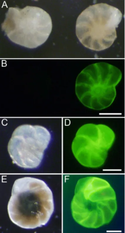
The surf clams only survived 1–3 weeks in the cores, but the quahogs remained ac-tive and grew, evidenced by the observations that many (∼25 %) of the quahogs had portions of their shell that were fluorescent (Fig. 2). The observation that the umbo
15
and other oldest parts of each quahog valve were not fluorescent is consistent with the fact that we seeded the cores with juveniles that were∼0.5 cm in length. Keeping in mind that bivalves add material to their valves in increasingly larger concentric annuli, somewhat akin to tree rings, it was noted that the fluorescence appeared as bands, where some portions fluoresced and others did not. Incorporation of calcein into newly
20
precipitated carbonate was confirmed by CLSM of bleached valves (Fig. 2g).
Because the system used during bivalve incubations was recirculating or lacking flow, it is important to consider the maximum concentration of calcein possible if all contents of the osmotic pump were dispensed into the seawater. We calculate that, at most, the recirculating seawater would have had 0.02 mg L−1 calcein. For mollusc 25
BGD
12, 9443–9463, 2015Technical Note: Towards resolving in situ, centimeter-scale
location
J. M. Bernhard et al.
Title Page
Abstract Introduction
Conclusions References
Tables Figures
◭ ◮
◭ ◮
Back Close
Full Screen / Esc
Printer-friendly Version
Interactive Discussion
Discussion
P
a
per
|
Discussion
P
a
per
|
Discussion
P
a
per
|
Discussion
P
a
per
label calcite (e.g., Kaehler and McQuaid, 1999; Klunzinger et al., 2014). Thus, the maximum possible calcein concentration in recirculating seawater during the first 3 weeks of bivalve incubations was far below the level required to fluorescently label calcite. Of course if the bivalves were near the concentrated source of calcein (i.e., pump port, 100 mg L−1calcein), then we predict the new calcite would be fluorescent, 5
as observed.
In the static (but aerated) setup, the maximum concentration of calcein possible if all contents of the osmotic pump were dispensed into the seawater was 0.2 mg L−1,
which again is far below the minimum mollusc threshold of 80 mg L−1. There are two
more lines of evidence that further support the inference that the lack of constant water
10
replacement did not cause artifactual calcein labeling in the bivalves: (1) the seawater overlying the cores did not have strong fluorescence when viewed with the appropriate optics using the stereo dissecting microscope (i.e., there was no significant background signal). (2) Not all bivalves were labeled with calcein. Some quahogs did not fluoresce in any part of their valves, indicating they were sufficiently removed from the point
15
source throughout the incubation.
Some of the calcareous benthic foraminifera in the cores exhibited bright fluores-cence while others did not (Fig. 3a, b). As established for the calcein labeling method, the non-fluorescent calcareous specimens either did not calcify during the incubation (Bernhard et al., 2004) or were too far from the osmotic pump port to incorporate
20
calcein. Some of the calcareous foraminiferal tests fully fluoresced (Fig. 3a–d), while others had only one or two brightly fluorescent chambers (Fig. 3e, f). It is possible that the fully fluorescent specimens were the result of reproduction during the experiment (Filipsson et al., 2010; Hintz et al., 2004). In contrast to brightly fluorescent rotalids, entire tests of miliolid (porcelaneous) calcareous foraminifera fluoresced dimly; no
ag-25
BGD
12, 9443–9463, 2015Technical Note: Towards resolving in situ, centimeter-scale
location
J. M. Bernhard et al.
Title Page
Abstract Introduction
Conclusions References
Tables Figures
◭ ◮
◭ ◮
Back Close
Full Screen / Esc
Printer-friendly Version
Interactive Discussion
Discussion
P
a
per
|
Discussion
P
a
per
|
Discussion
P
a
per
|
Discussion
P
a
per
|
et al., 2013). Such a growth habit explains the differential fluorescence patterns in some foraminiferal specimens, where 1–2 chambers are brightly fluorescent and remainder of the test has less intense fluorescence (Fig. 3f).
As noted for the non-recirculating bivalve incubations, we do not believe the calcein concentration of the overlying seawater would exceed the minimum labeling threshold
5
in the foraminiferal incubations even if the entire osmotic pump contents were released. For foraminiferal calcite labeling, a calcein concentration of 10 mg L−1 has been
typi-cally used previously (Bernhard et al., 2004; Denoyelle et al., 2012; Nardelli et al., 2014; Filipsson et al., 2010) although concentrations as low as 5 mg L−1 reportedly also
la-beled foraminiferal calcite (Dissard et al., 2009; Kurtarkar et al., 2015). The maximum
10
possible calcein concentration in overlying waters of our foraminiferal incubations was less than 1 mg L−1. As for bivalves, if growing calcareous foraminifera were near the
concentrated source of calcein (i.e., pump port, 100 mg L−1 calcein), then we predict
new calcite would be fluorescent, as observed.
Unfortunately, the calcein-labeled foraminiferal densities were insufficient to
deter-15
mine the vertical and horizontal extent of calcein diffusion into our muddy sediments. Specifically, calcein labeled foraminifera were absent from all small-volume radial sub-samples of the 0–1 cm interval of one core. Calcein-labeled foraminifera were found, however, in the remaining bulk 0–1 cm interval of the first multicore. Time and resource limitations prohibited full processing of additional multicores; spot checks in those
sam-20
ples did not yield convincing fluorescent foraminiferal calcite.
3.2 Notes and caveats
In the course of our method development, a number of lessons were learned. To assist future users of the method, these topics are discussed. Orienting the osmotic pump port downwards is problematic in fine-grained and/or low water-content sediments
be-25
BGD
12, 9443–9463, 2015Technical Note: Towards resolving in situ, centimeter-scale
location
J. M. Bernhard et al.
Title Page
Abstract Introduction
Conclusions References
Tables Figures
◭ ◮
◭ ◮
Back Close
Full Screen / Esc
Printer-friendly Version
Interactive Discussion
Discussion
P
a
per
|
Discussion
P
a
per
|
Discussion
P
a
per
|
Discussion
P
a
per
more compacted or consolidated sediments. Attempts to test the osmotic pump port at 4 cm depth did not result in any fluorescent specimens, but we do not know if that was due to lack of calcification or spatially limited calcein diffusion.
To document specifics regarding infaunal calcification horizons, it will be critical to determine the extent of calcein diffusion into sediments. Diffusion will vary with
sed-5
iment grain size, water content, compaction, hydrodynamics, and community compo-sition (e.g., presence or absence of bioturbators). Initial verification tests should be considered prior to initiating a lengthy or complicated experiment.
Calculations based on expected dispensation rate, temperature, and salinity can pro-vide estimated duration of calcein efflux. Osmotic pumps are single use; they will not
10
dispense if refilled. Per manufacturer’s instructions, osmotic pumps will not perform well if handled without clean gloves.
The cytoplasm of at least one benthic foraminiferal species autofluoresces using ex-citation and emission wavelengths similar to those for calcein (Apotheloz-Perret-Gentil et al., 2013). The foraminiferal species known to autofluoresce lacks a carbonate test,
15
so it cannot be confused with our calcein-labeling approach. If there are calcareous foraminifera with similarly autofluorescent cytoplasm, distinguishing between cytoplas-mic fluorescence (from viability indicators reliant on similar excitation and emission wavelengths) and carbonate fluorescence is not difficult if one considers the patterns and shapes of the signal (Nardelli et al., 2014).
20
3.3 Potential applications
The calcein-osmotic pump method can be used without modification to assess growth rates and calcification locations of juvenile and meiofaunal metazoans with calcareous hard parts (e.g., gastropods, echinoids, ostracods). These units can be deployed in shallow marine waters near shellfish fisheries and in reef areas with sediment pockets.
25
BGD
12, 9443–9463, 2015Technical Note: Towards resolving in situ, centimeter-scale
location
J. M. Bernhard et al.
Title Page
Abstract Introduction
Conclusions References
Tables Figures
◭ ◮
◭ ◮
Back Close
Full Screen / Esc
Printer-friendly Version
Interactive Discussion
Discussion
P
a
per
|
Discussion
P
a
per
|
Discussion
P
a
per
|
Discussion
P
a
per
|
As noted, our method will help to better understand foraminiferal microhabitats. Such knowledge will help to minimize uncertainty and increase precision in records of pale-oceanographic proxies preserved in foraminiferal tests. For instance, recently the dif-ference in theδ13C for epifaunalCibicides wuellerstorfiand for deep infaunal Globobu-liminaspp. was used to reconstruct bottom-water oxygen concentration (Hoogakker et
5
al., 2015), an approach that could be further improved by this method. Foraminiferal– based ecology studies under shifting environmental conditions, such as varying water oxygen concentration (Nardelli et al., 2014) or pH conditions would also benefit from our calcein-osmotic pump method. Of course, our method should be considered a first step given that most paleoceanographically-relevant foraminifera presumably are able
10
to migrate within the sediment column. It is unknown, however, if benthic foraminifera actually migrate vertically in situ and, if they do, where they calcify. Regardless, addi-tional refinements to the approach will be required to prevent or minimize foraminiferal migration during and/or after calcification.
Our osmotic pump method can be further modified to deploy these units in
deep-15
sea sediments using a Remotely Operated Vehicle (e.g.,Jason) or a Human Occupied Vehicle (e.g.,Alvin). Further, calcein-filled osmotic pumps can be installed in habitats that are spatially restricted, such as hydrocarbon seeps or brine pools, where we have little growth data for any sediment-dwelling species.
4 Conclusions
20
While calcein has been used in growth studies for a variety of organisms, to our knowl-edge, calcein has not been used as a point source to determine calcification in the en-vironment. Most studies using calcein to determine growth rates immerse entire speci-mens in the laboratory and then release them into nature for later recapture. Our new calcein-osmotic pump approach can help pinpoint where and when meiofaunal
organ-25
BGD
12, 9443–9463, 2015Technical Note: Towards resolving in situ, centimeter-scale
location
J. M. Bernhard et al.
Title Page
Abstract Introduction
Conclusions References
Tables Figures
◭ ◮
◭ ◮
Back Close
Full Screen / Esc
Printer-friendly Version
Interactive Discussion
Discussion
P
a
per
|
Discussion
P
a
per
|
Discussion
P
a
per
|
Discussion
P
a
per
Acknowledgements. We thank the Captain and crew of RV Endeavor, the science party of EN524, Megan Davis (UNCW) for lab and collecting assistance, Ellen Roosen for coring assis-tance, John Schriever (Bayfarm) for juvenile bivalves, and Caitlin Keating-Bitonti (Stanford) and Kamila Sztybor (Univ Tromsø) for lab assistance. This research was funded by WHOI’s Ocean Life Institute, WHOI’s Ocean and Climate Change Institute, by a Gori Fellowship (to FM), The
5
Investment in Science Fund at WHOI (to JMB) and the Robert W. Morse Chair for Excellence in Oceanography (to JMB). Ship time was provided by US NSF grant OCE-1219948 to JMB.
References
Allen, K. A. and Honisch, B.: The planktic foraminiferal B/Ca proxy for seawater carbonate chemistry: A critical evaluation, Earth Planet. Sci. Lett., 345, 203–211, 2012.
10
Apotheloz-Perret-Gentil, L., Holzmann, M., and Pawlowski, J.:Arnoldiellina fluorescensgen. et sp. nov. – A new green autofluorescent foraminifer from the Gulf of Eilat (Israel), Europ. J. Protistol., 49, 210–216, 2013.
Bernhard, J. M., Blanks, J. K., Hintz, C. J., and Chandler, G. T.: Use of the fluorescent calcite marker calcein to label foraminiferal tests, J. Foraminifer. Res., 34, 96–101, 2004.
15
Bernhard, J. M., Martin, J. B., and Rathburn, A. E.: Combined carbonate carbon isotopic and cellular ultrastructural studies of individual benthic foraminifera: 2. Toward an under-standing of apparent disequilibrium in hydrocarbon seeps, Paleoceanography, 25, Pa4206, doi:10.1029/2010pa001930, 2010.
Bijma, J., Spero, H. J., and Lea, D. W.: Reassessing foraminiferal stable isotopes
geochem-20
istry: Impact of the ocean carbonate system (experimental results), in: Uses of proxies in paleoceanography: Examples from the South Atlantic, edited by: Fischer, G. and Wefer, G., Springer-Verlag, Berlin, 489–512, 1999.
Bothner, M. H., Spiker, E. C., Johnson, P. P., Rendigs, R. R., and Aruscavage, P. J.: Geochem-ical evidence for modern sediment accumulation on the continental shelf offSouthern New
25
England, J. Sediment. Petrol., 51, 281–292, 1981.
Collin, R. and Voltzow, J.: Initiation, calcification, and form of larval ”archaeogastropod” shells, J. Morphol., 235, 77–89, 1998.
Corliss, B. H.: Microhabitats of benthic foraminifera within deep-sea sediments, Nature, 314, 435–438, 1985.
BGD
12, 9443–9463, 2015Technical Note: Towards resolving in situ, centimeter-scale
location
J. M. Bernhard et al.
Title Page
Abstract Introduction
Conclusions References
Tables Figures
◭ ◮
◭ ◮
Back Close
Full Screen / Esc
Printer-friendly Version
Interactive Discussion
Discussion
P
a
per
|
Discussion
P
a
per
|
Discussion
P
a
per
|
Discussion
P
a
per
|
de Nooijer, L. J., Hathorne, E. C., Reichart, G. J., Langer, G., and Bijma, J.: Variability in cal-citic Mg/Ca and Sr/Ca ratios in clones of the benthic foraminifer Ammonia tepida, Mar. Micropaleontol., 107, 32–43, 2014.
Denoyelle, M., Geslin, E., Jorissen, F. J., Cazes, L., and Galgani, F.: Innovative use of foraminifera in ecotoxicology: A marine chronic bioassay for testing potential toxicity of drilling
5
muds, Ecol. Indic., 12, 17–25, 2012.
Dissard, D., Nehrke, G., Reichart, G. J., Nouet, J., and Bijma, J.: Effect of the fluorescent indicator calcein on Mg and Sr incorporation into foraminiferal calcite, Geochem. Geophys. Geosyst., 10, Q11001, doi:10.1029/2009gc002417, 2009.
Elderfield, H., Yu, J., Anand, P., Kiefer, T., and Nyland, B.: Calibrations for benthic foraminiferal
10
Mg/Ca paleothermometry and the carbonate ion hypothesis, Earth Planet. Sci. Lett., 250, 633–649, 2006.
Erez, J.: The source of ions for biomineralization in foraminifera and their implications for pa-leoceanographic proxies, in: Biomineralization, edited by: Dove, P. M., DeYoreo, J. J., and Weiner, S., Mineral. Geochem., 54, 115–149, 2003.
15
Filipsson, H. L., Bernhard, J. M., Lincoln, S. A., and McCorkle, D. C.: A culture-based calibra-tion of benthic foraminiferal paleotemperature proxies: δ18O and Mg/Ca results, Biogeo-sciences, 7, 1335–1347, doi:10.5194/bg-7-1335-2010, 2010.
Healey, S. L., Thunell, R. C., and Corliss, B. H.: The Mg/Ca-temperature relationship of benthic foraminiferal calcite: New core-top calibrations in the<4◦C temperature range, Earth Planet. 20
Sci. Lett., 272, 523–530, 2008.
Hernaman, V., Munday, P. L., and Schlappy, M. L.: Validation of otolith growth-increment peri-odicity in tropical gobies, Mar. Biol., 137, 715–726, 2000.
Hintz, C. J., Chandler, G. T., Bernhard, J. M., McCorkle, D. C., Havach, S. M., Blanks, J. K., and Shaw, T. J.: A physicochemically constrained seawater culturing system for production
25
of benthic foraminifera, Limnol. Oceanogr. Meth., 2, 160–170, 2004.
Hoogakker, B. A. A., Elderfield, H., Schmiedl, G., McCave, I. N., and Rickaby, R. E. M.: Glacial-interglacial changes in bottom-water oxygen content on the Portuguese margin, Nat. Geosci., 8, 40–43, 2015.
Jorissen, F. J., deStigter, H. C., and Widmark, J. G. V.: A conceptual model explaining benthic
30
foraminiferal microhabitats, Mar. Micropaleontol., 26, 3–15, 1995.
BGD
12, 9443–9463, 2015Technical Note: Towards resolving in situ, centimeter-scale
location
J. M. Bernhard et al.
Title Page
Abstract Introduction
Conclusions References
Tables Figures
◭ ◮
◭ ◮
Back Close
Full Screen / Esc
Printer-friendly Version
Interactive Discussion
Discussion
P
a
per
|
Discussion
P
a
per
|
Discussion
P
a
per
|
Discussion
P
a
per
Katz, M. E., Cramer, B. S., Franzese, A., Hönisch, B., Miller, K. G., Rosenthal, Y., and Wright, J. D.: Traditional and emerging geochemical proxies in foraminifera, J. Foraminifer. Res., 40, 165–192, 2010.
Klunzinger, M. W., Beatty, S. J., Morgan, D. L., Lymbery, A. J., and Haag, W. R.: Age and growth in the Australian freshwater mussel, Westralunio carteri, with an evaluation of the
5
fluorochrome calcein for validating the assumption of annulus formation, Freshw. Sci., 33, 1127–1135, 2014.
Kurtarkar, S. R., Saraswat, R., Nigam, R., Banerjee, B., Mallick, R., Naik, D. K., and Singh, D. P.: Assessing the effect of calcein incorporation on physiological processes of benthic foraminifera, Mar. Micropaleontol., 114, 36–45, 2015.
10
Lea, D. W., Mashiotta, T. A., and Spero, H. J.: Controls on magnesium and strontium uptake in planktonic foraminifera determined by live culturing, Geochim. Cosmochim. Acta, 63, 2369– 2379, 1999.
McCorkle, D. C., Keigwin, L. D., Corliss, B. H., and Emerson, S. R.: The influence of microhabi-tats on the carbon isotopic composition of deep-sea benthic foraminifera, Paleoceanography,
15
5, 161–185, 1990.
McCorkle, D. C., Bernhard, J. M., Hintz, C. J., Blanks, J. K., Chandler, G. T., and Shaw, T. J.: The carbon and oxygen stable isotopic composition of cultured benthic foraminifera, in: Biogeochemical controls on palaeoceanographic environmental proxies, edited by: Austin, W. E. N. and James, R. H., The Geological Society Special Publication, London, 135–154,
20
2008.
Medeiros-Bergen, D. E. and Ebert, T. A.: Growth, fecundity and mortality rates of 2 intertidal brittlestars (Echinodermata, Ophiuroidea) with contrasting modes of development, J. Exp. Mar. Biol. Ecol., 189, 47–64, 1995.
Monaghan, J. P.: Comparison of calcein and tetracycline as chemical markers in summer
floun-25
der, T. Am. Fish. Soc., 122, 298–301, 1993.
Moran, A. L.: Calcein as a marker in experimental studies newly-hatched gastropods, Mar. Biol., 137, 893–898, 2000.
Moran, A. L. and Marko, P. B.: A simple technique for physical marking of larvae of marine bivalves, J. Shellfish Res., 24, 567–571, 2005.
30
BGD
12, 9443–9463, 2015Technical Note: Towards resolving in situ, centimeter-scale
location
J. M. Bernhard et al.
Title Page
Abstract Introduction
Conclusions References
Tables Figures
◭ ◮
◭ ◮
Back Close
Full Screen / Esc
Printer-friendly Version
Interactive Discussion
Discussion
P
a
per
|
Discussion
P
a
per
|
Discussion
P
a
per
|
Discussion
P
a
per
|
Nehrke, G., Keul, N., Langer, G., de Nooijer, L. J., Bijma, J., and Meibom, A.: A new model for biomineralization and trace-element signatures of Foraminifera tests, Biogeosciences, 10, 6759–6767, doi:10.5194/bg-10-6759-2013, 2013.
Raitzsch, M., Kuhnert, H., Groeneveld, J., and Bickert, T.: Benthic foraminifer Mg/Ca anoma-lies in South Atlantic core top sediments and their implications for paleothermometry,
5
Geochem. Geophys. Geosyst., 9, Q05010, doi:10.1029/2007gc001788, 2008.
Ravelo, A. C. and Hillaire-Marcel, C.: The Use of Oxygen and Carbon Isotopes of Foraminifera in Paleoceanography in: Proxies in Late Cenozoic Paleoceanography, edited by: Hillaire-Marcel, C., and De Vernal, A., Develop. Mar. Geol., 1, 735–764, 2007.
Rosenthal, Y., Lear, C. H., Oppo, D. W., and Linsley, B. K.: Temperature and carbonate ion
10
effects on Mg/Ca and Sr/Ca ratios in benthic foraminifera: Aragonitic speciesHoeglundina elegans, Paleoceanography, 21, PA1007, doi:10.1029/2005pa001158, 2006.
Russell, A. D., Hönisch, B., Spero, H. J., and Lea, D. W.: Effects of seawater carbonate ion concentration and temperature on shell U, Mg, and Sr in cultured planktonic foraminifera, Geochim. Cosmochim. Acta, 68, 4347–4361, 2004.
15
Spero, H. J., Bijma, J., Lea, D. W., and Bemis, B. E.: Effect of seawater carbonate concentration on foraminiferal carbon and oxygen isotopes, Nature, 390, 497–500, 1997.
Stott, L. D., Bunn, T., Prokopenko, M., Mahn, C., Gieskes, J., and Bernhard, J. M.: Does the ox-idation of methane leave an isotopic fingerprint in the geologic record?, Geochem. Geophys. Geosyst., 3, GC000196, doi:10.1029/2001gc000196, 2002.
20
BGD
12, 9443–9463, 2015Technical Note: Towards resolving in situ, centimeter-scale
location
J. M. Bernhard et al.
Title Page
Abstract Introduction
Conclusions References
Tables Figures
◭ ◮
◭ ◮
Back Close
Full Screen / Esc
Printer-friendly Version
Interactive Discussion
Discussion
P
a
per
|
Discussion
P
a
per
|
Discussion
P
a
per
|
Discussion
P
a
per
Figure 1. (a)ALZET®2ML4 osmotic pump filled with concentrated calcein. The visible calcein
BGD
12, 9443–9463, 2015Technical Note: Towards resolving in situ, centimeter-scale
location
J. M. Bernhard et al.
Title Page
Abstract Introduction
Conclusions References
Tables Figures
◭ ◮
◭ ◮
Back Close
Full Screen / Esc
Printer-friendly Version
Interactive Discussion
Discussion
P
a
per
|
Discussion
P
a
per
|
Discussion
P
a
per
|
Discussion
P
a
per
|
Figure 2.Paired micrographs of quahogs after calcein osmotic pump incubations. Reflected
(a, c, e)and epifluorescence (b, d, f)images of quahog after short bleach and air drying (e
BGD
12, 9443–9463, 2015Technical Note: Towards resolving in situ, centimeter-scale
location
J. M. Bernhard et al.
Title Page
Abstract Introduction
Conclusions References
Tables Figures
◭ ◮
◭ ◮
Back Close
Full Screen / Esc
Printer-friendly Version
Interactive Discussion
Discussion
P
a
per
|
Discussion
P
a
per
|
Discussion
P
a
per
|
Discussion
P
a
per
Figure 3.Paired micrographs of foraminifera after calcein osmotic pump incubations. Reflected