PONTIFÍCIA UNIVERSIDADE CATÓLICA DO RIO GRANDE DO SUL FACULDADE DE ODONTOLOGIA
PAULA CRISTINE GHIGGI
INFLUÊNCIA DO SELAMENTO DENTINÁRIO IMEDIATO NA POLIMERIZAÇÃO DE MATERIAIS DE MOLDAGEM E NA RESISTÊNCIA À FRATURA DE COROA
EM CERÂMICA
PAULA CRISTINE GHIGGI
IMMEDIATE DENTIN SEALING INFLUENCES THE POLYMERIZATION OF IMPRESSION MATERIALS
THICKNESS OF IMMEDIATE DENTIN SEALING MATERIALS AND ITS EFFECT ON THE FRACTURE LOAD OF A REINFORCED ALL-CERAMIC CROWN
Tese apresentada como requisito parcial para a obtenção do grau de Doutor pelo Programa de Pós-Graduação da Faculdade de Odontologia da Pontifícia Universidade Católica do Rio Grande do Sul.
Orientadora: Profa. Dra. Ana Maria Spohr
Dados Internacionais de Catalogação na Publicação (CIP)
G423i Ghiggi, Paula Cristine
Influência do selamento dentinário imediato na polimerização de materiais de moldagem e na resistência à fratura de coroa em cerâmica. / Paula Cristine Ghiggi. – Porto Alegre, 2013.
83 f. : il.
Tese (Doutorado) – Programa de Pós-Graduação em Odontologia, Faculdade de Odontologia, PUCRS.
Orientadora: Profa. Dra. Ana Maria Spohr
1. Odontologia. 2. Materiais Dentários. 3. Selamento Dentinário Imediato. 4. Resistência dos Materiais. I. Spohr, Ana Maria. II. Título.
CDD 617.695
PAULA CRISTINE GHIGGI
IMMEDIATE DENTIN SEALING INFLUENCES THE POLYMERIZATION OF IMPRESSION MATERIALS
THICKNESS OF IMMEDIATE DENTIN SEALING MATERIALS AND ITS EFFECT ON THE FRACTURE LOAD OF A REINFORCED ALL-CERAMIC CROWN
Tese apresentada como requisito parcial para a obtenção do grau de Doutor pelo Programa de Pós-Graduação da Faculdade de Odontologia da Pontifícia Universidade Católica do Rio Grande do Sul.
Aprovada em _____ de ___________________ de ________
BANCA EXAMINADORA
____________________________________________ Orientadora: Profa. Dra. Ana Maria Spohr - PUCRS __________________________________________
Profa. Dra. Andrea Brito Conceição - UFRGS _________________________________________
Prof. Dr. Cláudio Figueiró - UFSM
_________________________________________ Profa. Dra. Luciana Mayumi Hirakata - PUCRS _________________________________________
AGRADECIMENTOS
Aos meus pais Cesar e Marivone, os responsáveis pela minha formação, sempre me incentivaram e me apoiaram em cada etapa da minha vida!! Às minhas irmãs Gisele e Luciana que além de irmãs são minhas melhores amigas e vivem comigo cada momento da minha vida!!
Amo muito vocês!!!
À minha orientadora Profa. Dra. Ana Maria Spohr, pela paciência, dedicação, humildade, disposição, compreenção e grande sabedoria. Conviver com você durante esses 6 anos foram essenciais para a minha formação pessoal e profissional, você é um exemplo a ser seguido como orientadora e, acima de tudo, como pessoa. Obrigada por tudo!!!
Ao Prof. Dr. Luiz Henrique Burnett Jr. e ao Prof.Eduardo Mota pela amizade. Obrigada pelo carinho!!!
Ao Arno Kieling Steiger pela paciência, dedicação, disponibilidade e persistência na obtenção das imagens. Obrigada!!
À CAPES pela bolsa de estudos concedida.
Aos colegas Guilherme e Pati que dividiram comigo muitas dúvidas e angústias e sempre me incentivaram com palavras de tranqüilidade.
Aos funcionários da Secretaria de Pós-Graduação: Ana, Paulo e Davenir sempre muito atenciosos e dispostos!!!
LISTA DE ILUSTRAÇÕES
Artigo 1
Figure 1: Schematic design of the experimental groups……… 24 Figure 2: Control group (without IDS): a) Impression with vinyl polysiloxane; b) Impression with polyether. There is no impression material attached to the dentin
surface ……….. 29
Figure 3: Impression with vinyl polysiloxane: a) IDS with Clearfil SE Bond; b) IDS with Clearfil SE Bond and Protect Liner F. Small areas of unpolymerized impression material are attached to the surface of the resin materials ………... 29 Figure 4: IDS with Clearfil SE Bond and impression with vinyl polysiloxane: a) glycerin jelly; b) alcohol. There is no interaction between the impression material and
the resin materials ………..…. 29
Figure 5: IDS with Clearfil SE Bond and Protect Liner F and impression with vinyl polysiloxane: a) glycerin jelly; b) alcohol. Small areas of unpolymerized impression material are attached to the surface of the resin materials ………...…… 30 Figure 6: - Impression with polyether: a) IDS with Clearfil SE Bond; b) IDS with Clearfil SE Bond and Protect Liner F. There is polymerized impression material attached to the resin material surface ………..…… 30 Figure 7: IDS with Clearfil SE Bond and impression with polyether: a) glycerin jelly; b) alcohol. Polymerized impression material is attached to the Clearfil SE Bond
……….… 30
Figure 8: IDS with Clearfil SE Bond and Protect Liner F and impression with polyether: a) glycerin jelly; b) alcohol. There are no interactions between the impression material and the resin materials ………..…. 31
Artigo 2
Figure 1: Bucco-lingual section of the preparation. Ten positions were market and the thickness of resin cement / adhesive / low-viscosity microfilled resin were
measured………...… 70
LISTA DE TABELAS
Artigo 1
Table 1: Materials used in the study ………. 25
Table 2: Interactions between the impression materials and the resin materials……….... 28
Artigo 2 Table 1: Materials used in the study ………. 64
Table 2: Mean thickness (µm) and standard deviation of the resin cement, adhesive and low-viscosity microfilled resin of the experimental groups in the different positions ………...…….. 65
Table 3: Sum of thickness of resin material (µm) at different positions ……….. 67
Table 4: Mean fracture load (N) of the experimental groups ………...…. 68
LISTA DE ABREVIAÇÕES, SIGLAS E SÍMBOLOS
% Porcentagem
o Graus
0C Grau Celsius µm Micrometro
Bis-GMA Bisfenol glicidil metacrilato A CIO Camada inibida pelo oxigênio CSE Clearfil SE Bond
et al. Abreviatura de et allii (e outros) e.g. Exemplo
h Hora
HEMA 2- hidroxietil metacrilato IDS Immediate dentin sealing
KG Kilograma
MDP Metacriloxietil dihidrogênio fosfato
Min Minuto
mm Milímetro
N Newton
n0 Número
NMSA N-methacryloxyl-5-aminosalicylic acid. OIL Oxygen-inhibition layer
p Valor de probabilidade
PUCRS Pontifícia Universidade Católica do Rio Grande do Sul PLF Protect Liner F
r Correlação de Pearson
RESUMO
A primeira etapa deste estudo avaliou, in vitro, a interação entre materiais resinosos utilizados na técnica do selamento dentinário imediato (SDI) e materiais de moldagens associado a duas técnicas para reduzir/eliminar a camada inibida de oxigênio. A dentina oclusal de 35 terceiros molares humanos foi exposta, seguido de acabamento com lixa de carbeto de silício de granulação 400. Os dentes foram divididos aleatoriamente em 2 grupos: grupo 1- moldagem com silicone por adição Express XT; grupo 2 – moldagem com o poliéter Impregum. Os grupos 1 e 2 foram divididos em 14 subgrupos: grupo 1a e 2a: controle; grupos 1b e 2b: SDI com Clearfil SE Bond (CSE); grupos 1c e 2c: SDI with CSE + polimerização adicional com gel a base de glicerina; grupos 1d e 2d: SDI com CSE + álcool; grupos 1e e 2e: SDI com CSE e Protect Liner F (PLF); grupos 1f e 2f: SDI com CSE e PLF + polimerização adicional com gel a base de glicerina; grupos 1g e 2g: SDI com CSE e PLF + álcool. Cada superfície dentária foi fotografada com uma câmera digital. As imagens foram salvas e utilizadas para avaliar a presença de material de moldagem sobre a estrutura dentária. Por meio de uma análise qualitativa, observou-se que o SDI realizado com o CSE ou com PLF interagiram com o Express XT e com o Impregum. A aplicação do gel a base de glicerina e do álcool impediram a interação do CSE com o Express XT e do PLF com o Impregum; no entanto, estes mesmos tratamentos não foram totalmente efetivos para o CSE com o Impregum e para o PLF com o Express XT.
A segunda etapa deste estudo avaliou a espessura do sistema adesivo, resina de baixa viscosidade e cimento resinoso em preparos para coroas totais, e a influência na resistência à fratura de coroa total em cerâmica. Sessenta pré-molares superiores receberam prepares para coroa total e foram divididos em 3 grupos de acordo com o material aplicado na técnica do SDI: grupo 1 – controle; grupo 2 - CSE; G3 – CSE + PLF. Após moldagem com silicone por adição, os preparos receberam provisórios confeccionados com resina acrílica. As restaurações cerâmicas com IPS Empress 2 foram confeccionadas e cimentadas sobre os preparos com Panavia F. Dez amostras de cada grupo foram submetidos ao teste de resistência à fratura e 10 espécimes foram seccionados no sentido vestíbulo-lingual para avaliar a espessura do Panavia F, CSE e PLF em 10 posições diferentes com o auxílio de um microscópio. De acordo com ANOVA e teste de Tukey, a carga de fratura do grupo 3 (1300 N) foi estatisticamente superior ao grupo 1 (1001 N) (p<0.01) e o grupo 2 (1189 N) não apresentou diferença estatística com os grupos 1 e 3. A maior espessura de película do CSE Bond foi obtida na parte côncava dos prepares. O PLF apresentou a espessura mais uniforme em diferentes posições. A espessura do Panavia F foi maior na face oclusal dos preparos. A espessura da película formada pelo CSE e PLF aumentou a resistência à fratura de coroas cerâmicas confeccionadas com IPS Empress 2.
ABSTRACT
The first section of this study evaluated the interaction between resin materials used in the immediate dentin sealing (IDS) techniques and impression materials under two different techniques to reduce/eliminate the oxygen-inhibition layer. The occlusal dentin of 35 human molars was exposed and finished with 400 grit silicon carbide sandpaper. Teeth were randomly divided into 2 groups: group 1 – impression with vinyl polysiloxane Express XT, group 2 – impression with polyether Impregum. Groups 1 and 2 were divided into 14 subgroups: groups 1a e 2a: control groups; groups 1b e 2b: IDS with Clearfil SE Bond (CSE); groups 1c e 2c: IDS with CSE + additional polymerization with glycerine jelly; groups 1d e 2d: IDS with CSE + alcohol; groups 1e e 2e: IDS with CSE and Protect Liner F (PLF); groups 1f e 2f: IDS with CSE and PLF + additional polymerization with glycerin jelly; groups 1g e 2g: IDS with CSE and PLF + alcohol. Each tooth surface was photographed using a digital camera. The images saved were used to examine the presence of impression material left on the treated tooth surface. It was observed that IDS performed with CSE or with the PLF interacted with the Express XT and with the Impregum. The application of glycerine jelly and alcohol avoided the interaction of CSE with the Express XT and of the PLF with the Impregum; however, these treatments were not totally effective to avoid the interaction of CSE with the Impregum and of PLF with the Express XT.
The second section of this study evaluate, in vitro, the thickness of the adhesive, low-viscosity microfilled resin, and resin cement on full crown preparations, and its effect on the fracture load of a reinforced all-ceramic crown. Sixty maxillary premolars received full crown preparation and were divided in 3 groups according to the material applied for the immediate dentin sealing: G1 – control; G2 – CSE Bond; G3 – CSE Bond + PLF. After taking the impression with polyvinyl siloxane, the preparations were temporized with acrylic resin crowns. IPS Empress 2 restorations were fabricated, and cemented to the preparations with Panavia F. Ten specimens of each group were submitted to fracture load testing, and the other 10 specimens were sectioned buccolingually, and the thicknesses of Panavia F, CSE Bond and PLF were measured in 10 different positions using a microscope. According to ANOVA and Tukey’s test, the fracture load of group 3 (1300 N) was statistically higher than group 1 (1001 N) (p<0.01). Group 2 (1189 N) was not statistically different from groups 1 and 3. The higher thickness of the CSE Bond was obtained in the concave part of the preparation. The PLF presented a more uniform range of values at different positions. The thickness of Panavia F was higher in the occlusal portion of the preparation. The film thickness formed by CSE Bond and PLF increased the fracture load of the IPS Empress 2 ceramic crown.
SUMÁRIO
1 Introdução Geral ...14
2 Artigo 1 ...17
2.1 Abstract ...18
2.2 Introduction ...19
2.3 Materials and methods ...21
2.4 Results ... 26
2.5 Discussion ... 32
2.6 Conclusions ... 36
2.7 References ...37
3 Artigo 2 ... 40
3.1 Abstract ... 41
3.2 Introduction ... 42
3.3 Materials and methods ... 45
3.4 Results ... 49
3.5 Discussion ... 51
3.6 Conclusions ... 58
3.7 References ... 59
4 Discussão Geral ...75
5 Referências Bibliográficas...81
INTRODUÇÃO GERAL
As restaurações adesivas têm sido empregadas largamente como resultado
do desenvolvimento e aperfeiçoamento das resinas compostas, dos sistemas cerâmicos, dos sistemas adesivos e das técnicas operatórias. Porém, as
restaurações indiretas apresentam algumas vantagens em relação às restaurações diretas de resina composta, como estabilidade química, estabilidade de cor e maior resistência ao desgaste, sendo indicadas nos casos de maior perda da estrutura
dentária (VAN NOORT, 1994).
As restaurações indiretas requerem alguns passos clínicos adicionais e
críticos como moldagem, temporização e cimentação. Dependendo do material empregado, preferem-se os cimentos resinosos para a cimentação definitiva por apresentarem união à estrutura dental, baixa solubilidade e melhor resistência ao
desgaste que os cimentos convencionais (DIAZ-ARNOLD; VARGAS; HASELTON, 1999).
O preparo para restaurações indiretas pode induzir significativa exposição de dentina e, consequentemente, sensibilidade. A técnica convencional para restaurações indiretas consiste na moldagem do preparo imediatamente após este
ter sido concluído, seguido da confecção de um provisório. Quando a restauração definitiva está pronta, o provisório é removido e a cimentação adesiva é realizada.
Tem sido demonstrado que a dentina recém-cortada seria o substrato ideal para procedimentos adesivos; no entanto, a dentina contaminada com cimento provisório, sangue e saliva são fatores críticos que influenciam no potencial de adesão,
sensibilidade pós-operatória (BERTSCHINGER et al., 1996; PAUL; SCHÄRER, 1997).
Técnicas alternativas têm sido propostas para superar estes problemas, como o selamento dentinário imediato (SDI) com a aplicação de um sistema adesivo logo após a confecção do preparo e previamente ao procedimento de moldagem
(MAGNE et al., 2005). Outra técnica consiste na aplicação de um sistema adesivo e de uma resina microparticulada de baixa viscosidade (NIKAIDO et al., 1992). Estas
técnicas têm o objetivo de selar a ampla área de dentina exposta durante o preparo cavitário. Isto minimiza a irritação pulpar por estímulos térmicos e mecânicos, pela
infiltração de bactérias durante os procedimentos de moldagem, temporização e cimentação final (KITASAKO et al., 2002), e também aumenta a retenção para casos de coroas clínicas curtas (MAGNE; SO; CASCIONE, 2007).
O SDI com sistema adesivo e resina microparticulada de baixa viscosidade melhora a resistência adesiva(DE GOES et al., 2000), reduz a formação de fendas
na interface dentina/cimento resinoso (SUZUKI, 2000; KITASAKO et al., 2002) e, além disso, funciona como uma camada resiliente entre a restauração indireta e a dentina, absorvendo as tensões geradas durante a contração de polimerização do
cimento resinoso e dos efeitos mastigatórios (DIETSCHI et al., 2002; MONTES et al., 2003).
A etapa de moldagem, após a técnica do SDI, torna-se um procedimento crítico, visto que a camada superficial do material adesivo não polimeriza por entrar em contato com o oxigênio (CIO – camada inibida pelo oxigênio) (ELIADES;
sobrepolimerização do material adesivo com gel a base de glicerina, ou a aplicação de álcool podem ser utilizadas na tentativa de eliminar a CIO (BERGMANN; NOACK;
ROULET, 1991; NIKAIDO et al., 2003).
A avaliação da resistência de união utilizando as técnicas do SDI em restaurações indiretas de resina composta tem sido pesquisada (OKUDA et al.,
2007; UDO et al., 2007). No caso de preparos para coroa total, a região do término do preparo geralmente encontra-se em dentina ao nível radicular e muitos dentes
são vitais, justificando a técnica do SDI. No entanto, não há estudos evidenciando a influência da espessura dos materiais adesivos utilizados para a técnica do SDI na
resistência à fratura de coroas totais em cerâmica.
O presente estudo teve os seguintes objetivos: a) avaliar a interação entre os materiais adesivos utilizados na técnica do SDI e diferentes materiais de moldagem,
associado a duas técnicas utilizadas para eliminar a CIO; b) avaliar a influência da espessura de película dos materiais adesivos utilizados na técnica do SDI e do
IMMEDIATE DENTIN SEALING INFLUENCES THE POLYMERIZATION OF
IMPRESSION MATERIALS
Paula Cristine Ghiggi1 Ana Maria Spohr2
1Master degree student in Restorative Dentistry, Pontifical Catholic University of Rio Grande do Sul, Porto Alegre, Brazil.
2Department of Dental Materials, Pontifical Catholic University of Rio Grande do Sul, Porto Alegre, Brazil.
Corresponding author: Ana Maria Spohr
PUCRS – School of Dentistry, Block 6 Avenida Ipiranga, 6681, 90616-900 Porto Alegre, RS, Brazil.
ABSTRACT
This in vitro study evaluated the interaction between the resin materials used in immediate dentin sealing (IDS) techniques and impression materials with two different techniques to reduce/eliminate the oxygen-inhibition layer (OIL). The roots of 35 human molars were included in self-cured acrylic resin. The occlusal enamel was removed using a diamond saw, and the dentin surface was exposed. This step was followed by surface regularization with 400-grit silicon carbide sandpaper under water cooling. The teeth were randomly divided into 2 groups: group 1 – impression with Express XT vinyl polysiloxane and group 2 – impression with Impregum polyether. Groups 1 and 2 were divided into 14 subgroups: group 1a and 2a – control groups; 1b and 2b – IDS with Clearfil SE Bond (CSE); 1c and 2c – IDS with CSE + additional polymerization with glycerin jelly; 1d and 2d – IDS with CSE + alcohol; 1e and 2e – IDS with CSE and Protect Liner F (PLF); 1f e and 2f – IDS with CSE and PLF + additional polymerization with glycerin jelly; and 1g and 2g – IDS with CSE and PLF + alcohol. Each tooth surface was photographed using a digital camera, and the images were used to examine the interaction between the resin materials and the impression materials. IDS with either CSE or PLF interacted with Express XT and with Impregum. The application of glycerin jelly and alcohol prevented the interaction of CSE with Express XT and PLF with Impregum; however, these treatments were not completely effective in preventing the interaction of CSE with Impregum and PLF with Express XT.
INTRODUCTION
During tooth preparation for indirect restorations such as inlays, onlays,
veneers, and crowns, a significant area of dentin is exposed. To avoid problems such as dentin contamination by provisionalization (1, 2) and hybridization failure
sensitivity (3), a technique called immediate dentin sealing (IDS) was suggested in the early 1990s (4). This technique consists of the application of an adhesive system immediately after tooth preparation and before taking the impression. Another IDS
technique was developed in which a sealing film is applied to the dentin surface immediately after tooth preparation using an adhesive system and a low-viscosity
composite resin (5, 6). It is believed that this layer of low-viscosity composite resin isolates the underlying hybrid layer and, consequently, aids in preserving the dentin seal (7). Therefore, IDS techniques are based on the principle that adhesive systems
bond better to freshly prepared dentin (1, 2), thus protecting the dentin–pulp complex and preventing or decreasing sensitivity and bacterial leakage during the provisional
stage (8, 9).
When using the IDS techniques, the impression is taken after the application of the resin material on the dental substrate. This step is critical because the
impression material can interact with the outer resin layer (3), which is unpolymerized due to the oxygen inhibition of the radicals that initiate the polymerization reaction
(10, 11). Different techniques have been suggested to reduce or eliminate the oxygen-inhibition layer (OIL), such as the application of glycerin jelly followed by an additional light cure (12) or the use of a cotton pellet soaked in alcohol (13).
However, few studies have been published about this subject.
resin materials used in the IDS techniques and impression materials when two different techniques to reduce/eliminate the OIL are applied. This study was based
MATERIALS AND METHODS
Thirty-five unerupted human third molars, which were extracted for therapeutic
reasons, were obtained from the Tooth Bank after the approval of the Ethics Committee of the Pontifical Catholic University of Rio Grande do Sul (PUCRS). The
teeth were cleaned of gross debris and stored in distilled water at 4°C. The water was changed every week, and the teeth were used in the study within 6 months. The roots were mounted in self-cured acrylic resin, and the occlusal enamel surface was
removed with a diamond disc mounted in a low-speed laboratory cutting machine (Labcut 1010, Extec Corp., London, UK) under cooling conditions. The rest of the
enamel was removed with 400-grit silicon carbide abrasive paper in a polishing machine (DPU-10, Panambra, São Paulo, SP, Brazil) under water. The superficial dentin was exposed and finished with 600-grit silicon carbide abrasive paper in the
polishing machine, and a flat dentin surface was obtained.
After polishing, the teeth were randomly divided into 14 groups (Figure 1)
according to the materials used (Table 1). As a nondestructive methodology, the teeth were used again after remotion of the resin material with 600-grit silicon carbide abrasive paper.
Express XT vinyl polysiloxane impression material was used in group 1, and the following subgroups were assigned (Figure 1):
Group 1a: Unsealed tooth surface.
and light cured for 10 s using a conventional halogen light-curing unit.
Group 1c: IDS with CSE and glycerin jelly. The adhesive system was applied as
described for group 1b. The polymerization of the adhesive was followed by the application of an air-blocking barrier with glycerin jelly and was then light cured for an
additional 10 s. The glycerin jelly was rinsed under running tap water.
Group 1d: IDS with CSE and alcohol. The adhesive system was applied as described for group 1b. The surface of the adhesive system was wiped with a cotton
pellet soaked in 70% alcohol for 10 s.
Group 1e: IDS with CSE and Protect Liner F (PLF). The adhesive system was
applied as described for group 1b. After application of the adhesive, PLF was placed on the adhesive surface using a brush-on technique and was then light cured for 20 s.
Group 1f: IDS with CSE and PLF + glycerin jelly. Both materials were applied as described for group 1e. The polymerization of the cured low-viscosity composite resin
was followed by the application of an air-blocking barrier with glycerin jelly and light curing for an additional 10 s. The glycerin jelly was rinsed under running tap water.
Group 1g: IDS with CSE and PLF + alcohol. Both materials were applied as
described for group 1e. The surface of the cured low-viscosity composite resin was wiped with a cotton pellet soaked in 70% alcohol for 10 s.
A monophase polyether Impregum was used in group 2 (2a, 2b, 2c, 2d, 2e, 2f, 2g) and subgroups similar to group 1 (1a, 1b, 1c, 1d, 1e, 1f, 1g) were assigned based on the materials and techniques used (Figure 1).
Brazil) were prepared. The adhesive was applied to the tray and was permitted to dry for 10 minutes. In groups 1a to 1g, the putty/wash one-step technique was applied
using Express XT. The light-body material was injected over the tooth surface. The tray was filled with the heavy-body material, and then the tray was placed over the tooth. In groups 2a to 2g, the one-step technique was applied using Impregum. The
medium-body material was injected over the tooth and in the tray, and the tray was placed over the tooth. The impression materials were allowed to set for 10 minutes
before being removed from the tooth. Five impressions were taken for each group.
Each tooth surface was photographed using a digital camera Nikon Coolpix
Table 1: Materials used in the study.
Materials Composition Manufacturer
Clearfil SE Bond
Self-etch primer: 10-MDP, HEMA, hydrophilic
dimethacrylate, photo-initiator, water.
Adhesive: 10-MDP, Bis-GMA, HEMA, hydrophilic
dimethacrylate, microfiller
Kuraray Medical Inc., Tokyo, Japan
Protect Liner F TEG-DMA, Bis-GMA, methacryloyl fluoride-methyl, methacrylate copolymer Inc., Tokyo, Japan Kuraray Medical
Express XT
Heavy paste - Base Paste: alumina, cristobalite, vinyl polidimetilsiloxane, hydrocarbons, dimethyl polysiloxane copolymer, quartz, amorphous silica.
Paste Catalyst: alumina, cristobalite, vinyl polidimetilsiloxane, hydrocarbons, amorphous silica.
Slurry viscosity average - Paste Base: vinyl polidimetilsiloxane, cristobalite, dimethyl polysiloxane copolymer, silicon-treated silica, polyethylene, siloxane. Paste Catalyst: vinyl polydimethylsiloxane, cristobalite, silica treated with silicon, polydimethylsiloxane, blue pigment.
3M ESPE, Saint Paul, Minessota,
USA
Impregum Soft Medium Body
Base paste: copolymer of ethylene oxide and tetramethylene oxide, diatomaceous earth, triglycerides, dibenzyl toluene, substituted imidazole, copolymer of ethylene oxide and propylene oxide, flavorings and colorings.
Paste Catalyst: salt sulfonic ester, citric acid, silica, diatomaceous earth, copolymer of ethylene oxide and propylene oxide, and ethyl polymeric dye.
3M ESPE, Saint Paul, Minessota,
USA
Glycerin jelly Water, hydroxyethylcellulose, glycerin, monobasic propylene sodium glycol, phosphate, methylparaben, dibasic sodium phosphate, propylparaben.
Johnson & Johnson, New Brunswick, New
Jersey, USA
Alcohol 70% Ethyl alcohol, water
HEMA=hydroxyethylmethacrylate; TEGDMA= triethylene glycol dimethacrylate; Bis-GMA= bisphenol-glycidyl methacrylate; 10-MDP = 10-methacryloyloxydecyl dihydrogen phosphate; 5-NMSA: N
RESULTS
The interactions between the impression materials and the resin materials are
described in Table 2.
In the control groups (groups 1a and 2a), no interaction was observed
between the impression materials and the tooth structure (Figure 2).
For the vinyl polysiloxane impression material, the group that received IDS with the CSE (group 1b) and the group with PLF (group 1e) had observable
interactions in 3 and 5 impressions, respectively. A small quantity of unpolymerized impression material remained attached to the adhesive system or to the low-viscosity
composite resin (Figure 3). The application of glycerin jelly followed by an additional light cure (group 1c) and the use of cotton pellet soaked in alcohol (group 1d) prevented the interaction between the vinyl polysiloxane and the adhesive system
(Figure 4). However, neither treatment was completely effective with the low-viscosity composite resin, as a small quantity of unpolymerized impression material remained
attached to the PLF in one impression (Figure 5).
For the polyether impression material, the group that received IDS with CSE (group 2b) and the group with PLF (group 2e) had observable interactions in 5 and 3
impressions, respectively. A small quantity of polymerized impression material remained attached to the adhesive system or to the low-viscosity composite resin
(Figure 6). The same interaction was observed for the groups that received IDS with CSE and the application of glycerin jelly (group 2c) and a cotton pellet soaked in alcohol (group 2d), occurring in 3 and 2 impressions, respectively (Figure 7). When
Table 2 – Interactions between the impression materials and the resin materials Group 1 Vinyl polysiloxane (Express XT) Group 2 Polyether (Impregum)
Control (without IDS) Group 1a - No
interactions Group 2a - No interactions
IDS with CSE Group 1b - Interaction in 3 impressions
(unpolymerized material attached to the
CSE)
Group 2b - Interaction in 5 impressions (polymerized material
attached to the CSE)
IDS with CSE + Jelly Group 1c - No
interactions Group 2c - Interaction in 3 impressions (polymerized material
attached to the CSE)
IDS with CSE + Alcohol Group 1d - No
interactions Group 2d - Interaction in 2 impressions (polymerized material
attached to the CSE)
IDS with CSE + PLF Group 1e - Interaction in 5 impressions
(unpolymerized material attached to the
PFL)
Group 2e - Interaction in 3 impressions (polymerized material
attached to the PLF)
IDS with CSE + PLF + Jelly Group 1f - Interaction in 1 impression (unpolymerized material attached to the
PFL)
Group 2f - No interactions
IDS with CSE + PLF + Alcohol Group 1g - Interaction in 1 impression (unpolymerized material attached to the
PFL)
Group 2g – No interactions
* Five impressions were obtained for each group.
CSE – Clearfil SE Bond PLF – Protect Liner F
Figure 2 - Control group (without IDS): a) Impression with vinyl polysiloxane; b) Impression with polyether. There is no impression material attached to the dentin surface.
Figure 3 – Impression with vinyl polysiloxane: a) IDS with Clearfil SE Bond; b) IDS with Clearfil SE Bond and Protect Liner F. Small areas of unpolymerized impression material are attached to the surface of the resin materials.
Figure 4 – IDS with Clearfil SE Bond and impression with vinyl polysiloxane: a) glycerin jelly; b) alcohol. There is no interaction between the impression material and the resin materials.
a b
a b
Figure 5 – IDS with Clearfil SE Bond and Protect Liner F and impression with vinyl polysiloxane: a) glycerin jelly; b) alcohol. Small areas of unpolymerized impression material are attached to the surface of the resin materials.
Figure 6 - Impression with polyether: a) IDS with Clearfil SE Bond; b) IDS with Clearfil SE Bond and Protect Liner F. There is polymerized impression material attached to the resin material surface.
Figure 7 – IDS with Clearfil SE Bond and impression with polyether: a) glycerin jelly; b) alcohol. Polymerized impression material is attached to the Clearfil SE Bond.
a b
a b
Figure 8 - IDS with Clearfil SE Bond and Protect Liner F and impression with polyether: a) glycerin jelly; b) alcohol. There are no interactions between the impression material and the resin materials.
DISCUSSION
The hypothesis of this study was partially rejected, as the prevention of
interactions following the proposed treatments depended on the impression material and the resin materials employed.
In the control groups, in which the IDS was not applied, there was no interaction between the impression material and the tooth structure. However, in the groups in which the IDS was applied, there was an observable interaction of vinyl
polysiloxane and the polyether with the resin materials, either with the adhesive systems or the low-viscosity composite resin. This finding is related to the presence
of the OIL (3). The CSE adhesive system and the PLF low-viscosity composite resin are composed of methacrylates (14) and, when light cured, these materials present a superficial layer of approximately 40 µm that does not polymerize with air-oxygen
contact (11, 15). The OIL has a jelly-like consistency and is composed mainly of residual monomers that do not react after the polymerization of the resin material
(16). These non-reacting monomers may go on to interact with the impression materials (17, 18).
Different interaction types occurred between the resin materials and the
impression materials. For the vinyl polysiloxane, unpolymerized impression material remained over the resin materials. For the polyether, polymerized impression
material remained joined to the resin materials. It is likely that the difference between the chemical composition of the vinyl polysiloxane and the polyether caused the impression materials to react in the different ways to the resin materials.
adhesive system was observed in 3 impressions and was also observed in every impression with the PLF low-viscosity composite resin. It is speculated that the
monomers presented in the OIL may have reacted with the platinum salt, which is the catalyst in the reaction of polymerization of the vinyl polysiloxane (19), and a small portion of the light impression material remained unpolymerized over the resin
materials. However, the amount of unpolymerized material is negligible and is likely functionally irrelevant; therefore, it is believed that this interaction does not render the
use of the impressions impractical.
For the polyether, the polymerized material remained joined to the resin
materials in all the impressions with CSE and in 3 impressions with PLF. This interaction may have occurred due to the polymerization reaction of the polyether (ionic polymerization), in which the initiator agent of the reaction is an ion (cation) that
can react with the free radicals of the monomers from the resin materials on the surface. Moreover, the hydrophilicity, as well as the higher stiffness and the low
resistance to the tearing of the polyether when compared to the vinyl polysiloxane, may have favored the superficial adhesion and the tearing of the impression material (20). This type of interaction renders the use of the impressions impractical.
Taking into account the principles of molecular interaction, it is believed that the OIL was critical for the adhesion between the increments of composite resin and
with the adhesive system (16). However, currently, it is known that OIL is not essential for the chemical adhesion of the layers of the composite resin (16, 21).
Additional polymerization with glycerin jelly over the layer of resin material
OIL. Both procedures were effective when applied over the CSE adhesive system and the impression was made with vinyl polysiloxane, as no interaction was observed
between the adhesive and impression material. However, it is speculated that there is a certain amount of residual unpolymerized monomers on the surface of the adhesive after the application of glycerin jelly or alcohol because the polyether
remained joined to the adhesive surface, subsequently tearing and rendering the impression unusable. These findings agree with the results of a study by Magne and
Nielsen (2009) (24).
PLF has a higher percentage of filler than CSE and, consequently, should
present fewer residual monomers after the light curing. Therefore, the application of glycerin jelly and alcohol were effective in preventing interactions with PLF when the polyether was used, as there was no interaction of this impression material with the
viscosity composite resin. The application of glycerin jelly and alcohol on the low-viscosity composite resin was effective in 4 impressions with vinyl polysiloxane, and
an interaction was observed only in 1 impression. The interaction was characterized by incomplete polymerization and the permanence of unpolymerized impression material adhering to the surface of the PLF. However, the amount of unpolymerized
material was insignificant, and therefore the resulting impressions remain usable. Despite using standardized procedures, small variations, such as the final thickness
of the low-viscosity composite resin, the thickness of the glycerin jelly applied on the low-viscosity composite resin, and the application pressure of the cotton pellet soaked in alcohol, likely contributed to the lack of effectiveness when removing or
Both the application of glycerin jelly and alcohol affected the OIL, as the results were similar for the same impression material when comparing the two
techniques. This result indicates that professional discretion must be used when selecting a technique.
This in vitro study presents some limitations, such as the shape of the
samples. The application of IDS was evaluated using flat dental surfaces. However, the preparations for indirect restorations are geometric and irregular, making it
difficult to apply resin materials and remove OIL. Thus, additional studies are necessary to determine the nature of the interactions between the resin materials
CONCLUSION
Within the limitations of this study, it was possible to conclude the following:
- The IDS using the Clearfil SE Bond adhesive system or the Protect Liner F low-viscosity composite resin produces interactions with the Express XT vinyl
polysiloxane and with the Impregum polyether.
- The application of glycerin jelly and alcohol prevented the interactions between the Clearfil SE Bond and the Express XT and between the Protect Liner F and the
Impregum. However, these treatments were not completely effective in preventing the interactions of Clearfil SE Bond with the Impregum or the interactions of the
REFERENCES
1. Terata R. Characterization of enamel and dentin surfaces after removal of temporary cement. Study on removal of temporary cement. Dent Mater J. 1993;12:18-28.
2. Watanabe EZ, Yamashita A, Imai M, Yatani H, Suzuki K. Temporary cement remnants as a adhesion inhibiting factor in the interface between resin cements and bovine dentin. Int J Prosthodont. 1997;10:440-452.
3. Paul SJ, Schärer P. The Dual Bonding Technique: A Modified Method to Improve Adhesive Luting Procedures. J Periodont Res. 1997b;17:537-545.
4. Pashley EL, Comer RW, Simpson MD, Horner JA, Pashley DH, Caughman WF. Dentin permeability: sealing the dentin in crown preparations. Oper Dent. 1992;17:13-20.
5. Nikaido T, Takada T, Burrow MF, Tagami J. The early bond strength of dual cured resin cement to enamel and dentin. J Jpn Dent Mater. 1992;11:910-915.
6. Otsuki M, Yamada T, Inokoshi S. Establishment of a composite resin inlay technique. Part 7. Use of low viscous resin. J Jpan Conserv Dent. 1993;36:1324-1330.
7. Duarte RM, Goes MF, Montes MAJR. Effect of time on tensile bond strength of resin cement bonded to dentin and low-viscosity composite. J Dent. 2006;34:52-61.
9. Stavridakis M, Krejci I, Magne P. Immediate Dentin Sealing of Onlay Preparations: Thickness of Pre-cured Dentin Bonding Agent and Effect of Surface Cleaning. Oper Dent. 2005;30:747-757.
10. Eliades GC, Caputo AA. The Strength of Layering Technique in Visible Light-Cured Composites. J Prosthet Dent. 1989;61:31-38.
11. Rueggeberg FA, Margeson DH. The effect of oxygen inhibition on an unfilled/filled composite system. J Dent Res. 1990;69:1652–1658.
12. Magne P, Kim TH, Cascione D, Donavan TE. Immediate dentin sealing improves bond strength of indirect restorations J Prosthet Dent. 2005; 94:511-519.
13. Udo T, Nikaido T, Ikeda M, Weerasingue D, Harada N, Foxton M, Tagami J. Enhancement of adhesion between resin coating materials and resin cements Dent Mater J. 2007;26:519-525.
14. Gauthier MA, Stangel I, Ellis TH, Zhu XX. Oxygen inhibition in dental resins. J Dent Res. 2005;84:725-729.
15. Stansbury JW. Curing dental resins and composites by photopolymerization. J Esthet Dent. 2000;12:300-308.
16. Suh BI. Oxygen-inhibited layer in adhesion dentistry. J Esthet Restor Dent. 2004;16:316-323.
17. Hannig C, Laubach S, Hahn P, Attin T. Shear bond strength of repaired adhesive filling materials using different repair procedures. J Adhes Dent 2006;8:35-40.
19. Anusavice KJ – Phillips – dental materials. 10th ed. Rio de Janeiro: Guanabara; 1998.
20. Lawson NC, Burgess JO, Litaker M. Tear strength of five elastomeric impression materials at two setting times and two tearing rates. J Esthet Restor Dent. 2008;20:186-93.
21. Papacchini F et al. Composite-to-composite microtensile bond strength in the repair of a microfilled hybrid resin: effect of surface treatment and oxygen inhibition. J Adhes Dent. 2007;9:25-31.
22. Nikaido T, Cho E, Nakajima M, Tashiro H, Toba S, Burrow M et al. Tensile Bond strength of resin cements to bovine dentin using resin coating. Am J Dent. 2003;16:41A–46A.
23. Sultana S, Nikaido T, Matin K, Ogata M, Foxton RM, Tagami J. Effect of resin coating on dentin bonding of resin cement in class II cavities. Dent Mater J. 2007;26:506–513.
Thickness of immediate dentin sealing materials and its effect on the fracture load of a reinforced all-ceramic crown
Paula Cristine Ghiggi1 Ana Maria Spohr2
1Master degree student in Restorative Dentistry, Pontifical Catholic University of Rio Grande do Sul, Porto Alegre, Brazil.
2Department of Dental Materials, Pontifical Catholic University of Rio Grande do Sul, Porto Alegre, Brazil.
Corresponding author: Ana Maria Spohr
PUCRS – School of Dentistry, Block 6 Avenida Ipiranga, 6681, 90616-900 Porto Alegre, RS, Brazil.
ABSTRACT
The aim was to evaluate in vitro the thickness of the adhesive, low-viscosity microfilled resin, and resin cement on full crown preparations, and its effect on the fracture load of a reinforced all-ceramic crown. Sixty maxillary premolars received full crown preparation and were divided in 3 groups according to the material applied for the immediate dentin sealing: G1 – control; G2 – Clearfil SE Bond; G3 – Clearfil SE Bond and Protect Liner F. After taking the impression with polyvinyl siloxane, the preparations were temporized with acrylic resin crowns. IPS Empress 2 restorations were fabricated, and cemented to the preparations with Panavia F. Ten specimens of each group were submitted to fracture load testing, and the other 10 specimens were sectioned buccolingually, and the thicknesses of Panavia F, Clearfil SE Bond and Protect Liner F were measured in 10 different positions using a microscope. According to ANOVA and Tukey’s test, the fracture load of group 3 (1300 N) was statistically higher than group 1 (1001 N) (p<0.01). Group 2 (1189 N) was not statistically different from groups 1 and 3. The higher thickness of the Clearfil SE Bond was obtained in the concave part of the preparation. The Protect Liner F presented a more uniform range of values at different positions. The thickness of Panavia F was higher in the occlusal portion of the preparation. The film thickness formed by Clearfil SE Bond and Protect Liner F increased the fracture load of the IPS Empress 2 ceramic crown.
INTRODUCTION
The traditional technique for indirect esthetic restorations consists of taking an
impression of the tooth immediately after preparation followed by luting of a provisional restoration. After indirect restoration fabrication, the provisional material is
removed, an adhesive system applied to the tooth followed by using a resin luting agent for the adhesive luting procedure (1).
Some studies have shown that adhesive systems bond better to freshly prepared
dentin than to dentin contaminated by provisionalization (2, 3), which may lead to microleakage (4), hybridization failure and sensitivity (5). To avoid these problems,
the immediate dentin sealing (IDS) technique was suggested in the early 1990s (6). This technique consists of the application of an adhesive system immediately after tooth preparation, and before taking the impression. Another technique was
developed in which a sealing film is produced on the dentin surface with an adhesive system and a low-viscosity composite resin also immediately after tooth preparation
(7, 8). It is believed that this layer of low-viscosity composite resin isolates the underlying hybrid layer, and consequently, aids in preserving the dentin seal (9).
IDS techniques have the clinical advantages of covering the prepared dentin with a
resinous agent immediately after cavity preparation, sealing and protecting the dentin–pulp complex, and preventing or decreasing sensitivity and bacterial leakage
Most studies on IDS techniques have evaluated the efficacy of the bond strength between resin cement and dentin. It has been shown that there is good bonding of
the resin used in IDS (11) and an increased resin bond strength in IDS with an adhesive system and an additional low-viscosity microfilled resin (12, 13). Fewer gaps were observed at the internal dentin–restoration interface in the specimens
coated with an adhesive system and a low-viscosity microfilled resin compared with non-coated specimens (14).
Due to the demand for tooth-colored restorations, ceramic or composite resin materials have been widely used. Ceramic biocompatibility and mechanical
properties (e.g., high-elastic modulus and hardness) make them attractive for use as biomechanical prostheses. Thus, ceramics are used widely for cusp replacement restorations, as well as for esthetics. Despite their many advantages, ceramics are
fragile under tensile strain. This weakness can be attributed to the presence and propagation of microflaws present on the surface of the material, making the ceramic
susceptible to fracture during the luting procedure and under occlusal force (15, 16). To increase retention (17) and fracture strength of the restored tooth (18), resin luting materials are commonly used to join ceramic crowns to the prepared hard tissue
foundation.
The cement layer may act as a cushion between the crown and dentin substrate
(19) and the effect of this on the fracture strength of all-ceramic restorations is not well established. Molin et al. (20) verified the influence of the film thickness of resin luting agents on the joint bond strength of the ceramic–dentin interface showing that
µm and 200µm films. Scherrer et al. (21) reported the effect of cement film thickness on the fracture resistance of glass ceramic plates loaded under compression using a
spherical indenter. They found that the fracture resistance of glass ceramic cemented with zinc phosphate cement was not dependent on film thickness. When resin cement was used, a gradual decrease in the fracture strength was observed with
increasing cement thickness. Prakki et al. (22) evaluated the fracture resistance of ceramic plates (1mm and 2mm thick) cemented to dentin as a function of the resin
cement film thickness. These authors concluded that higher cement film thickness resulted in increased fracture resistance only for 1mm ceramic plates.
The materials used in the IDS can create a film thickness covering a vast range of values, depending on the type of resin material and the topography of the tooth preparation (23). There is no information available about this film thickness in a full
crown preparation and its influence on the fracture load of all-ceramic crowns.
Therefore, the aim of this in vitro study was to evaluate the thickness of an
adhesive, a low-viscosity microfilled resin, and an resin cement under full crown preparations, and its influence on the compressive fracture load of a reinforced all-ceramic crown luted to human teeth. This study investigated the following
hypotheses: (a) there are differences in the thickness of the resin materials at different positions under crowns; (b) the thickness of the resin materials does not
MATERIALS AND METHODS
Sixty sound maxillary premolars, extracted for therapeutic indications, were
obtained from the Tooth Bank after the approval of the Ethics Committee of the Pontifical Catholic University of Rio Grande do Sul (PUCRS). The teeth were cleaned
and disinfected by immersion in 10% thymol for 24 h. After this period, they were stored in distilled water at 4°C for a maximum period of 6 months. These teeth had the following coronal dimensions: buccal–lingual distance of 9.0–9.6 mm; mesiodistal
distance of 7.0–7.4 mm; and cervical–occlusal distance of 7.7–8.8 mm. A variation of 0.5 mm was associated with each measurement.
The roots were mounted in acrylic resin approximately 2 mm below the cementoenamel junction of the tooth. Tooth preparation was done using a standardized preparation machine. This device consisted of a high-speed hand piece
(Kavo, Joinville, SC, Brazil) coupled to a mobile base. The mobile base moves
vertically and horizontally, in increments of 3 m, with the aid of a micrometer
(Mitutoyo, Tokyo, Japan). Cusps were removed and the long axis of the tooth was
positioned vertically on the preparation machine. Then a no. 3139 diamond wheel bur (Sorensen, Cotia, SP, Brazil) was attached to a high-speed hand piece and all lateral
convex surfaces were levelled. Each tooth was prepared for a full crown using a no. 2135 diamond wheel bur (KG Sorensen, Cotia, SP, Brazil). The cervical margin was situated below the cementoenamel junction. Water spray was used throughout the
materials used (Table 1):
– Group 1: control, without IDS technique.
– Group 2: IDS technique with Clearfil SE Bond. SE Primer was first applied to the
cavity for 20 s and gently air dried. SE Bond was then applied, mildly air dried,
and light cured for 10 s using a conventional halogen light curing unit. Polymerization of the adhesive was followed by the application of an air-blocking barrier (glycerin jelly) and light cured for a further 10 s to polymerize the oxygen
inhibition layer. The glycerine jelly was rinsed under running tap water.
– Group 3: IDS with Clearfil SE Bond and Protect Liner F. Clearfil SE Bond was
applied as described in group 2, without the air-blocking barrier. After application of the adhesive, Protect Liner F was placed on the adhesive surface using a brush-on technique and light cured for 20 s. The surface of the cured
low-viscosity microfilled resin was wiped with a cotton pellet soaked in alcohol for 10 s to remove the unpolymerized layer on the surface.
An impression of each prepared tooth was taken using a polyvinyl siloxane impression material (Express, 3M/ESPE, St. Paul, MN, USA) and a custom-made impression tray fabricated with acrylic resin. The impressions were then cast in a type
IV stone (Durone, Dentsply, York, PA, USA) to produce dies. After taking the impression, the preparations were temporized with self-curing acrylic resin crowns
cemented with a non-eugenol provisional cement (TempBond NE, Kerr, Orange, CA, USA). Tooth specimens were stored in distilled water at 37 °C for 2 months.
lithium disilicate core was made, and IPS Empress dentin ceramic was applied to the core creating a crown thickness of 1.5mm.
After storage, the provisional restoration was removed and the preparation was cleaned using a pumice slurry until all the provisional cement was removed. Trial
insertion before luting was performed to ensure an adequate fit for each crown. The intaglio surface of the crown was etched with 10% hydrofluoric acid for 20 s, rinsed and dried. A layer of silane (Clearfil Ceramic Primer, Kuraray Medical Inc., Tokyo,
Japan) was applied, followed by gently air drying for 5 s. The coated surfaces of the preparation (except group 1) were then acid etched with 37% phosphoric acid for 10
s and rinsed and dried to remove debris. A mixture of ED Primer A and B was applied for 30 s and gently air dried for 5 s. The base and catalyst of Panavia F resin cement were mixed according to the manufacturer’s instructions. The crowns were
seated using a 2kg standard load for 2 min. Excess cement was removed with a microbrush and each surface (buccal, lingual, mesial, distal, and occlusal) was light
cured for 40 s. The margins were finished with polishing disc sand silicone tips (Soft-Lex,3M Espe, Saint Paul, MN). After 2 months of storage in distilled water at 37°C, each specimen was seated in a jig placed on the base of a universal testing machine.
A compressive load was applied through a 3.2 mm diameter hardened steel sphere attached to the moving head of the testing machine (model 1123, Instron Corp.,
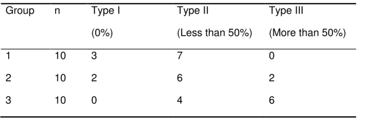
Canton, MA, USA). Load was applied at a crosshead speed of 0.5 mm per minute until failure occurred and the maximum load before failure was recorded. The remnant ceramic on the prepared tooth was determined as type I (zero%), type II
In the other 10 specimens for each group, only a lithium disilicate core was made, without veneer ceramic. The crowns were luted to their respective preparation as
described above. After storage in 37oC distilled water for 2 months, each crown was sectioned buccolingually, in the centre of the crown, with a diamond blade in an Isomet Saw (Buehler, Lake Bluff, IL, USA), obtaining two portions. One portion of
each specimen was placed under a measuring microscope (Profile Projector V-16D, Nikon, Tokyo, Japan) and the thickness of the adhesive system, low-viscosity
microfilled resin and resin cement was measured at the 10 points shown in Fig. 1. All sections were measured under 100x magnification.
The final thickness of the resin material (adhesive, low-viscosity microfilled resin, and resin cement) at the different positions in each group was compared by the Friedman and Wilcoxon signed-rank non-parametric tests. The Kruskal–Wallis and
Mann-Whiney U non-parametric tests were used to compare the final thickness between the groups in each position. Fracture loads were analyzed using one-way
RESULTS
The mean film thickness of the adhesive, low-viscosity microfilled resin, and resin
cement in each position for the different groups is shown in Table 2 and Fig. 2, 3, and 4. The thickness of the resin cement was higher in positions 5 and 6 compared with
the other positions. The thickness of adhesive was higher in positions 2 and 9 and lower in positions 1 and 10. Intermediate values were obtained in the other positions. The thickness of the low-viscosity microfilled resin was higher in positions 5 and 6,
and lower in positions 1 and 10.
The sum of resin materials in each position is presented in Table 3. According to
the Friedmann non-parametric test, statistically significant differences were noted between the positions (p<0.01). In group 1, statistically higher resin cement thickness was obtained in positions 5 and 6. In group 2 (adhesive + resin cement) and group 3
(adhesive + low-viscosity microfilled resin + resin cement), statistically lower resin thickness was obtained in positions 1 and 10. Intermediate values were found in
positions 2, 3, 7, and 8. Although there was not necessarily a statistical difference between these positions and positions 5 and 6 in groups 2 and 3, a higher thickness of resin material was observed at the occlusal surface (positions 5 and 6).
According to Kruskal–Wallis, the thickness of resin material differed statistically between the groups in all positions (p<0.01). The highest values were obtained in
group 3, differing statistically from group 2. The lowest values were obtained in group 1, differing statistically from group 2 (Table 3).
N) (p<0.01). Group 2 (1189 N) was not statistically different from groups 1 and 3 (Table 4). All fractures occurred through the veneer and the core materials. In group
1, 3 specimens presented with type I failure and 7 specimens with type II failure. In group 2, 2 specimens presented with type I failure, 6 with type II, and 2 with type III. In group 3, 4 specimens presented with type II failure and 6 specimens with type III
failure (Table 5).
According to Pearson’s correlation coefficient there was a regular positive
DISCUSSION
The first hypothesis was accepted, because the film thickness of the 3 resin
materials (adhesive, low-viscosity microfilled resin, and resin cement) was different and it was influenced by the position under the crown. In groups 2 and 3, the Clearfil
SE Bond adhesive system was applied to seal the dentin immediately after tooth preparation. The film thickness of this material presented a vast range of values at different positions of the adhesive layer, in accordance with other studies (6, 23, 24).
Higher thickness was obtained in positions 2 and 9 (concave parts of the preparation), and is consistent with the tendency of the adhesive to pool at the inner
angles of the preparation (23, 24). The minimum thickness in both groups was observed in positions 1 and 10 (borders of the preparation). The thinner film of adhesive at the borders is fortunate because a thicker film would expose more
adhesive to the degradation process in the oral cavity.
In group 2, the thickness of adhesive could be measured in practically all positions,
probably because the application of the glycerine gel allowed the polymerization of the outer layer. In some positions, e.g., positions 1, 4, and 10 (Figure 3), the film
thickness was less than 40 m, which corresponds to the inhibition layer associated
with oxygen inhibition of the radicals that initiate the polymerization reaction (25). Without the glycerine gel layer, the adhesive would not be polymerized and would be removed during cleaning of the adhesive interface, resulting in many areas of
exposed dentin. In fact, in group 2, the adhesive film could not be seen or measured at one of the borders of the preparation in 6 specimens. The film thickness was
Panavia F (23).
Comparing the adhesive film thickness of groups 2 and 3, there was a trend toward
higher thickness in group 3, probably due to the application of the Protect Liner F over the adhesive, protecting the adhesive layer during the cleaning procedure. The
cleaning of the adhesive interface was done with pumice slurry to remove all remnants of provisional cement. During this procedure, part of the adhesive layer is likely removed and the thickness of the adhesive reduced (23).
The film thickness of the Protect Liner F (group 3) presented a more uniform range of values at different positions compared with the adhesive layer. This material has a
higher percentage of filler compared with Clearfil SE Bond, and it has less tendency to pool at the inner angles of the preparation. Using a microbrush, the material was applied over the adhesive as thinly as possible visually. At the borders, a clean
microbrush was applied to remove part of the material and avoid a thicker layer, which could considerably increase the amount of material exposed to the oral cavity.
The minimum thickness was obtained in positions 1 and 10 (marginal areas of the
preparation), with a range from 19 to 67 m. Glycerine gel was not used, but the
surface of the cured low-viscosity microfilled resin was wiped with a cotton pellet
soaked in alcohol to remove the unpolymerized layer on the surface (26). Without this procedure, the film thickness would be higher. In addition, the surface of the low-viscosity microfilled resin was cleaned with a pumice slurry to remove the cement
remnants, and some micrometers of the material could have also been removed.
The thickness of the resin cement is influenced by many factors, such as the
a shoulder bevel facilitates better seating than a shoulder (27), but the preparation for a lithium disilicate ceramic requires a shoulder or a pronounced chamfer, and a
shoulder was used in the present study. The omission of a die spacer affects the proper seating of the restoration, and an excessive layer can also enlarge the luting space (28). The best crown seating was found when 20–40 µm of cement space was
provided (29). In the present study, 2 coats of die spacer were applied, which corresponds to a thickness of approximately 30 µm (30). However, the thickness of
the resin cement was higher in positions 5 and 6 (the occlusal portion of the preparation). This finding corroborates previous reports on marginal fit and cement
distribution under all-ceramic restorations that showed the highest cement film thickness is usually located at the occlusal surface underneath the crown (31).
IDS with Clearfil SE Bond and Protect Liner F (group 3) had the highest film
thickness of resin material in all positions compared with the other groups (Table 3). At the borders of the preparation (positions 1 and 10), the median thickness of the
resin materials exposed to the oral environment corresponded to 120 µm, 85 µm, and 56 µm for groups 3, 2 and 1, respectively. The marginal and internal fit of all-ceramic crowns is still very important for conventional and adhesive luted restorations (32,
33). However, the marginal fit is one of the crucial criteria for the clinical decision of whether a restoration should be inserted or not. Opinions on the clinical relevance of
the size of marginal discrepancies are controversial. Most authors agree that discrepancies in the range of 100 µm seem to be clinically acceptable with regard to longevity of the restorations (34, 35). For other authors, marginal discrepancies up to
For the luting procedure with Panavia F, ED Primer was applied on the Clearfil SE Bond adhesive (group 2) and on the low-viscosity microfilled resin (group 3). It is
likely that this material contributed to the final thickness of resin materials. However, it was not possible to visualise the layer of ED Primer. In relation to the luting procedure, ED Primer contains water, as well as the hydrophilic monomer HEMA; it
would be more appropriate to apply a hydrophobic adhesive that did not contain water. Nevertheless, according to the study of Okuda et al. (38), ED Primer did not
negatively influence the bond strength when it was applied on Protect Liner F for luting with Panavia F, and higher bond strength was obtained in the study of Udo et
al. (26). The reason for this finding is not clear, but it may be related to the polymerization of Panavia F in the presence of ED Primer (26). ED Primer contains an aromatic sulfinate salt, and it is believed that this accelerates interfacial
polymerization between the sealed dentin surface and the resin cement (38).
The second study hypothesis was rejected because a significant upward trend of
the fracture load with increasing thickness of resin material was noted. This finding is not in accordance with other studies that observed a downward trend of the fracture load with increasing thickness of resin cement (21). Kim et al. (39) observed that
increased cement thickness can have an effect on reducing flexural failure load. In that study, the load to failure of silicon bonded to glass with variation in the thickness
of the bonding epoxy layer indicated that by increasing this layer from 20 to 200 µm, there was a 50% reduction in strength. Burke and Watts (40) evaluated the resin cement thickness of 2-mm ceramic crowns that were submitted to compressive
tested was similar to the group that did not perform as well. However, these studies evaluated the influence of the thickness of the resin cement on the ceramic strength,
not taking into consideration the film thickness formed by IDS techniques. It is difficult to make direct associations between studies, because they used different specimen dimensions, types of ceramic, and resin cement systems. These are factors that can
affect ceramic fracture resistance behaviour (41).
In the present study, the load was applied to the occlusal region of the crowns,
corresponding to positions 5 and 6. It was at these positions that the highest final thickness of resin material was recorded for all groups (approximately 130 µm, 250
µm, and 360 µm for groups 1, 2, and 3, respectively). Because the resin cement thickness was similar for all groups in positions 5 and 6 (approximately 150 µm), it is thought that the thickness of the Clearfil SE Bond and Protect Liner F influenced the
values of the compressive fracture load.
During the curing process, the resin cement is transformed from a liquid to a solid
state, and this causes a volume change and shrinkage of the material. Studies have shown that shrinkage stress may cause rupture of the bonded interfaces (42, 43). The additional film thickness formed by the adhesive and the low-viscosity microfilled
resin may have favored greater absorption of stresses generated by shrinkage of the resin cement (42, 44), contributing to greater stress relief at the interfaces. According
to Rees and Jacobsen (45), high shrinkage stress, even over a small area of an interface, is sufficient to induce crack formation. This becomes an area of stress concentration and it is liable to induce further failure under occlusal loading. The