AVALIAÇÃO DOS NÍVEIS PLASMÁTICOS
E URINÁRIOS DAS CITOCINAS EM
CRIANÇAS E ADOLESCENTES
PORTADORES DE HIPERCALCIÚRIA
IDIOPÁTICA
Augusto César Soares dos Santos Junior
Universidade Federal de Minas Gerais Belo Horizonte
Augusto César Soares dos Santos Junior
AVALIAÇÃO DOS NÍVEIS PLASMÁTICOS
E URINÁRIOS DAS CITOCINAS EM
CRIANÇAS E ADOLESCENTES
PORTADORES DE HIPERCALCIÚRIA
IDIOPÁTICA
Dissertação de Mestrado apresentada ao Programa de Pós-Graduação em Ciências da Saúde - Área de Concentração Saúde da Criança e do Adolescente - da Faculdade de Medicina da Universidade Federal de Minas Gerais sob a orientação da Profª. Dra. Ana Cristina Simões e Silva.
Belo Horizonte
UNIVERSIDADE FEDERAL DE MINAS GERAIS FACULDADE DE MEDICINA
AVALIAÇÃO DOS NÍVEIS PLASMÁTICOS E URINÁRIOS DAS CITOCINAS EM CRIANÇAS E ADOLESCENTES PORTADORES
DE HIPERCALCIÚRIA IDIOPÁTICA
Augusto César Soares dos Santos Junior
Dissertação de Mestrado apresentada ao Programa de Pós-Graduação em Ciências da Saúde - Área de Concentração Saúde da Criança e do Adolescente - da Faculdade de Medicina da Universidade Federal de Minas
Gerais, como requisito parcial para obtenção do grau Mestre.
Orientadora: Profª. Ana Cristina Simões e Silva Professora Titular do Departamento de Pediatria
Faculdade de Medicina da Universidade Federal de Minas Gerais
Co-Orientadora: Profª. Maria Goretti Moreira Guimarães Penido Professora Adjunta do Departamento de Pediatria
Faculdade de Medicina da Universidade Federal de Minas Gerais
UNIVERSIDADE FEDERAL DE MINAS GERAIS
Reitor: Prof. Clélio Campolina Diniz
Vice-Reitor: Profª. Rocksane de Carvalho Norton
Pró-Reitor de Pós Graduação: Prof. Ricardo Santiago Gomez Pró-Reitor de Pesquisa: Prof. Renato de Lima dos Santos
FACULDADE DE MEDICINA
Diretor: Prof. Francisco José Penna Vice-Diretor: Prof. Tarcizo Afonso Nunes Coordenador do Centro de Pós-Graduação: Prof. Manoel Otávio da Costa Rocha
Sub-coordenadora do Centro de Pós-Graduação: Profª. Teresa Cristina de Abreu Ferrari
Chefe do Departamento de Pediatria: Profª. Maria Aparecida Martins
PROGRAMA DE PÓS-GRADUAÇÃO EM CIÊNCIAS DA SAÚDE –
ÁREA DE CONCENTRAÇÃO SAÚDE DA CRIANÇA E DO ADOLESCENTE
Coordenadora: Profª. Ana Cristina Simões e Silva Sub-coordenador: Prof. Eduardo Araújo Oliveira
COLEGIADO DO PROGRAMA DE PÓS-GRADUAÇÃO EM
CIÊNCIAS DA SAÚDE – ÁREA DE CONCENTRAÇÃO EM SAÚDE
DA CRIANÇA E DO ADOLESCENTE
Profª. Ana Cristina Simões e Silva Prof. Cassio da Cunha Ibiapina Prof. Eduardo Araújo Oliveira Prof. Francisco José Penna Profª. Ivani Novato Silva Prof. Jorge Andrade Pinto
Prof. Marcos José Burle de Aguiara
À minha esposa, Ana Carolina, pelo estímulo, apoio e presença.
À minha querida filha, Ana Luísa, que participou comigo dessa
jornada desde a sua gestação.
AGRADECIMENTOS
À Profa. Dra. Ana Cristina Simões e Silva, presente desde os meus
primeiros passos na iniciação científica, pela amizade, ensinamentos e confiança.
À Profa. Dra. Maria Goretti Moreira Guimarães Penido, pela amizade,
convivência e colaborações que fizeram despertar em mim o gosto pela nefrologia.
Aos meus familiares por compreenderem meus momentos de ausência.
Ao Prof. Mauro Martins Teixeira pela grande colaboração na execução do trabalho.
À Kátia Daniela da Silveira pelo apoio no árduo trabalho de laboratório.
Meus pais, Augusto e Maria da Conceição, pelo amor, confiança, dedicação, e por estarem sempre ao meu lado, investindo na minha formação mesmo nos momentos de dificuldade.
À minha irmã Rosana, companheira de todos os momentos.
“Viver sem filosofar é o que se chama ter os olhos fechados
sem nunca os haver tentado abrir”
NOTA EXPLICATIVA
A apresentação da presente dissertação foi organizada sob a forma de artigos científicos, de acordo com a resolução 03/2010, aprovada pelo Programa de Pós-graduação em Ciências da Saúde, Área de concentração Saúde da Criança e do Adolescente, da Faculdade de Medicina da Universidade Federal de Minas Gerais, disponível em http://www.medicina.ufmg.br/cpg/programas/ saude_crianca/arquivos/2010/Resolucao03-2010.pdf.
O primeiro artigo consiste em uma revisão da literatura, na qual são discutidos os principais aspectos, achados recentes e controvérsias sobre o papel das citocinas no remodelamento ósseo e na hipercalciúria idiopática (HI). O segundo artigo avalia os níveis plasmáticos e urinários de citocinas e quimiocinas associadas à regulação do metabolismo ósseo em crianças e adolescentes com HI em acompanhamento ambulatorial na Unidade de Nefrologia Pediátrica do Hospital das Clínicas da Universidade Federal de Minas Gerais, entre 2009 e 2010.
LISTA DE ABREVIATURAS E SIGLAS
COEP – Comitê de Ética em Pesquisa DMO – Densidade mineral óssea DP – Desvio padrão
ELISA – Enzyme-linked immunosorbent assay
GM-CSF - Granulocyte-macrophage colony-stimulating factor HI – Hipercalciúria idiopática
IL-1β – Interleucina 1 beta IL-6 - Interleucina 6
IL-8 - Interleucina 8
MCP-1 - Proteína de quimiotaxia de monócitos-1 M-CSF - Macrophage colony-stimulating factor OPG - Osteoprotegerina
RANK - Receptor activator of NF-κβ
RANKL - Receptor activator of NF-κβ ligand
TGF-β - Fator de crescimento e transformação β
SUMÁRIO
1. INTRODUÇÃO 15
2. REVISÃO DA LITERATURA 20
2.1. ABSTRACT 21
2.2. INTRODUCTION 22
2.3. BRIEFOVERVIEWOFCYTOKINEFUNCTIONINBONEMETABOLISM 23
2.4. BONEDISEASEANDCYTOKINESINIDIOPATHICHYPERCALCIURIA 28
2.5. CONCLUDINGREMARKS 33
2.6. ABBREVIATIONS 34
3. OBJETIVOS 47
4. PACIENTES E MÉTODOS 49
4.1. CRITÉRIOSDEINCLUSÃO 49
4.2. CRITÉRIOSDEEXCLUSÃO 49
4.3. ASPECTOSÉTICOS 50
4.4. PROTOCOLODOESTUDO 51
4.5. COLETAEPROCESSAMENTODEAMOSTRAS 52
4.6. ENSAIOSIMUNOENZIMÁTICOS 53
4.7. ANÁLISEESTATÍSTICA 56
5. RESULTADOS E DISCUSSÃO 58
5.1. ABSTRACT 59
5.2. INTRODUCTION 60
5.3. PATIENTSANDMETHODS 61
5.3.1. PATIENTS 61
5.3.2. STUDYPROTOCOL 62
5.3.3. BLOODSAMPLING 63
5.3.4. URINESAMPLING 64
5.3.5. CYTOKINESMEASUREMENT 64
5.3.6. STATISTICALANALYSIS 65
5.3.7. ETHICALASPECTS 65
5.4. RESULTS 66
6. COMENTÁRIOS FINAIS 89
7. ANEXO 1- PARECER DO COEP 96 8. ANEXO 2 - TERMO DE CONSENTIMENTO 97
9. ANEXO 3 - COMPROVANTE DE APROVAÇÃO PARA APRESENTAÇÃO EM CONGRESSO INTERNACIONAL
1. INTRODUÇÃO
A hipercalciúria idiopática (HI), descrita por Albright et al (1), é a principal alteração metabólica responsável pela formação de cálculos urinários (2-4). Acomete todas as faixas etárias (5) e caracteriza-se pela hiperexcreção urinária de cálcio na ausência de estados hipercalcêmicos ou de qualquer outra doença primária (6-8). A hipercalciúria é definida como excreção de cálcio igual ou maior a 4mg/kg/24h em crianças e adolescentes. Em adultos, entende-se por hipercalciúria, excreção urinária de cálcio igual ou maior que 300 mg/24h e 250mg/24h, para homens e mulheres respectivamente. (9-10)
Apesar de muito estudada, a fisiopatologia da HI permanece obscura. Atualmente, se sabe que a HI é uma entidade complexa associada a modificações em fatores regulatórios envolvidos na absorção intestinal, no remodelamento ósseo e na excreção urinária de cálcio. (11)
quimiocinas constituem um grupo de citocinas de baixo peso molecular cuja principal ação é o recrutamento e ativação de leucócitos em vários modelos de inflamação (15).
Diversos autores relataram redução da densidade mineral óssea em adultos e crianças portadores de HI, sugerindo a necessidade de uma investigação mais aprofundada sobre os mecanismos regulatórios do remodelamento ósseo na HI (16-26). Estudos clínicos e experimentais demonstraram a participação das citocinas e quimiocinas no processo que regula o remodelamento ósseo, controlando tanto a formação quanto a reabsorção óssea (25, 27-28). No entanto, a participação dessas substâncias na fisiopatologia da HI em crianças e adolescentes ainda não foi esclarecida.
REFERÊNCIAS BIBLIOGRÁFICAS
1. Albright F, Henneman P, Benedict PH, Forbes AP. Idiopathic hypercalciuria: a preliminary report. Proc R Soc Med. 1953 Dec;46(12):1077-81.
2. Stapleton FB. Idiopathic hypercalciuria: association with isolated hematuria and risk for urolithiasis in children. The Southwest Pediatric Nephrology Study Group. Kidney Int. 1990 Feb;37(2):807-11.
3. Stapleton FB, Miller LA. Renal function in children with idiopathic hypercalciuria. Pediatr Nephrol. 1988 Apr;2(2):229-35. 4. Coe FL, Parks JH, Asplin JR. The pathogenesis and treatment of kidney stones. N Engl J Med. 1992 Oct 15;327(16):1141-52.
5. Vezzoli G, Soldati L, Gambaro G. Hypercalciuria revisited: one or many conditions? Pediatr Nephrol. 2008 Apr;23(4):503-6.
6. Ammenti A, Neri E, Agistri R, Beseghi U, Bacchini E. Idiopathic hypercalciuria in infants with renal stones. Pediatr Nephrol. 2006 Dec;21(12):1901-3.
7. Moore ES, Coe FL, McMann BJ, Favus MJ. Idiopathic hypercalciuria in children: prevalence and metabolic characteristics. J Pediatr. 1978 Jun;92(6):906-10.
8. Spivacow FR, Negri AL, del Valle EE, Calvino I, Zanchetta JR. Clinical and metabolic risk factor evaluation in young adults with kidney stones. Int Urol Nephrol. 2010 Jun;42(2):471-5.
9. Coe FL, Evan A, Worcester E. Kidney stone disease. J Clin Invest. 2005 Oct;115(10):2598-608.
10. Srivastava T, Schwaderer A. Diagnosis and management of hypercalciuria in children. Curr Opin Pediatr. 2009 Apr;21(2):214-9. 11. Srivastava T, Alon US. Pathophysiology of hypercalciuria in children. Pediatr Nephrol. 2007 Oct;22(10):1659-73.
13. Pacifici R. The immune system and bone. Arch Biochem Biophys. 2010 Nov 1;503(1):41-53.
14. Borish LC, Steinke JW. 2. Cytokines and chemokines. J Allergy Clin Immunol. 2003 Feb;111(2 Suppl):S460-75.
15. Segerer S, Alpers CE. Chemokines and chemokine receptors in renal pathology. Curr Opin Nephrol Hypertens. 2003 May;12(3):243-9.
16. Penido MG, Lima EM, Marino VS, Tupinamba AL, Franca A, Souto MF. Bone alterations in children with idiopathic hypercalciuria at the time of diagnosis. Pediatr Nephrol. 2003 Feb;18(2):133-9. 17. Penido MG, Lima EM, Souto MF, Marino VS, Tupinamba AL, Franca A. Hypocitraturia: a risk factor for reduced bone mineral density in idiopathic hypercalciuria? Pediatr Nephrol. 2006 Jan;21(1):74-8.
18. Vezzoli G, Rubinacci A, Bianchin C, Arcidiacono T, Giambona S, Mignogna G, et al. Intestinal calcium absorption is associated with bone mass in stone-forming women with idiopathic hypercalciuria. Am J Kidney Dis. 2003 Dec;42(6):1177-83.
19. Polito C, Iolascon G, Nappi B, Andreoli S, La Manna A. Growth and bone mineral density in long-lasting idiopathic hypercalciuria. Pediatr Nephrol. 2003 Jun;18(6):545-7.
20. Garcia-Nieto V, Ferrandez C, Monge M, de Sequera M, Rodrigo MD. Bone mineral density in pediatric patients with idiopathic hypercalciuria. Pediatr Nephrol. 1997 Oct;11(5):578-83.
21. Tasca A, Cacciola A, Ferrarese P, Ioverno E, Visona E, Bernardi C, et al. Bone alterations in patients with idiopathic hypercalciuria and calcium nephrolithiasis. Urology. 2002 Jun;59(6):865-9; discussion 9.
22. Freundlich M, Alonzo E, Bellorin-Font E, Weisinger JR. Reduced bone mass in children with idiopathic hypercalciuria and in their asymptomatic mothers. Nephrol Dial Transplant. 2002 Aug;17(8):1396-401.
patients with idiopathic hypercalciuria. Nephrology (Carlton). 2005 Apr;10(2):99-102.
24. Giannini S, Nobile M, Sartori L, Calo L, Tasca A, Dalle Carbonare L, et al. Bone density and skeletal metabolism are altered in idiopathic hypercalciuria. Clin Nephrol. 1998 Aug;50(2):94-100. 25. Weisinger JR, Alonzo E, Bellorin-Font E, Blasini AM, Rodriguez MA, Paz-Martinez V, et al. Possible role of cytokines on the bone mineral loss in idiopathic hypercalciuria. Kidney Int. 1996 Jan;49(1):244-50.
26. Jaeger P, Lippuner K, Casez JP, Hess B, Ackermann D, Hug C. Low bone mass in idiopathic renal stone formers: magnitude and significance. J Bone Miner Res. 1994 Oct;9(10):1525-32.
27. Ghazali A, Fuentes V, Desaint C, Bataille P, Westeel A, Brazier M, et al. Low bone mineral density and peripheral blood monocyte activation profile in calcium stone formers with idiopathic hypercalciuria. J Clin Endocrinol Metab. 1997 Jan;82(1):32-8.
2. REVISÃO
DA
LITERATURA
BONE
DISEASE
AND
CYTOKINES
IN
IDIOPATHIC HYPERCALCIURIA: A REVIEW
Augusto C. S. Santos Jr
1, Ana Cristina Simões e Silva
11Pediatric Nephrology Unit, Department of Pediatrics, Faculty of
Medicine, Federal University of Minas Gerais (UFMG), Belo Horizonte, MG, Brazil
Key words: biomarkers, cytokines, MCP-1, idiopathic hypercalciuria, mineral bone disease
Conflicts of interest: none
Correspondence: Ana Cristina Simões e Silva, MD, PhD Avenida Bernardo Monteiro 1300 / 1104 Belo Horizonte - Minas Gerais
Postal Code: 30150-281
2.1. ABSTRACT
2.2. INTRODUCTION
Idiopathic hypercalciuria (IH) was first described by Albright et al. and is characterized by normal serum calcium levels and excessive urine calcium loss. It is the most common metabolic abnormality in patients with nephrolithiasis accounting for 30-50% of calcium-oxalate stone formers. (1-4)
IH is defined by a daily urinary calcium excretion equal or superior to 4mg/kg or >300 mg Ca/d (7.5 mmol) in men and >250mg Ca/d (6.25 mmol) in women. (5) The diagnosis of IH depends on the exclusion of other causes of hypercalciuria such as sarcoidosis, malignancy, Paget's disease, high calcium or vitamin D intake, renal tubular acidosis and thyrotoxicosis. The most frequent clinical findings in IH are hematuria, abdominal and flank pain, urinary tract infections, nephrolithiasis, dysuria, urinary frequency, nocturnal enuresis and osteopenia. (6, 7)
The purpose of this review is to summarize the recent published evidence on the possible mechanisms by which cytokines may be associated to the pathogenesis of IH.
2.3. BRIEF
OVERVIEW
OF
CYTOKINE
FUNCTION
IN
BONE
METABOLISM
Cytokines are redundant secreted proteins with growth, differentiation, and activation functions that regulate and determine the nature of immune responses and control the immune cell trafficking and the cellular arrangement of immune organs. These mediators are involved in virtually every facet of immunity and inflammation, including innate immunity, antigen presentation, bone marrow differentiation, cellular recruitment and activation, and adhesion molecule expression. A cascade of responses is triggered in response to cytokines, and several cytokines acting together are required to express their optimal function. Numerous cytokines have both inflammatory and anti-inflammatory properties (15). Chemokines constitute a large family of low molecular-weight cytokines whose main action is the recruitment and activation of leukocyte subsets in various models of inflammation—the word
―chemokine‖ is a contraction of the terms ―chemoattractant‖ and
Bone remodeling is a continuous and dynamic process of skeletal destruction and renewal. It consists of two distinct stages: resorption and formation. A complex regulatory mechanism with the participation of several cytokines precisely defines the role of osteoclasts or osteoblasts in the chain of events leading to bone resorption or formation (17, 18).
The receptor activator of NF-κβ ligand (RANKL) is a member of the tumor necrosis factor (TNF) superfamily critically important in the differentiation of osteoclast precursor cells. It exists in membrane-bound and soluble forms. The interaction between RANKL and its receptor, the receptor activator of NF-κβ (RANK), induce bone-resorbing activity in mature osteoclasts and the formation of osteoclasts from precursor cells. (17-19) In vitro studies
have demonstrated that low levels or absence of either or both RANKL and RANK cause osteopetrosis and reduce mature osteoclast concentrations. On the other hand, an excess of either or both RANKL and RANK results in osteoporosis and rapid bone loss as a consequence of increased osteoclastic activity.(20)
M-CSF corrected the defect in osteoclast formation and bone resorption. (22) The granulocyte-macrophage colony-stimulating factor (GM-CSF) is also implicated in bone loss, but this factor can stimulate human osteoblastic cells as an autocrine proliferative factor. (23)
Osteoprotegerin (OPG), a soluble secreted receptor of the TNF superfamily, acts as a decoy receptor for RANKL by preventing RANK activation (24). Therefore, OPG is a potent inhibitor of osteoclast formation. (19, 25) Overexpression of OPG in transgenic mice results in severe osteopetrosis, characterized by increased bone turnover and the inhibition of osteoclastogenesis. (24) On the other hand, OPG-deficient mice develop osteoporosis due to unopposed RANKL activity. (26)
Interleukin 1 (IL-1) refers to two different polypeptides: IL-1α
Interleukin 6 (IL-6) is also known to induce osteoclast formation and bone resorption. IL-6 directly induces increased expression of RANKL and OPG in osteoblasts and regulates osteoclast progenitor cell differentiation into mature osteoclasts in states of increased bone turnover. (30-32)
Interleukin 8 (IL-8) is a chemokine produced by osteoclasts and serve as an important mediator in bone remodeling. The mechanism of action of this chemokine is independent of the RANKL pathway. It involves the expression and activation of the specific IL-8 receptor (CXCR1) on the surface of osteoclasts and their precursors. (33, 34)
The monocyte-derived TNF, as IL-1, also refers to two polypeptides with potent bone resorption induction capacity: TNF-α
and TNF-β. TNF directly stimulates bone marrow osteoclastogenesis by increasing the expression of c-fms, the receptor for M-CSF. As a consequence of M-CSF stimulation, the differentiation and proliferation of progenitor osteoclasts cells occur.(35) TNF also acts directly on the osteoclast precursor by enhancing RANK signaling mechanisms, even in the absence of elevated levels of RANKL. (36)
inhibition of osteoclast differentiation. TGF-β strongly decreases messenger RNA (mRNA) expression for RANKL in cultured osteoblasts. Low TGF-β levels stimulate osteoclast differentiation by changing the RANKL/OPG ratio, while high TGF-β levels suppress osteoclast differentiation by alternative pathways independent of the RANKL/OPG ratio or M-CSF expression regulation.(37, 38)
Monocyte chemoattractant protein-1 (MCP-1) is a chemokine mainly involved with the recruitment of monocytes to areas of both bone formation and resorption during bone remodeling. Monocyte products are potential regulators of bone cell activity, since growth factors produced by these cells may stimulate bone formation (39, 40). In vitro and in vivo studies indicated that MCP-1 induces the
recruitment of monocytes to bone, which, in turn, is associated with an increase in osteoblast number (39, 40). This is likely to occur via indirect mechanisms, because MCP-1 did not directly enhance DNA synthesis in osteoblastic cells in vitro (40). Thus, activated
mononuclear phagocytes may play an important role in bone metabolism by stimulating proliferation of osteoblastic cells. MCP-1 is typically not expressed in normal bone or by normal osteoblasts in vitro. Upon stimulation by inflammatory mediators, MCP-1 is
inflammation and during developmentally regulated bone remodeling (40). Indeed, monocytes seem to have different functional roles in areas of bone formation and resorption. The recruitment of monocytes in areas of bone formation was associated with a decrease in the number of osteoclasts, while in bone-resorbing areas, recruitment of cells of the monocytic lineage is associated with formation of osteoclasts (44). The receptor activator of NF-κβ ligand (RANKL) seems to be a key variable in this process, once MCP-1 stimulates the formation of osteoclasts in the presence of RANKL. MCP-1 is also induced by RANKL during osteoclast differentiation (41). Receptors for MCP-1 (CCR2 and CCR4) are induced by RANKL, providing evidence for an autocrine loop for MCP-1 in human osteoclasts (45).
2.4. BONE
DISEASE
AND
CYTOKINES
IN
IDIOPATHIC
HYPERCALCIURIA
Insert Figure 1
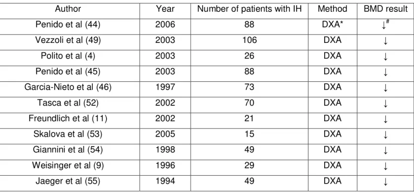
To date, several studies have been performed to evaluate bone mass density (BMD) in patients with IH, as summarized in Table 1. (4, 13, 14, 46-49) These studies reported significant bone loss in patients with IH regardless of age. Despite the increased risk for reduced BMD, children with long-lasting IH tend to have normal growth curves. (4) This bone loss in patients with IH mainly involves areas of trabecular bone in the axial skeleton, such as vertebral bodies. (50) Malluche et al reported an increased osteoid volume and surface in line with a reduced osteoblastic activity. (51) Steiniche et al. showed decreased bone formation, increased mineralization time and resorption surfaces in a large series of patients with IH. (52)
Insert table 1
Insert Figure 2
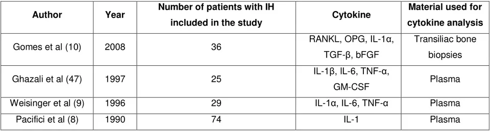
There are multiple mechanisms underlying the regulation of bone remodeling, which may be involved in the pathogenesis of IH. In fact, decreased bone formation or excessive bone resorption or both are possible mechanisms to explain the decreased BMD in patients with IH. Experimental studies demonstrated the importance of RANKL, OPG, TGF-β, IL-1α, IL-1β, IL-6, TNF-α and GM-CSF in the regulation of resorption and calcium release from bones in IH, as summarized in Table 2. (8-10, 48)
Insert table 2
Gomes et al. performed immunohistochemical analysis in undecalcified bone samples from transiliac bone biopsies of patients with IH (10). The authors reported a higher expression of RANKL in bone tissue of patients with IH, suggesting that increased bone resorption in IH is mediated by RANKL. In this study, OPG was also increased possibly as an attempt to counteract the bone resorption triggered by RANKL. TGF-β was reduced, thus justifying a delayed mineralization process observed in these patients. The levels of IL-1α
Pacifici et al described an association, but not a cause-effect relationship, between IL-1 activity and bone resorption (8). An increased production of IL-1 by cultured peripheral blood monocytes in line with a decreased vertebral BMD was described in patients with fasting hypercalciuria. The authors also found an association between IL-1 and urinary calcium excretion. (8)
Likewise, Weisinger et al detected an increased expression of IL-1α, IL-6 and TNF-α mRNA in unstimulated blood monocytes obtained from hypercalciuric patients when compared with normal subjects (9). It was found a correlation between basal production of IL-1α, but not IL-1β, and decreased trabecular bone density. The authors suggested a link between the high mRNA expression for IL-1α from unstimulated peripheral blood mononuclear cells with spinal bone loss in IH. The results indicated that cytokines could play an important role in bone resorption in IH either by direct activation of osteoclasts or by cell recruitment. (9)
The authors hypothesized that monocyte activation is directly involved in the bone loss of calcium stone formers with IH (48).
and decreased bone mineral density could be the clinical expression of this phenomenon in patients with IH. Figure 3 displays the hypothetical mechanism by which cytokines release induce osteoclasts differentiation.
Insert Figure 3
2.5. CONCLUDING
REMARKS
2.6. ABBREVIATIONS
BMD - Bone mass density
GM-CSF - Granulocyte-macrophage colony-stimulating factor IH - Idiopathic hypercalciuria
IL-1 - Interleukin 1 IL-6 - Interleukin 6 IL-8 - Interleukin 8
MCP-1 - Monocyte chemoattractant protein-1 M-CSF - Macrophage colony-stimulating factor OPG - Osteoprotegerin
RANK - Receptor activator of NF-κβ
RANKL - Receptor activator of NF-κβ ligand TGF-β - Transforming growth factor β
BIBLIOGRAPHY
1. Stapleton FB. Idiopathic hypercalciuria: association with isolated hematuria and risk for urolithiasis in children. The Southwest Pediatric Nephrology Study Group. Kidney Int. 1990 Feb;37(2):807-11.
2. Vachvanichsanong P, Malagon M, Moore ES. Recurrent abdominal and flank pain in children with idiopathic hypercalciuria. Acta Paediatr. 2001 Jun;90(6):643-8.
3. Stojanovic VD, Milosevic BO, Djapic MB, Bubalo JD. Idiopathic hypercalciuria associated with urinary tract infection in children. Pediatr Nephrol. 2007 Sep;22(9):1291-5.
4. Polito C, Iolascon G, Nappi B, Andreoli S, La Manna A. Growth and bone mineral density in long-lasting idiopathic hypercalciuria. Pediatr Nephrol. 2003 Jun;18(6):545-7.
5. Coe FL, Evan A, Worcester E. Kidney stone disease. J Clin Invest. 2005 Oct;115(10):2598-608.
6. Ammenti A, Neri E, Agistri R, Beseghi U, Bacchini E. Idiopathic hypercalciuria in infants with renal stones. Pediatr Nephrol. 2006 Dec;21(12):1901-3.
7. Moore ES, Coe FL, McMann BJ, Favus MJ. Idiopathic hypercalciuria in children: prevalence and metabolic characteristics. J Pediatr. 1978 Jun;92(6):906-10.
8. Pacifici R, Rothstein M, Rifas L, Lau KH, Baylink DJ, Avioli LV, et al. Increased monocyte interleukin-1 activity and decreased vertebral bone density in patients with fasting idiopathic hypercalciuria. J Clin Endocrinol Metab. 1990 Jul;71(1):138-45.
9. Weisinger JR, Alonzo E, Bellorin-Font E, Blasini AM, Rodriguez MA, Paz-Martinez V, et al. Possible role of cytokines on the bone mineral loss in idiopathic hypercalciuria. Kidney Int. 1996 Jan;49(1):244-50.
11. Freundlich M, Alonzo E, Bellorin-Font E, Weisinger JR. Reduced bone mass in children with idiopathic hypercalciuria and in their asymptomatic mothers. Nephrol Dial Transplant. 2002 Aug;17(8):1396-401.
12. Misael da Silva AM, dos Reis LM, Pereira RC, Futata E, Branco-Martins CT, Noronha IL, et al. Bone involvement in idiopathic hypercalciuria. Clin Nephrol. 2002 Mar;57(3):183-91.
13. Penido MG, Lima EM, Marino VS, Tupinamba AL, Franca A, Souto MF. Bone alterations in children with idiopathic hypercalciuria at the time of diagnosis. Pediatr Nephrol. 2003 Feb;18(2):133-9. 14. Penido MG, Lima EM, Souto MF, Marino VS, Tupinamba AL, Franca A. Hypocitraturia: a risk factor for reduced bone mineral density in idiopathic hypercalciuria? Pediatr Nephrol. 2006 Jan;21(1):74-8.
15. Borish LC, Steinke JW. 2. Cytokines and chemokines. J Allergy Clin Immunol. 2003 Feb;111(2 Suppl):S460-75.
16. Segerer S, Alpers CE. Chemokines and chemokine receptors in renal pathology. Curr Opin Nephrol Hypertens. 2003 May;12(3):243-9.
17. Lorenzo J, Horowitz M, Choi Y. Osteoimmunology: interactions of the bone and immune system. Endocr Rev. 2008 Jun;29(4):403-40.
18. Pacifici R. The immune system and bone. Arch Biochem Biophys. 2010 Nov 1;503(1):41-53.
19. Perez-Sayans M, Somoza-Martin JM, Barros-Angueira F, Rey JM, Garcia-Garcia A. RANK/RANKL/OPG role in distraction osteogenesis. Oral Surg Oral Med Oral Pathol Oral Radiol Endod. 2010 May;109(5):679-86.
20. Grimaud E, Soubigou L, Couillaud S, Coipeau P, Moreau A, Passuti N, et al. Receptor activator of nuclear factor kappaB ligand (RANKL)/osteoprotegerin (OPG) ratio is increased in severe osteolysis. Am J Pathol. 2003 Nov;163(5):2021-31.
factor kappaB (RANK) receptors. J Exp Med. 1999 Dec 20;190(12):1741-54.
22. Felix R, Cecchini MG, Fleisch H. Macrophage colony stimulating factor restores in vivo bone resorption in the op/op osteopetrotic mouse. Endocrinology. 1990 Nov;127(5):2592-4.
23. Takahashi N, Udagawa N, Akatsu T, Tanaka H, Shionome M, Suda T. Role of colony-stimulating factors in osteoclast development. J Bone Miner Res. 1991 Sep;6(9):977-85.
24. Simonet WS, Lacey DL, Dunstan CR, Kelley M, Chang MS, Luthy R, et al. Osteoprotegerin: a novel secreted protein involved in the regulation of bone density. Cell. 1997 Apr 18;89(2):309-19.
25. Hofbauer LC, Neubauer A, Heufelder AE. Receptor activator of nuclear factor-kappaB ligand and osteoprotegerin: potential implications for the pathogenesis and treatment of malignant bone diseases. Cancer. 2001 Aug 1;92(3):460-70.
26. Mizuno A, Amizuka N, Irie K, Murakami A, Fujise N, Kanno T, et al. Severe osteoporosis in mice lacking osteoclastogenesis inhibitory factor/osteoprotegerin. Biochem Biophys Res Commun. 1998 Jun 29;247(3):610-5.
27. Dinarello CA. Interleukin-1 and interleukin-1 antagonism. Blood. 1991 Apr 15;77(8):1627-52.
28. Jimi E, Nakamura I, Duong LT, Ikebe T, Takahashi N, Rodan GA, et al. Interleukin 1 induces multinucleation and bone-resorbing activity of osteoclasts in the absence of osteoblasts/stromal cells. Exp Cell Res. 1999 Feb 25;247(1):84-93.
29. Hofbauer LC, Lacey DL, Dunstan CR, Spelsberg TC, Riggs BL, Khosla S. Interleukin-1beta and tumor necrosis factor-alpha, but not interleukin-6, stimulate osteoprotegerin ligand gene expression in human osteoblastic cells. Bone. 1999 Sep;25(3):255-9.
31. Franchimont N, Wertz S, Malaise M. Interleukin-6: An osteotropic factor influencing bone formation? Bone. 2005 Nov;37(5):601-6.
32. Ishimi Y, Miyaura C, Jin CH, Akatsu T, Abe E, Nakamura Y, et al. IL-6 is produced by osteoblasts and induces bone resorption. J Immunol. 1990 Nov 15;145(10):3297-303.
33. Rothe L, Collin-Osdoby P, Chen Y, Sunyer T, Chaudhary L, Tsay A, et al. Human osteoclasts and osteoclast-like cells synthesize and release high basal and inflammatory stimulated levels of the potent chemokine interleukin-8. Endocrinology. 1998 Oct;139(10):4353-63.
34. Bendre MS, Margulies AG, Walser B, Akel NS, Bhattacharrya S, Skinner RA, et al. Tumor-derived interleukin-8 stimulates osteolysis independent of the receptor activator of nuclear factor-kappaB ligand pathway. Cancer Res. 2005 Dec 1;65(23):11001-9. 35. Yao Z, Li P, Zhang Q, Schwarz EM, Keng P, Arbini A, et al. Tumor necrosis factor-alpha increases circulating osteoclast precursor numbers by promoting their proliferation and differentiation in the bone marrow through up-regulation of c-Fms expression. J Biol Chem. 2006 Apr 28;281(17):11846-55.
36. Lam J, Takeshita S, Barker JE, Kanagawa O, Ross FP, Teitelbaum SL. TNF-alpha induces osteoclastogenesis by direct stimulation of macrophages exposed to permissive levels of RANK ligand. J Clin Invest. 2000 Dec;106(12):1481-8.
37. Quinn JM, Itoh K, Udagawa N, Hausler K, Yasuda H, Shima N, et al. Transforming growth factor beta affects osteoclast differentiation via direct and indirect actions. J Bone Miner Res. 2001 Oct;16(10):1787-94.
38. Karst M, Gorny G, Galvin RJ, Oursler MJ. Roles of stromal cell RANKL, OPG, and M-CSF expression in biphasic TGF-beta regulation of osteoclast differentiation. J Cell Physiol. 2004 Jul;200(1):99-106.
40. Graves DT, Jiang Y, Valente AJ. Regulated expression of MCP-1 by osteoblastic cells in vitro and in vivo. Histol Histopathol. 1999 Oct;14(4):1347-54.
41. Valente AJ, Xie JF, Abramova MA, Wenzel UO, Abboud HE, Graves DT. A complex element regulates IFN-gamma-stimulated monocyte chemoattractant protein-1 gene transcription. J Immunol. 1998 Oct 1;161(7):3719-28.
42. Rahimi P, Wang CY, Stashenko P, Lee SK, Lorenzo JA, Graves DT. Monocyte chemoattractant protein-1 expression and monocyte recruitment in osseous inflammation in the mouse. Endocrinology. 1995 Jun;136(6):2752-9.
43. Williams SR, Jiang Y, Cochran D, Dorsam G, Graves DT. Regulated expression of monocyte chemoattractant protein-1 in normal human osteoblastic cells. Am J Physiol. 1992 Jul;263(1 Pt 1):C194-9.
44. Volejnikova S, Laskari M, Marks SC, Jr., Graves DT. Monocyte recruitment and expression of monocyte chemoattractant protein-1 are developmentally regulated in remodeling bone in the mouse. Am J Pathol. 1997 May;150(5):1711-21.
45. Kim MS, Day CJ, Morrison NA. MCP-1 is induced by receptor activator of nuclear factor-{kappa}B ligand, promotes human osteoclast fusion, and rescues granulocyte macrophage colony-stimulating factor suppression of osteoclast formation. J Biol Chem. 2005 Apr 22;280(16):16163-9.
46. Schwaderer AL, Cronin R, Mahan JD, Bates CM. Low bone density in children with hypercalciuria and/or nephrolithiasis. Pediatr Nephrol. 2008 Dec;23(12):2209-14.
47. Garcia-Nieto V, Ferrandez C, Monge M, de Sequera M, Rodrigo MD. Bone mineral density in pediatric patients with idiopathic hypercalciuria. Pediatr Nephrol. 1997 Oct;11(5):578-83.
49. Asplin JR, Bauer KA, Kinder J, Muller G, Coe BJ, Parks JH, et al. Bone mineral density and urine calcium excretion among subjects with and without nephrolithiasis. Kidney Int. 2003 Feb;63(2):662-9. 50. Vezzoli G, Rubinacci A, Bianchin C, Arcidiacono T, Giambona S, Mignogna G, et al. Intestinal calcium absorption is associated with bone mass in stone-forming women with idiopathic hypercalciuria. Am J Kidney Dis. 2003 Dec;42(6):1177-83.
51. Malluche HH, Tschoepe W, Ritz E, Meyer-Sabellek W, Massry SG. Abnormal bone histology in idiopathic hypercalciuria. J Clin Endocrinol Metab. 1980 Apr;50(4):654-8.
52. Steiniche T, Mosekilde L, Christensen MS, Melsen F. A histomorphometric determination of iliac bone remodeling in patients with recurrent renal stone formation and idiopathic hypercalciuria. APMIS. 1989 Apr;97(4):309-16.
53. Tasca A, Cacciola A, Ferrarese P, Ioverno E, Visona E, Bernardi C, et al. Bone alterations in patients with idiopathic hypercalciuria and calcium nephrolithiasis. Urology. 2002 Jun;59(6):865-9; discussion 9.
54. Skalova S, Palicka V, Kutilek S. Bone mineral density and urinary N-acetyl-beta-D-glucosaminidase activity in paediatric patients with idiopathic hypercalciuria. Nephrology (Carlton). 2005 Apr;10(2):99-102.
TABLE 1 - STUDIES ON BONE MINERAL DENSITY (BMD) IN PATIENTS WITH IDIOPATHIC
HYPERCALCIURIA
Author Year Number of patients with IH Method BMD result Penido et al (44) 2006 88 DXA* ↓#
Vezzoli et al (49) 2003 106 DXA ↓
Polito et al (4) 2003 26 DXA ↓
Penido et al (45) 2003 88 DXA ↓
Garcia-Nieto et al (46) 1997 73 DXA ↓
Tasca et al (52) 2002 70 DXA ↓
Freundlich et al (11) 2002 21 DXA ↓
Skalova et al (53) 2005 15 DXA ↓
Giannini et al (54) 1998 49 DXA ↓
Weisinger et al (9) 1996 29 DXA ↓
Jaeger et al (55) 1994 49 DXA ↓ * DXA = dual energy X-ray absorptiometry
TABLE 2 - STUDIES ON CYTOKINES IN PATIENTS WITH IDIOPATHIC HYPERCALCIURIA
Author Year Number of patients with IH
included in the study Cytokine
Material used for cytokine analysis
Gomes et al (10) 2008 36 RANKL, OPG, IL-1α, TGF-β, bFGF
Transiliac bone biopsies Ghazali et al (47) 1997 25 IL-1β, IL-6, TNF-α,
FIGURE LEGENDS
FIGURE 1 –Hypothetical mechanism by which increased bone resorption may contribute to hypercalciuria.
FIGURE 2 –Hypothetical mechanism by which increased bone resorbing cytokines trigger calcium release from bones.
FIGURE 1
FIGURE 2
(a) Resting bone;
(b) Differentiation and activation;
FIGURE 3
3. OBJETIVOS
O objetivo principal desse trabalho foi avaliar, em crianças e adolescentes com HI, os níveis plasmáticos e urinários de citocinas e quimiocinas associadas à regulação do metabolismo ósseo.
Objetivos específicos:
a. Verificar se há diferença entre as concentrações das citocinas após dividir os pacientes em grupos estratificados de acordo com a excreção urinária de cálcio (< 4 mg/Kg/dia versus 4 mg/Kg/dia);
b. Verificar se há diferença entre as concentrações das citocinas após dividir os pacientes em grupos estratificados de acordo com a densidade mineral óssea (Z-score ≤ -1 desvio padrão versus Z-score > - 1 desvio padrão);
c. Verificar se há diferença entre as concentrações das citocinas após dividir os pacientes em grupos estratificados de acordo com a idade dos pacientes (≤12
anos versus > 12 anos);
4. PACIENTES
E
MÉTODOS
4.1. CRITÉRIOS
DE
INCLUSÃO
Foram inicialmente incluídas 81 crianças e adolescentes com diagnóstico estabelecido de HI e que estavam em acompanhamento regular na Unidade de Nefrologia Pediátrica do Hospital das Clínicas da UFMG, durante o período de coleta (2009 a 2010). Desses 81 pacientes, 11 se recusaram em participar do estudo, totalizando, portanto, 70 pacientes com IH avaliados.
O diagnóstico de HI foi realizado de acordo com critérios diagnósticos estabelecidos pela literatura internacional (1-2), que consideram portadores de HI os pacientes que apresentam aumento persistente da excreção urinária de cálcio na ausência de estados hipercalcêmicos ou de qualquer outra doença primária. De acordo com os valores de referência estabelecidos para excreção urinária de cálcio em crianças e adolescentes (3-4), a hipercalciúria foi definida como excreção de cálcio igual ou maior a 4 mg/Kg/dia.
4.2. CRITÉRIOS
DE
EXCLUSÃO
como: hiperparatireoidismo, hipertireoidismo, sarcoidose, doenças oncológicas, acidose tubular renal, doença inflamatória aguda, estados febris, tratamento com estrogênio, progesterona, corticosteróides, anticonvulsivantes, bifosfonados, calcitonina ou vitamina D. (5)
4.3. ASPECTOS
ÉTICOS
4.4. PROTOCOLO
DO
ESTUDO
Trata-se de um estudo transversal, com amostra de conveniência, reunindo um total de 70 pacientes.
Os pacientes foram avaliados quanto à calciúria em 24h, Z-score e conteúdo mineral ósseo da densitometria óssea.
Os pacientes foram divididos em 2 grupos de acordo com os níveis de calciúria na data da coleta das amostras: calciúria descompensada (n=23) e calciúria compensada (n=47). A calciúria descompensada foi definida como excreção de cálcio igual ou superior a 4mg/kg/24h.
Os pacientes também foram estratificados levando-se em consideração a idade ( 12 anos, n=18; > 12 anos, n=52) e o Z-score da densidade mineral óssea (Z-Z-score >-1 desvio padrão, n=28; Z-score ≤-1 desvio padrão, n=18), quando disponível.
citrato, magnésio, ácido úrico, cistina, oxalato e creatinina. Os exames foram realizados para afastar causas secundárias de hipercalciúria (4, 6).
Após confirmação do diagnóstico de HI e assinatura do termo de consentimento, os pacientes foram submetidos, em única ocasião, a coletas simultâneas de amostras de sangue e urina para determinação de citocinas e quimiocinas conforme detalhado a seguir.
4.5. COLETA
E
PROCESSAMENTO
DE
AMOSTRAS
As amostras de urina foram coletadas no mesmo momento em que foi realizada a coleta das amostras de sangue. Após homogeneização, 10 ml de urina foram recondicionados em tubo estéril. As amostras foram, então, centrifugadas a 1300 g por 20 minutos. Alíquotas de 1,5 ml foram também conservadas em freezer a -80ºC.
4.6. ENSAIOS
IMUNOENZIMÁTICOS
Os níveis plasmáticos e urinários de IL-1β, IL-6, IL-8, TNF-α,
TGF-β1 e MCP-1 foram medidos usando kits de ensaio
imuno-enzimático (enzyme-linked immunoassay - ELISA) produzidos pelo laboratório R&D Systems (Minneapolis, MN, EUA).
Foram seguidas as instruções do fabricante para cada kit. As amostras foram analisadas em duplicata. O protocolo de ELISA utilizou um anticorpo monoclonal específico para determinação de cada citocina ou quimiocina estudada, fornecido pelo fabricante.
em temperatura ambiente. Um novo procedimento de lavagem foi feito, como descrito acima. Em seguida, as amostras foram adicionadas às placas e incubadas por 12 horas a 4ºC, sendo depois submetidas a novos ciclos de lavagem. Os anticorpos de detecção específicos para cada citocina, diluídos em PBS, foram adicionados, e foi feita incubação por duas horas em temperatura ambiente, seguida de novo procedimento de lavagem. A seguir, o reagente de cor (fenilenediamina) foi adicionado a cada poço e as placas deixadas no escuro por 15 minutos. A reação foi parada com a adição de 1M H2SO4 aos poços. A absorbância foi lida em um leitor
de placas (Emax, Molecular Devices, MN, EUA), ajustado no comprimento de onda de 492nm.
Especificamente para a determinação das concentrações de TGF-β1, foi utilizado kit do tipo Quantikine® (R&D Systems). O kit
fornece placas de poliestireno já preenchidas com anticorpo monoclonal anti-TGF-β1. As amostras de plasma e de urina foram submetidas inicialmente ao procedimento específico de ativação do
TGF-β1, da seguinte forma: foi adicionado a cada amostra 20μL HCl
e incubado por 10 minutos em temperatura ambiente. Em seguida, a amostra acidificada foi neutralizada usando 20μL de NaOH/HEPES.
temperatura ambiente por duas horas. Em seguida, os poços foram submetidos a quatro ciclos de lavagem com tampão de lavagem. O conjugado específico, que consiste de anticorpo anti-TGF-β1
conjugado a peroxidase, foi adicionado a cada poço, sendo a placa incubada por uma hora em temperatura ambiente. Foi realizado, em seguida, novo ciclo de lavagens. O próximo passo foi adicionar aos poços a solução de substrato, contendo peróxido de hidrogênio e tetra-metil-benzidina. As placas foram, então, deixadas por 15 minutos em temperatura ambiente, protegidas da luz. Em seguida, foi adicionada a cada poço a solução de parada, e foi medida a densidade óptica usando leitor de placas, ajustado no comprimento de onda de 450nm (Emax, Molecular Devices, MN, EUA).
4.7. ANÁLISE
ESTATÍSTICA
REFERÊNCIAS BIBLIOGRÁFICAS
1. Ammenti A, Neri E, Agistri R, Beseghi U, Bacchini E. Idiopathic hypercalciuria in infants with renal stones. Pediatr Nephrol. 2006 Dec;21(12):1901-3.
2. Moore ES, Coe FL, McMann BJ, Favus MJ. Idiopathic hypercalciuria in children: prevalence and metabolic characteristics. J Pediatr. 1978 Jun;92(6):906-10.
3. Srivastava T, Schwaderer A. Diagnosis and management of hypercalciuria in children. Curr Opin Pediatr. 2009 Apr;21(2):214-9. 4. Butani L, Kalia A. Idiopathic hypercalciuria in children--how valid are the existing diagnostic criteria? Pediatr Nephrol. 2004 Jun;19(6):577-82.
5. Zerwekh JE. Bone disease and hypercalciuria in children. Pediatr Nephrol. 2010 Mar;25(3):395-401.
5. RESULTADOS
E
DISCUSSÃO
PLASMA
AND
URINARY
LEVELS
OF
CYTOKINES IN PATIENTS WITH IDIOPATHIC
HYPERCALCIURIA
Augusto C. S. Santos Jr1, Katia D. Silveira2, Maria Goretti M. G. Penido1, Eleonora Moreira Lima1, Mauro M. Teixeira2, Eduardo A.
Oliveira1, Ana Cristina Simões e Silva1
1Pediatric Nephrology Unit, Department of Pediatrics, Faculty of
Medicine, Federal University of Minas Gerais (UFMG), Belo Horizonte, MG, Brazil
2
Laboratory of Immunopharmacology, Department of
Biochemistry and Immunology, Institute of Biological Sciences, UFMG, Belo Horizonte, MG, Brazil
Acknowledgements: This study was partially supported by CNPq and FAPEMIG
Key words: biomarkers, cytokines, MCP-1, idiopathic hypercalciuria, mineral bone disease
Conflicts of interest: none
Correspondence: Ana Cristina Simões e Silva, MD, PhD Avenida Bernardo Monteiro 1300 / 1104 Belo Horizonte - Minas Gerais
Postal Code: 30150-281
5.1. ABSTRACT
Several studies suggest that cells involved in bone formation and resorption take part in the mechanisms of increased urinary calcium excretion. Cytokines have been implicated in this process by modulating osteoclast function towards bone resorption. Therefore, this study aimed to identify noninvasive biomarkers in patients with uncontrolled idiopathic hypercalciuria (IH). Plasma and spot-urine
levels of interleukin (IL) 1β, IL-6, IL-8, tumor necrosis factor alpha (TNF-α), transforming growth factor beta1 (TGF-β1) and monocyte chemoattractant protein (MCP-1) were measured in 70 children and adolescents with IH. Patients were divided in two groups according to their calciuria levels at the time of sample collection: equal or superior to 4mg/kg/day (uncontrolled IH, n=27) and below 4mg/kg/day (controlled IH, n=43). Cytokines were determined by specific enzyme-linked immunoassay kits. Plasma and urinary levels of MCP-1 and TGF-β1 were detected in patients IH, but without
differences between controlled and uncontrolled hypercalciuria. Plasma and urinary concentrations of IL-1β, IL-6, IL-8, TNF-α were
5.2. INTRODUCTION
Idiopathic hypercalciuria (IH) was first described by Albright et al (1), which defined as an excessive urinary calcium loss accompanied by normal serum calcium levels. IH is the most common metabolic abnormality in patients with nephrolithiasis, accounting for 30-50% of calcium-oxalate stone formers (2-4).
Although the pathogenesis of IH is not fully understood, it is generally due to an alteration in calcium homeostasis at sites where large amounts of calcium must be precisely controlled (5). Several studies have shown decreased bone mineral density (BMD) in patients with IH (6-18). The progressive decrease in bone mineral content suggest that cells involved in bone formation and resorption could play a key role in the chain of events leading to hypercalciuria. Recent studies indicate that cytokines are key factors in the pathogenesis of IH, mostly by the modulation of the balance between bone formation/resorption (19-24).
increased calcium excretion we evaluated plasma and urinary levels of interleukin 1 beta (IL-1β), interleukin 6 (IL-6), interleukin 8 (IL-8), tumor necrosis factor α (TNF-α), transforming grow factor β1 (TGF
-β1) and monocyte chemoattractant protein (MCP-1) in children and adolescents with IH.
5.3. PATIENTS
AND
METHODS
5.3.1. PATIENTS
This cross-sectional study consisted of a sample of patients with confirmed diagnosis of IH followed at the Pediatric Nephrology Unit of our institution from 2009 to 2010.
concentrations of calcium, citrate, uric acid, oxalate, cystine and creatinine (25, 27).
Therefore, patients with known diseases or use of medication that could affect calcium excretion, bone remodeling or monocyte function such as hyperparathyroidism, hyperthyroidism, sarcoidosis, malignancy, Paget's disease, renal tubular acidosis, acute inflammatory disease, febrile infections, treatment with estrogen, progesterone, corticosteroids, anticonvulsants, bisphosphonate, calcitonin or vitamin D were excluded. (6)
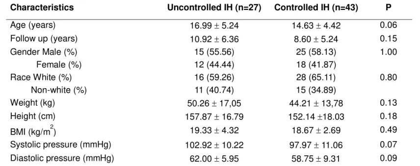
A total of 81 patients with confirmed IH were invited to participate in the study. However, 11 patients refused to participate. The remaining 70 patients were then divided in two subgroups according to their urinary calcium excretion at the time of urine and blood sample collection. Patients with calcium excretion equal or superior to 4mg/kg/day were allocated to the uncontrolled IH group (n=27). Patients with calcium excretion below 4mg/kg/day were allocated at the controlled group (n=43) (25). Figure 1 shows a flow diagram of eligible, excluded and included patients.
5.3.2. STUDY
PROTOCOL
diastolic blood pressure, serum creatinine, calciuria, citraturia, phosphaturia, magnesuria, use of potassium citrate and hydrochlorothiazide, bone mineral density (BMD), familiar history for nephrolithiasis, presence of calculus, past history for extracorporeal shock lithotripsy and symptoms were analyzed.
BMD was assessed by dual energy x-ray absorptiometry at lumbar spine (L1-L4) using a Lunar Prodigy Primo DXA System (GE Healthcare Lunar Corp., Madison, WI, USA). Bone density was stratified as Z-score >-1 SD and ≤-1 SD (12, 17, 18, 28, 29).
5.3.3. BLOOD
SAMPLING
After informed consent, all subjects were submitted to blood collection. Blood sampling occurred at only one occasion, simultaneously to other routine exams. The samples were collected into sterile citrate tubes, which were immediately immersed in ice, and processed within 30 min after collection. Cells were sedimented by centrifugation at 700 g for 10 min at 4oC. Then the supernatant
5.3.4. URINE
SAMPLING
A single urine sample was obtained from all patients at the same day of blood collection from 7.30 AM to 9.00 AM. After homogenization, 10 mL of the collected urine were centrifuged at 4oC
for 20 min at 1300 g. Cell-free urine was aliquoted into 0.5 mL tubes and stored at -80oC until measurements.
5.3.5. CYTOKINES
MEASUREMENT
Plasma and urinary levels of IL-1β, IL-6, IL-8, TNF-α, TGF-β1
and MCP-1 were measured by specific enzyme-linked immunoassay (ELISA) kits (R&D Systems, Minneapolis, MN), following the
manufacturer’s instructions, as described elsewhere (31). Urine
5.3.6. STATISTICAL
ANALYSIS
The values are expressed as medians and interquartile range (percentile 25, percentile 75) or means and standard deviation (SD), when appropriate. The Mann-Whitney test was used to compare nonparametric continuous variables. Dichotomous variables were compared by the two-sided Fisher's exact test. Correlation between plasma cytokines, urinary cytokines and BMD was performed using a nonparametric test (Spearman rank correlation test). The level of significance was set at p < 0.05.
5.3.7. ETHICAL
ASPECTS
5.4. RESULTS
General clinical characteristics at baseline
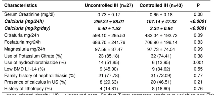
A total of 70 patients were included in the analysis. The baseline clinical characteristics are summarized in Table 1. Except for the increased use of hydrochlorothiazide in the uncontrolled group (p<0.05), there were no differences between patients with uncontrolled and controlled IH. All patients were normotensive and had normal serum creatinine levels at the time of sample collections. The most common signs and symptoms at the time of diagnosis were recurrent abdominal pain (45.7%) and macroscopic hematuria (27.1%). Other findings at baseline were microscopic hematuria (14.3%), urinary tract infection (11.4%) and nephrolithiasis (1.4%).
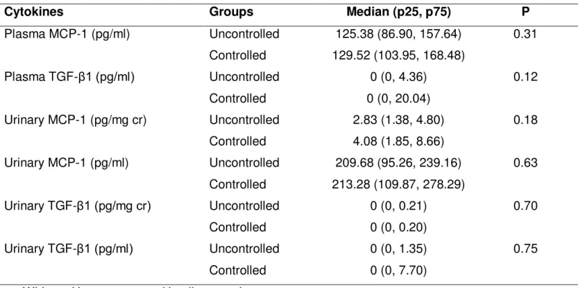
Association of plasma and urinary cytokine concentrations
with urinary calcium excretion
Both in uncontrolled and controlled IH groups, plasma and urinary concentrations of IL-1β, IL-6, IL-8, TNF-α were under the
cr) also revealed similar values for urinary TGF-β1/cr and MCP-1/cr between the two studied groups (table 2).
There was also a trend toward a positive correlation between plasma and urinary levels of MCP-1 standardized to creatinine (r=0.24, p=0.08), as shown in Figure 2.
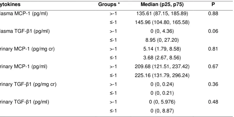
Association of plasma and urinary cytokine concentrations
with bone mineral density (BMD)
Patients were also stratified according to their BMD Z-score in two groups: >-1 SD, n=28; ≤-1SD; n=18, as shown in Table 3. The comparison between these groups did not reveal differences in general clinical findings as well as in 24 hour urinary calcium excretion.
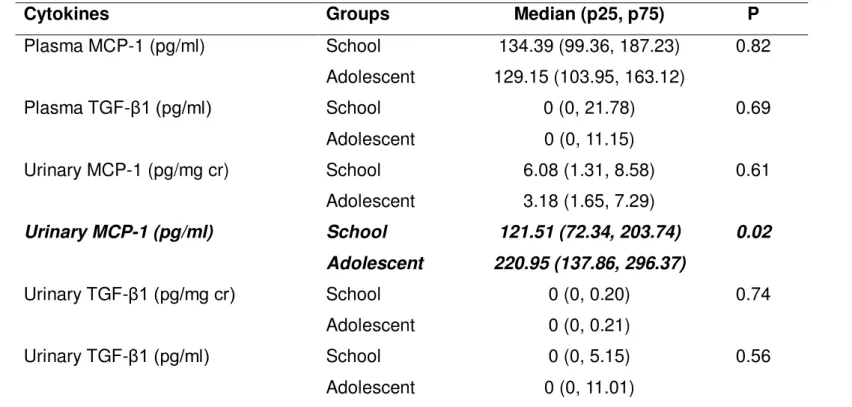
Association of plasma and urinary cytokine concentrations
with age groups
In order to detect possible changes in cytokine levels related to age, the patients were stratified in the following age groups: school (age ≤ 12 year; n=18) and adolescent (age> 12 year; n=52). The absolute levels of MCP-1 (pg/ml) were significantly higher in adolescents than in school age children (p=0.02). However, this difference was not observed when values were standardized to creatinine (p=0.61, Table 6).
5.5. DISCUSSION
patients with high urinary calcium excretion or reduced BMD Z-score. However, there was a significant positive correlation between urinary MCP-1 levels and the BMC.
There is very little information concerning the role of MCP-1 in bone metabolism. The principal function of MCP-1 is the recruitment of monocytes (33, 34). Monocyte products are potential regulators of bone cell activity, since growth factors produced by these cells may stimulate bone formation (35, 36). In vitro and in vivo
studies indicated that MCP-1 induces the recruitment of monocytes to bone, which, in turn, is associated with an increase in osteoblast number (35, 36). This is likely to occur via indirect mechanisms, because MCP-1 did not directly enhance DNA synthesis in osteoblastic cells in vitro (36). Thus, activated mononuclear
phagocytes may play an important role in bone metabolism by stimulating proliferation of osteoblastic cells. MCP-1 is typically not expressed in normal bone or by normal osteoblasts in vitro. Upon
formation was associated with a decrease in the number of osteoclasts, while in bone-resorbing areas, recruitment of cells of the monocytic lineage is associated with formation of osteoclasts (33). The receptor activator of NF-κβ ligand (RANKL) seems to be a key
variable in this process, once MCP-1 stimulates the formation of osteoclasts in the presence of RANKL. MCP-1 is also induced by RANKL during osteoclast differentiation. (37) Despite the absence of differences in MCP-1 levels according to urinary calcium excretion and according to the presence of lower BMD Z-score, the positive correlation between urinary MCP-1 and the BMC might indicate a role of this chemokine in bone remodeling in patients with IH. In IH, the mechanisms involved in bone formation and bone resorption were probably activated in spite of the levels of urinary calcium excretion. Therefore, MCP-1 could be locally produced by osteoblasts and signaling towards both events depending on the area of expression and on the interactions with other mediators.
TGF-β is known to stimulate bone formation, mineralization,
and inhibiting bone resorption through a proapoptotic effect on mature osteoclasts and by the inhibition of RANKL expression in osteoblasts (22, 40-42). Our measurements showed low levels of
observed similar results with a significantly lower immunostaining for
TGF-β in patients with IH when compared with control subjects (22).
In our study, plasma and urinary levels of IL-1β, IL-6, IL-8,
TNF-α were under detectable limits. Other authors, by using different
methodologies, were able to evaluate these cytokines in IH (20, 22, 43). Pacifici et al described an association, but not a cause-effect relationship, between IL-1β activity and bone resorption. In his study, cultured peripheral blood monocytes were used to show an increased production of IL-1β in IH. (43) Weisinger et al used unstimulated blood monocytes to show an increased expression of
IL-1α, IL-6 and TNF-α mRNA in patients with IH. (18) These authors
also described a correlation between basal production of IL-1α, but
not IL-1β, and decreased trabecular bone (20)
The main possible weakness of our study was that our patients were not on standard diets during sample collections. Diet rich in proteins and salt can significantly affect calciuria by different mechanisms from those involved in bone remodeling in IH. (45) Parathyroid hormone and vitamin D are also important variables not concomitantly measured with cytokines in our research protocol. (46) In this study, age was addressed as a possible confounder. Bone remodeling regulatory mechanisms may vary according to age, once children and adolescents experience different stages in the skeletal development. (47) In spite of that, no significant difference in cytokine measurements were found in the comparison between school age and adolescents with IH.
BIBLIOGRAPHY
1. Albright F, Henneman P, Benedict PH, Forbes AP. Idiopathic hypercalciuria: a preliminary report. Proc R Soc Med. 1953 Dec;46(12):1077-81.
2. Ammenti A, Neri E, Agistri R, Beseghi U, Bacchini E. Idiopathic hypercalciuria in infants with renal stones. Pediatr Nephrol. 2006 Dec;21(12):1901-3.
3. Moore ES, Coe FL, McMann BJ, Favus MJ. Idiopathic hypercalciuria in children: prevalence and metabolic characteristics. J Pediatr. 1978 Jun;92(6):906-10.
4. Spivacow FR, Negri AL, del Valle EE, Calvino I, Zanchetta JR. Clinical and metabolic risk factor evaluation in young adults with kidney stones. Int Urol Nephrol. 2010 Jun;42(2):471-5.
5. Frick KK, Bushinsky DA. Molecular mechanisms of primary hypercalciuria. J Am Soc Nephrol. 2003 Apr;14(4):1082-95.
6. Zerwekh JE. Bone disease and hypercalciuria in children. Pediatr Nephrol. 2010 Mar;25(3):395-401.
7. Zerwekh JE. Bone disease and idiopathic hypercalciuria. Semin Nephrol. 2008 Mar;28(2):133-42.
8. Weisinger JR, Alonzo E, Carlini RG, Paz-Martinez V, Martinis R, Bellorin-Font E. Bone disease in hypercalciuria: a new form of osteodystrophy? Nephrol Dial Transplant. 1998;13 Suppl 3:88-90. 9. Weisinger JR. New insights into the pathogenesis of idiopathic hypercalciuria: the role of bone. Kidney Int. 1996 May;49(5):1507-18. 10. Tasca A, Dalle Carbonare L, Nigro F, Giannini S. Bone disease in patients with primary hypercalciuria and calcium nephrolithiasis. Urology. 2009 Jul;74(1):22-7.
12. Schwaderer AL, Cronin R, Mahan JD, Bates CM. Low bone density in children with hypercalciuria and/or nephrolithiasis. Pediatr Nephrol. 2008 Dec;23(12):2209-14.
13. Polito C, Iolascon G, Nappi B, Andreoli S, La Manna A. Growth and bone mineral density in long-lasting idiopathic hypercalciuria. Pediatr Nephrol. 2003 Jun;18(6):545-7.
14. Giannini S, Nobile M, Sella S, Dalle Carbonare L. Bone disease in primary hypercalciuria. Crit Rev Clin Lab Sci. 2005;42(3):229-48.
15. Garcia-Nieto V, Ferrandez C, Monge M, de Sequera M, Rodrigo MD. Bone mineral density in pediatric patients with idiopathic hypercalciuria. Pediatr Nephrol. 1997 Oct;11(5):578-83.
16. Garcia-Nieto V, Navarro JF, Ferrandez C. Bone loss in children with idiopathic hypercalciuria. Nephron. 1998;78(3):341-2. 17. Penido MG, Lima EM, Souto MF, Marino VS, Tupinamba AL, Franca A. Hypocitraturia: a risk factor for reduced bone mineral density in idiopathic hypercalciuria? Pediatr Nephrol. 2006 Jan;21(1):74-8.
18. Penido MG, Lima EM, Marino VS, Tupinamba AL, Franca A, Souto MF. Bone alterations in children with idiopathic hypercalciuria at the time of diagnosis. Pediatr Nephrol. 2003 Feb;18(2):133-9. 19. Pfeilschifter J, Chenu C, Bird A, Mundy GR, Roodman GD. Interleukin-1 and tumor necrosis factor stimulate the formation of human osteoclastlike cells in vitro. J Bone Miner Res. 1989 Feb;4(1):113-8.
20. Weisinger JR, Alonzo E, Bellorin-Font E, Blasini AM, Rodriguez MA, Paz-Martinez V, et al. Possible role of cytokines on the bone mineral loss in idiopathic hypercalciuria. Kidney Int. 1996 Jan;49(1):244-50.
21. Gowen M, Mundy GR. Actions of recombinant interleukin 1, interleukin 2, and interferon-gamma on bone resorption in vitro. J Immunol. 1986 Apr 1;136(7):2478-82.
23. Freundlich M, Alonzo E, Bellorin-Font E, Weisinger JR. Reduced bone mass in children with idiopathic hypercalciuria and in their asymptomatic mothers. Nephrol Dial Transplant. 2002 Aug;17(8):1396-401.
24. Misael da Silva AM, dos Reis LM, Pereira RC, Futata E, Branco-Martins CT, Noronha IL, et al. Bone involvement in idiopathic hypercalciuria. Clin Nephrol. 2002 Mar;57(3):183-91.
25. Butani L, Kalia A. Idiopathic hypercalciuria in children--how valid are the existing diagnostic criteria? Pediatr Nephrol. 2004 Jun;19(6):577-82.
26. Coe FL, Evan A, Worcester E. Kidney stone disease. J Clin Invest. 2005 Oct;115(10):2598-608.
27. Hughes P. The CARI guidelines. Kidney stones: metabolic evaluation. Nephrology (Carlton). 2007 Feb;12 Suppl 1:S31-3.
28. Lewiecki EM, Gordon CM, Baim S, Leonard MB, Bishop NJ, Bianchi ML, et al. International Society for Clinical Densitometry 2007 Adult and Pediatric Official Positions. Bone. 2008 Dec;43(6):1115-21. 29. Brandao CM, Camargos BM, Zerbini CA, Plapler PG, Mendonca LM, Albergaria BH, et al. [2008 official positions of the Brazilian Society for Clinical Densitometry--SBDens]. Arq Bras Endocrinol Metabol. 2009 Feb;53(1):107-12.
30. Reinhold D, Bank U, Buhling F, Junker U, Kekow J, Schleicher E, et al. A detailed protocol for the measurement of TGF-beta1 in human blood samples. J Immunol Methods. 1997 Dec 1;209(2):203-6.
31. Sousa-Pereira SR, Teixeira AL, Silva LC, Souza AL, Antunes CM, Teixeira MM, et al. Serum and cerebral spinal fluid levels of chemokines and Th2 cytokines in Schistosoma mansoni myeloradiculopathy. Parasite Immunol. 2006 Sep;28(9):473-8.
32. Heilberg IP, Weisinger JR. Bone disease in idiopathic hypercalciuria. Curr Opin Nephrol Hypertens. 2006 Jul;15(4):394-402.
protein-1 are developmentally regulated in remodeling bone in the mouse. Am J Pathol. 1997 May;150(5):1711-21.
34. Yadav A, Saini V, Arora S. MCP-1: chemoattractant with a role beyond immunity: a review. Clin Chim Acta. 2010 Nov 11;411(21-22):1570-9.
35. Posner LJ, Miligkos T, Gilles JA, Carnes DL, Taddeo DR, Graves DT. Monocyte chemoattractant protein-1 induces monocyte recruitment that is associated with an increase in numbers of osteoblasts. Bone. 1997 Oct;21(4):321-7.
36. Graves DT, Jiang Y, Valente AJ. Regulated expression of MCP-1 by osteoblastic cells in vitro and in vivo. Histol Histopathol. 1999 Oct;14(4):1347-54.
37. Valente AJ, Xie JF, Abramova MA, Wenzel UO, Abboud HE, Graves DT. A complex element regulates IFN-gamma-stimulated monocyte chemoattractant protein-1 gene transcription. J Immunol. 1998 Oct 1;161(7):3719-28.
38. Rahimi P, Wang CY, Stashenko P, Lee SK, Lorenzo JA, Graves DT. Monocyte chemoattractant protein-1 expression and monocyte recruitment in osseous inflammation in the mouse. Endocrinology. 1995 Jun;136(6):2752-9.
39. Williams SR, Jiang Y, Cochran D, Dorsam G, Graves DT. Regulated expression of monocyte chemoattractant protein-1 in normal human osteoblastic cells. Am J Physiol. 1992 Jul;263(1 Pt 1):C194-9.
40. Lorenzo J, Horowitz M, Choi Y. Osteoimmunology: interactions of the bone and immune system. Endocr Rev. 2008 Jun;29(4):403-40.
41. Quinn JM, Itoh K, Udagawa N, Hausler K, Yasuda H, Shima N, et al. Transforming growth factor beta affects osteoclast differentiation via direct and indirect actions. J Bone Miner Res. 2001 Oct;16(10):1787-94.
42. Pacifici R. The immune system and bone. Arch Biochem Biophys. 2010 Nov 1;503(1):41-53.