INTRODUÇÃO
Linfoma é neoplasia maligna resultante da prolifera-ção de células do sistema linfóide, com variado potencial de agressão orgânica. Da linhagem linfóide originam-se as leucemias linfocíticas, os plasmocitomas, o linfoma de Hodgkin e os linfomas não Hodgkin.
Os linfomas não Hodgkin constituem um grupo de neoplasias derivadas de clones de linfócitos em seus difer-entes estágios evolutivos. Podem originar-se primaria-mente nos linfonodos (linfomas nodais) ou em tecidos
lin-INTRODUCTION
Lymphoma is a malign neoplasia resulting from the proliferation of cells from the lymphoid system, which has a varying potential for organic aggression. Lymphocytic leukemias, plasmocytomas, Hodgkin's lymphoma and non Hodgkin's lymphomas stem from this lymphoid lineage.
The non Hodgkin's lymphomas comprise a neopla-sia group derived from lymphocyte clones in their different evolutive stages. They may originate primarily in the lymph nodes (nodal lymphomas) or in lymphoid tissue connected
Linfoma cutâneo de células B: relato de caso
*
Cutaneous B-cell lymphoma: a case report
*
Mariana de Gusmão Nunes
1Ana Paula Soares de Moura Pierro
2Maria Fernanda Villela Coutinho
3José Carlos Oliveira de Morais
4Sueli Coelho da Silva Carneiro
5David Rubem Azulay
6Resumo: Os linfomas cutâneos são classificados em linfomas de células B ou de células T, sendo os primeiros menos freqüentes. O linfoma cutâneo de células B (LCCB) pode ser primário ou secundário, tendo o último história natural mais agressiva, com pior prognósti-co. Os autores apresentam um caso de LCCBsecundário com três anos de evolução, curso indolente, sem envolvimento de outros órgãos além da pele, com ótima resposta ao trata-mento quimioterápico. A distinção entre LCCBprimário e secundário é muito difícil de ser realizada, uma vez que eles são clínica e histopatologicamente indistinguíveis. Deve-se sem-pre pesquisar o acometimento de órgãos internos para então definir o prognóstico.
Descritores : linfoma de células B; manifestações cutâneas; prognóstico.
Abstract:Cutaneous lymphomas are classified as either type B cell or T cell lymphoma, the former type being less frequent. Cutaneous B cell lymphoma (CBCL) may be primary or sec-ondary. The latter has a more aggressive natural history with a worse prognostic. In this paper, the authors present a secondary CBCL case with a three-year evolution, indolent course, without involvement of other organs beside the skin and with optimal response to chemotherapeutic treatment. The distinction between primary and secondaryCBCLis very difficult to determine insofar as they are clinically and histopathologically indistinguish-able. It is always necessary to investigate whether internal organs have been affected prior to defining the prognosis.
Key words: lymphoma, B-Cell; skin manifestations; prognosis.
Caso Clínico /
Case Report
Recebido em 02.05.2002. / Received on May 02, 2002.
Aprovado pelo Conselho Consultivo e aceito para publicação em 29.11.2002. / Approved by the Consultive Council and accepted for publication on November 29, 2002. * Trabalho realizado na Disciplina/Serviço de Dermatologia do Hospital Universitário Clementino Fraga Filho (HUCFF), Universidade Federal do Rio de Janeiro (UFRJ). /
Work performed at the Dermatology Service, Hospital Universitário Clementino Fraga Filho (HUCFF) and the Universidade Federal do Rio de Janeiro (UFRJ).
1Mestranda do Serviço de Dermatologia - HUCFF/UFRJ. / Studying for masters degree of Dermatology Service, HUCFF/UFRJ.
2Médica estagiária do Serviço de Dermatologia - HUCFF/UFRJ. / MD Trainee Dermatology of Dermatology Service, HUCFF/UFRJ.
3Médica especialista em Dermatologia pela UFRJ / MD Specialist Dermatology at UFRJ
4Professor Adjunto do Departamento de Patologia - FM/UFRJ / Adjunct Professor, Department of Pathology, FM/UFRJ.
5Professora do Curso de Pós-Graduação em Dermatologia - FM/UFRJ; Professora Adjunta de Dermatologia - FCM/UERJ; Médica do Serviço de Dermatologia - HUCFF/UFRJ /
Professor, Post-Graduation Program in Dermatology, FM/UFRJ; Adjunct Professor of Dermatology, FCM/UERJ; Physician, Dermatology Service, HUCFF/UFRJ.
6Professor Assistente de Dermatologia - FM/ UFRJ; Médico do Serviço de Dermatologia - HUCFF/UFRJ; Professor Titular de Dermatologia - PUC; Professor Adjunto de Dermatologia -
FTESM/ Assistant Professor of Dermatology, FM/ UFRJ; M.D. Dermatology Service, HUCFF/UFRJ; Titular Professor of Dermatology, PUC; Adjunct Professor of Dermatology, FTESM.
fóides associados a mucosas, pele ou outras estruturas (lin-fomas extranodais).1
Os linfomas cutâneos são classificados de acordo com sua origem celular em linfoma de células T e linfoma de células B.2Sua freqüência média é de 0,3/100.000
habi-tantes/ ano, sendo 65% de células T, 25% de células B e 10% linfomas histiocíticos verdadeiros ou de tipos celulares raros.2,3 Do ponto de vista dermatológico, o de células B
caracteriza-se por lesões pouco numerosas, em geral nódu-los ou infiltrações, de crescimento relativamente rápido, e, ao contrário daquele de células T, não apresenta prurido.
Do ponto de vista histopatológico, os primeiros são monomorfos (de pequenas ou grandes células), e o infiltra-do está separainfiltra-do da epiderme pela faixa de colágeno de Unna; enquanto os de células T são epidermotrópicos.4
Atualmente para diferenciá-los são usados anticorpos monoclonais. A expressão de CD3, CD4, CD8, CD43 ou
CD45 confirma o caráter de células T, e a expressão de
CD20, principalmente, CD19,CD23eCD79, além deCD5,
CD10,CD32ouCD38confirma o caráter de células B.3
O linfoma cutâneo de células B (LCCB) pode ser considerado primário ou secundário. O primário é aquele com apresentação cutânea, sem qualquer evidência de lesão extracutânea no momento do diagnóstico e até seis meses após.5-9Apesar de serem idênticos na aparência
mor-fológica, possuem comportamentos clínicos diferentes. O primário apresenta história natural mais indolente do que a do secundário, com bom prognóstico, recorrências locais em 25-68% dos casos e rara disseminação extracutânea. Sua sobrevida média em cinco anos varia de 89% a 96%.10
RELATO DE CASO
Homem negro, de 46 anos, natural e procedente do Rio de Janeiro, RJ, desempregado (era entregador), procurou o serviço de Dermatologia do Hospital Universitário Clementino Fraga Filho (HUCFF) em dezem-bro de 2000 com queixa de tumor na pele. Relatava história de aparecimento de pequenas pápulas na região torácica há três anos, que associava com picada de inseto ocorrida no mesmo local 15 anos antes. Evoluiu com aumento do tamanho das lesões, que se tornaram nódulos e, há aproxi-madamente um ano, tumoração de 8cm de diâmetro. Referia dor de forte intensidade no local e aumento de volume na axila esquerda, também doloroso. Chegou a procurar outro serviço, onde foram feitas drenagem e curetagem da lesão, sem melhora. Encaminhado a esse hospital, permaneceu internado por 17 dias para investigação diagnóstica.
O exame físico revelou lesão nodular com aproxi-madamente 8cm de diâmetro, de consistência endurecida, aderida aos planos profundos, com superfície lisa e multi-lobulada, e com ulceração em sua porção inferior, localiza-da na região torácica anterior direita. Havia ainlocaliza-da pequenas pápulas cor da pele próximo à lesão (Figura 1). Observou-se também aumento de volume na região axilar esquerda, que correspondia a linfadenomegalia dolorosa, de
con-to mucouses, skin or other structures (extranodal lym-phomas).1
Cutaneous lymphomas are classified according to their cellular origin in T-cell lymphomas or B-cell lym-phomas.2
Their average frequency is 0.3/100,000 inhabi-tants yearly, with 65% T-cell, 25% B-cell and 10% true his-tiocytic lymphomas or rare cell types.2,3
From the dermato-logical point of view, B-cell types are characterized by only a few lesions that are usually nodules or infiltrations, and are relatively fast growing. As opposed to T-cell types, they do not show pruritus.
From the histopathological point of view, B-cell types are monomorphic (small and large cells), and the infiltrate is separated from the epidermis by the so-called Unna colla-gen strip; whereas T-cell types are epidermotropic.4In order
to differentiate them, monoclonal antibodies are currently used. The expression of CD3,CD4,CD8,CD43orCD45 con-firms their T-cell character. And the expression of CD20, mainly, and CD19, CD23 and CD79, in addition to CD5, CD10,CD32orCD38, confirms the B-cell character.3
B-cell cutaneous lymphoma (BCCL) may be considered primary or secondary. The primary type is the one with a cuta-neous presentation but with no evidence of extracutacuta-neous lesions at the moment of the diagnosis, or up to six months thereafter.5-9In spite of being identical in morphological
appear-ance, they have different clinical behaviors. The primary type has a more indolent natural history than the secondary one. It has a good prognosis, local relapses in 25% to 68% of cases, and rare extracutaneous dissemination. A patient's average sur-vival rate over five years varies from 89% to 96%.1010
CASE REPORT
A 46-year-old Afro-Brazilian man, born and living in Rio de Janeiro city, Rio de Janeiro state, and unemployed (he used to be a delivery man), sought assistance at the Dermatology Service of the Hospital Universitário Clementino Fraga Filho (HUCFF) in December 2000, com-plaining of a skin tumor. He reported having a history of small papules that appeared in the thoracic region three years prior. The latter associated with an insect sting that had occurred on the same spot 15 years earlier. It progressed as the lesion increased in size, turning into nodules. Approximately a year ago, the tumor grew to 8 cm in size. The patient referred to high-intensity pain at the localization and to an increased volume in the left axilla, which was also painful. He ended up seeking out another service at which draining and curettage of the lesion were done, without any improvement. He was referred to this hospital, where he was interned for 17 days for a diagnostic investigation.
sistência endurecida, aderida aos planos profundos, com aproxi-madamente 4cm de diâmetro (Figura 2). O restante do exame físico era normal.
As hipóteses diagnósticas feitas foram dermatofibrossarcoma protuberans, tumor de anexo, carcino-ma espinocelular e linfocarcino-ma cutâneo.
Os exames laboratoriais mostraram hemograma normal; VDRL, anti-HIV e anti-HTLV-1 negativos; radi-ografia do tórax sem alterações, e USGabdominal sem vis-ceromegalias ou linfadenomegalias. A tomografia com-putadorizada do tórax mostrou lesão expansiva com impregnação irregular por meio de contraste, localizada na pele e no tecido subcutâneo da parede torácica anterior direita, sem plano de clivagem definido com o músculo grande peitoral; linfadenomegalia axilar esquerda e linfon-odo reacional à direita. Ausência de acometimento pul-monar ou mediastinal (Figura 3).
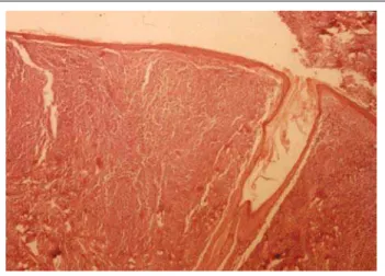
O exame histopatológico da lesão mostrou denso infiltrado na derme superficial e profunda, separa-do da epiderme por estreita faixa de colágeno (Figura 4). O infiltrado era composto por células mononucleares com núcleos grandes, nucléolos evidentes e com pouco citoplasma, entremeadas por linfócitos reacionais (Figura 5). A imuno-histoquímica foi positiva para CD20 nas grandes células e CD3 nas
pequenas, confirmando o diagnóstico de linfoma cutâneo de grandes células B (Figura 6). O aspirado e a biópsia da medula óssea foram normais.
O Serviço de Hema-tologia instituiu tratamento
in volume was also observed in the left axillary region, which corresponded to a painful lymphadenomegaly. It had a hardened surface, adhered to the deep planes, and was roughly 4 cm in diameter (Figure 2). The rest of the physical examination was normal.
The diagnostic hypotheses were dermatofibrosarcoma protuberans, a tumor of the annex, spinocellular carcinoma and cutaneous lymphoma. The laboratory investigation revealed a normal hemogram; VDRL, anti-HIV and anti-HTLV-2 were nega-tive; the X-ray of the thorax was without alteration, and abdominal USG without visceromegalies or lymphade-nomegalies. Computerized tomography of the thorax showed the lesion to be expansive and, by means of con-trast, with irregular impregnation, localized in the skin and the subcutaneous tissue of the right anterior thoracic wall, and without a defined cleavage plane with the large pec-toral muscle; left axilla lymphadenopathy and reactional lymph node on the right. There was an absence of pul-monary or mediastinal affection (Figure 3).
The histopathologic examination of the lesion showed dense infiltrate in the superficial and deep dermis, separated from the epidermis by a collagen strip (Figure 4). The infil-trate consisted of mononuclear cells with large nuclei, evi-dent nucleoli and sparse cytoplasm, intermixed with reac-tional lymphocytes (Figure 5). The immunohistochem-istry was positive for CD20 in large cells and CD3 in small ones. This confirmed the diagnosis of cutaneous large B-cell lymphoma (Figure 6). Bone marrow aspirate and biopsy were normal.
Figura 1: Lesão tumoral de 8cm de diâmetro, de con-sistência endurecida, aderi-da aos planos profundos, com superfície lisa e multi-lobulada e com ulceração em sua porção inferior, localizada em região torácica anterior direita. Observam-se pequenas pápulas cor da pele próximo à lesão.
Figure 1: A tumoral lesion 8 cm in diameter, with a hard-ened consistency, adhering to the deep planes. It has a smooth and multilobulated surface, with ulceration in its lower part. It is localized in the right anterior thoracic region. Small skin-colored papules may be observed close to the lesion.
Figure 2: Increased volume on the left axilla.
quimioterápico combinado com regime CHOP (ciclofos-famida, adriamicina, vimentina e prednisona). Programaram-se seis ciclos com intervalo de três sem-anas entre eles. O paciente evoluiu com ótima resposta ao tratamento, redução do tamanho da lesão cutânea após o primeiro ciclo e regressão completa das lesões cutânea e axilar após o quarto ciclo. Entretanto, por considerar-se curado e pela intensidade dos efeitos colaterais, recusou-se a continuar o tratamento e com-pletar o número de ciclos programados, apesar do esclarecimento da equipe médica a respeito dessa necessidade.
DISCUSSÃO
Embora os linfomas de células B sejam mais fre-qüentes do que os de células T nos linfonodos, na pele ocorre justamente o contrário. Atualmente sabe-se que a pele atua como órgão efetor imunológico e apresenta pop-ulação linfocitária T-residente na derme, semelhante ao
The Hematologic Service instituted chemotherapeu-tic treatment in combination with CHOPregimen (cyclophos-phamide, adriamycin, vimentin and prednisone). Six cycles were programmed at three-week intervals between them. The patient progressed with excellent response to treatment, a reduction of the size of the cutaneous lesion after the first cycle and complete regression of the cutaneous and axillary lesions after the fourth cycle. On the other hand, the patient considered himself cured. Due to this, as well as the intensi-ty of the side effects, he refused to continue the treatment and complete the programmed number of cycles-despite the med-ical staff's clarifications on the need to complete it.
DISCUSSION
B-cell lymphomas are more frequent in the lymph nodes than T-cell lymphomas. Regarding the skin, the former occur in the exact opposite way. It is now known that the skin acts like an immunologic effector organ. Moreover, it presents a resident T-lymphocytic population in the dermis similar to the lymphoid Figura 3: Tomografia
com-putadorizada do tórax: lesão expansiva com impregnação irregular por meio de contraste, localiza-da na pele e no tecido sub-cutâneo da parede torácica anterior direita, sem plano de clivagem definido com o músculo grande peitoral. Presença de linfadenome-galia axilar esquerda e lin-fonodo reacional à direita.
Figure 3: Computerized tomography of the thorax: expansive lesion shown by means of contrast to be with irregular impregnation. It is localized on the skin and sub-cutaneous tissue of the right anterior thoracic wall. It has no plane cleavage defined with the large pectoral mus-cle. Presence of left axillary lymphadenomegaly and reac-tive lymph node on the right.
Figura 4: Aspecto histológico da lesão nodular: denso infiltrado na derme superficial e profunda, separado da epiderme por estreita faixa de colágeno (HE, X40). / Figure 4: Histologic aspect
of the nodular lesion: dense infiltrate in the superficial and deep dermis, separated from the epidermis by a narrow strip of
collagen (X40 HE).
Figura 5: Infiltrado composto por células mononucleares com núcleos grandes, nucléolos evidentes e com pouco citoplasma, entremeadas por linfócitos reacionais (HE, X100). / Figure 5: Infiltrate consisting of mononuclear cells with large nuclei,
tecido linfóide associado às mucosas. Sendo assim, não existem dúvidas quanto à origem cutânea primária de linfomas de células T, tanto epidermotrópicos como não e p i d e r m o t r ó p i c o s . Entretanto, muito se discute
a respeito da origem cutânea de alguns processos linfo-matosos de células B. Poderiam ser processos primários verdadeiros da pele ou infiltrações linfomatosas dérmicas a partir de neoplasias nodais ou viscerais não diagnosticadas. Discute-se ainda a possibilidade de o LCCBoriginar-se de infiltrações linfocitárias benignas persistentes da pele, como pseudolinfomas.1 Sabe-se apenas que o LCCB
primário tem melhor prognóstico, com rara disseminação extracutânea.
O paciente apresentava no momento do diagnóstico, além da lesão cutânea, aumento de linfonodo axilar esquer-do. Questionou-se na época se a adenomegalia seria rea-cional ou tumoral, porém os aspectos clínicos levaram a considerá-la tumoral, classificando o LCCB como secundário. Não havia acometimento de outros sítios lin-fonodais nem de órgãos internos ou de medula óssea, ape-sar dos três anos de evolução, evidenciando, portanto, um curso indolente. Por outro lado, apresentou bom prognósti-co, com excelente resposta ao tratamento instituído. Para o tratamento de LCCB CD20+ com baixa resposta às te-rapêuticas convencionais, foi aprovado em 1998 pelo Food and Drug Administrationo uso de rituximab, um anticorpo monoclonal antiCD20.11 Este se liga à molécula CD20,
desencadeando o processo de lise celular. Nos três casos descritos na literatura com utilização desse anticorpo mon-oclonal houve excelente resposta inicial, com posterior recidiva de LLCB CD20-.11,12,13 Dados o baixo índice de
efeitos colaterais e sua eficácia inicial, o rituximab apre-senta-se como um tratamento promissor, restando avaliar se variações na dose e na administração, além da combi-nação com outros agentes, poderiam evitar a seletividade de células CD20- que leva à recidiva.
Não se pode esquecer ainda da relação que o paciente fez com picada de inseto no local muitos anos antes, o que poderia ter transmitido um fator carcinogênico e evoluído para um pseudolinfoma. Alguns autores relatam o achado da Borrelia burgdorferi em lesões de LCCB e questionam seu possível papel etiopatogênico.14,15 Para
pesquisá-la ressaltam a necessidade de realizarPCRe cul-tura das lesões, além da sorologia. Relatam também a
pos-tissue associated with mucous-es. This being the case, there is no doubt about the primary cutaneous origin of either epi-dermotropic T-cell lymphomas or non epidermotropic ones. On the other hand, there is a lot of debate about the cuta-neous origin of some lymphomatous B cell processes. These may be real primary skin processes, or dermal lymphomatous infiltrations from the nodal or visceral neoplasias that were not diagnosed. There is even a debate on whether BCCLmay possi-bly stem from persistent benign lymphocytic infiltrations of the skin, such as pseudolymphomas.1The only thing known for sure
is that primary BCCLhas a better prognosis with respect to rare extracutaneous dissemination.
At the time of diagnosis, the patient showed a n increase of the left axillary lymph node in addition to the cutaneous lesion. At the time, there was speculation on whether the adenomegaly would be reactive or tumoral. But the clinical aspects led to considering them as tumoral, which thereby classified BCCLas secondary. There was no affection of the other lymph node sites, not even of the internal organ or bone marrow despite three years of progression that had nonetheless evidenced an indolent course. On the other hand, the affection showed good prognosis and an excellent response to the treatment instituted. For treatment ofBCCL CD 20-positive having low response rate to conventional therapies, the Food and Drug Administration approved the use of rituximab in 1998, a monoclonal antiCD20 antibody.11
The latter links up to molecule CD20 so as to trigger the cel-lular lysis process. In the three cases described in the litera-ture in which this monoclonal antibody was used, there was an excellent initial response, with a subsequent relapse of BCLLCD20-negative.11,12,13Given the low rate of side effects
and its initial effectiveness, rituximab showed itself to be a promising treatment. It remains to be evaluated whether dose and administration variations, in addition to combinations with other agents, might prevent the selectivity of CD20-neg-ative which had led to the relapse.
Still, the link the patient made to an insect sting that had occurred on the same localization several years earlier must not be forgotten. The sting may have transmitted a car-cinogenic factor and progressed into a pseudolymphoma. Some authors report the finding of Borrelia burgdorferiin BCCLlesions and question whether it has a possible etiopath-ogenic role.14,15To examine this finding, they emphasize the
need to perform PCRand lesion cultures, in addition to serol-Figura 6:
Imuno-histo-química mostrando posi-tividade para CD20 nas grandes células.
sibilidade de tratamento com doxiciclina oral ou cefalospo-rina parenteral nos estágios iniciais da doença, como tenta-tiva de estimular a regressão da lesão.
Os autores concluem que a diferenciação entre origem cutânea primária ou secundária do LCCBé muito difícil, uma vez que eles são clínica e histopatologica-mente indistinguíveis. Torna-se extremahistopatologica-mente importante pesquisar acometimento extracutâneo nesses pacientes, e, em sua presença, deve-se sempre que possível realizar biópsia para confirmação. O estagiamento irá contribuir para a avaliação do prognóstico e para a escolha da
tera-pêutica a ser adotada.
ogy. They also report the possibility of using oral doxycycline or parenteral cephalosporin as a treatment of the disease in its initial stage in an attempt to stimulate lesion regression.
The authors conclude that differentiating between the primary or secondary cutaneous origins of BCCL is very difficult indeed, since they are clinically and histopathologically indistinguishable. It has become extremely important to examine extracutaneous afflictions in these patients. Furthermore, in the patients' presence, it must always be possible to perform a biopsy for confirma-tion. The staging will contribute to an assessment of the prognosis and to the choice of therapy to be adopted.
10. Pandolfino TL, Siegel RS, Kuzel TM, Rosen ST, Guitart J. Primary cutaneous B-cell lymphoma: review and current concepts. J Clin Oncol. 2000;18:2152-68.
11. Massengale WT, McBurney E, Gurtler J. CD20-nega-tive relapse of cutaneous B-cell lymphoma after anti-CD20 monoclonal antibody therapy. J Am Acad Dermatol. 2002;46:441-3.
12. Kinoshita T, Nagai H, Murate T, Saito H. CD20-nega-tive relapse in B-cell lymphoma after treatment with Rituximab. J Clin Oncol. 1998;16:3916-9.
13. Maloney DG, Davis T, Levy R. In reply (letter). J Clin Oncol. 1998; 16:1636-7.
14. Slater DN. Borrelia burgdorferi-associated primary cuta-neous B-cell lymphoma. Histophathology. 2001;38:73-7. 15. Goodlad JR, Davidson MM, Hollowood K, Baststone P, Ho-Yen DO. Borrelia burgdorferi-associated cutaneous marginal zone lymphoma: a clinicopathological study of two cases illustrating the temporal progression of Borrelia-associated B-cell proloferation in the skin. Histopathology. 2000;37:501-8.
REFERÊNCIAS / REFERENCES
1. Sampaio SAP, Rivitti EA. Leucemias, Linfomas e Pseudolinfomas. In: Dermatologia. 2a ed. São Paulo: Artes Médicas; 2000. p. 887-913.
2. Odom RB, James WD, Berger TG. Cutaneous Lymphoid Hyperplasia, Cutaneous T-Cell Lymphoma, Other Malignant Lymphomas, and Allied Diseases. In: Andrews` Diseases of the Skin, Clinical Dermatology. 9th ed. Philadelphia: W.B. Saunders Company; 2000. p. 918-42. 3. Burg G, Kempf W, Haeffner AC et al. Cutaneous lym-phomas. Curr Probl Dermatol. 1997; 9:137-204.
4. Azulay RD, Azulay DR. Oncologia Dermatológica. In: Dermatologia. 2a ed. Rio de Janeiro: Guanabara Koogan; 1997. p. 320-77.
5. Heald PW, Edelson RL. Cutaneous T-Cell Lymphomas. In: Freedberg I.M., Eisen A.Z, Wolff K et al. Fitzpatrick`s: Dermatology in General Medicine. 5th ed. New York: McGraw-Hill; 1999. p. 1227-50.
6. Sterry W, Jahn S. Other Systemic Lymphomas with Skin Infiltrations. In: Freedberg I.M., Eisen A.Z, Wolff K et al. Fitzpatrick`s: Dermatology in General Medicine. 5th ed. New York: McGraw-Hill; 1999. p. 1250-8.
7. Suchi T et al. Histopathology and immunohistochemistry of peripheral T-cell lymphomas - a proposal for classifica-tion of T-cell lymphomas. J Clin Pathol. 1987;40:995. 8. Harris N et al. A revised European-American Classification of Lymphoma neoplasms: A proposal from the International Lymphoma Study Group. Blood. 1994;84:1361.
9. Kerl H, Cerroni L. The morphologic spectrum of cuta-neous B-cell lymphomas. Arch Dermatol. 1996;132:1376.
ENDEREÇO PARA CORRESPONDÊNCIA: / MAILINGADDRESS: