Assessment of Neuroprotective Properties
of
Melissa officinalis
in Combination With
Human Umbilical Cord Blood Stem Cells
After Spinal Cord Injury
Seyed Ruhollah Hosseini
1, Gholamreza Kaka
1, Mohammad
Taghi Joghataei
2, Mehdi Hooshmandi
3, Seyed Homayoon Sadraie
4,
Kayvan Yaghoobi
1, and Alireza Mohammadi
1Abstract
Introduction:The pathophysiology of spinal cord injury (SCI) has a classically bad prognosis. It has been demonstrated that human umbilical cord blood stem cells (hUCBSCs) andMelissa officinalis(MO) are useful for the prevention of neurological disease.
Methods: Thirty-six adult male rats were randomly divided into intact, sham, control (SCI), MO, hUCBSC, and MO-hUCBSC groups. Intraperitoneal injection of MO (150 mg/kg) was commenced 24 hr post-SCI and continued once a day for 14 days. Intraspinal grafting of hUCBSCs was commenced immediately in the next day. The motor and sensory functions of all animals were evaluated once a week after the commencement of SCI. Electromyography (EMG) was per-formed in the last day in order to measure the recruitment index. Immunohistochemistry, reverse transcription-polymerase chain reaction, and transmission electron microscopy evaluations were performed to determine the level of astrogliosis and myelination.
Results:The results revealed that motor function (MO-hUCBSC: 150.3, SCI: 8.20.37,p<.001), sensory function
(MO-hUCBSC: 3.570.19, SCI: 6.380.23, p<.001), and EMG recruitment index (MO-hUCBSC: 3.710.18, SCI: 1.60.1,
p<.001) were significantly improved in the MO-hUCBSC group compared with SCI group. Mean cavity area (MO-hUCBSC:
0.030.03, SCI: 0.070.004, p<.001) was reduced and loss of lower motor neurons (MO-hUCBSC: 7.60.43, SCI:
30.12,p<.001) and astrogliosis density (MO-hUCBSC: 3.10.15, SCI: 6.251.42,p<0.001) in the ventral horn of spinal
cord were prevented in MO-hUCBSC group compared with SCI group.
Conclusion:The results revealed that the combination of MO and hUCBSCs in comparison with the control group has neuroprotective effects in SCI.
Keywords
astrogliosis, human umbilical cord blood,Melissa officinalis, myelination, spinal cord injury
Received March 13, 2016; Received revised May 17, 2016; Accepted for publication July 6, 2016
Introduction
The spinal cord injury (SCI) is very common and has poor prognosis (Webb et al., 2010). SCI could cause severe damage to motor, sensory, and autonomic nervous system and their functions which may lead to severe dis-abilities (Coutts and Keirstead, 2008). The pathophysi-ology of SCI after the primary trauma has a vital role in the initial structural disruption. In addition, the sec-ondary process includes the cascades of cellular and
1Neuroscience Research Center, Baqiyatallah University of Medical Sciences, Tehran, Iran
2Department of Anatomy, School of Medicine, Iran University of Medical Sciences, Tehran, Iran
3Neuroscience Research Center, Shahid Beheshti University of Medical Sciences, Tehran, Iran
4Department of Anatomy, School of Medicine, Baqiyatallah University of Medical Sciences, Tehran, Iran
Corresponding Author:
Gholamreza Kaka, Neuroscience Research Center, Baqiyatallah University of medical sciences, Tehran, Iran.
Email: [email protected]
ASN Neuro
November-December 2016: 1–17
!The Author(s) 2016
DOI: 10.1177/1759091416674833 asn.sagepub.com
Creative Commons CC-BY: This article is distributed under the terms of the Creative Commons Attribution 3.0 License
biological disruptions which can lead to long-term spinal deficits (Dasari et al., 2014). Increased oxidative stress (Fatima et al., 2015), redox transcription factors activity as well as elevated expression of inflammatory mediators play important roles (Popovich and Jones, 2003) in the promotion of the secondary process after the initial injury. SCI results in structural deformation such as degeneration of axons, disruption of neural tissue, neural and glial cell death, and demyelination around the lesion site. Axonal regeneration is inhibited by myelin-associated inhibitors in the central nervous system (CNS; Geoffroy and Zheng, 2014) and formation of astrogliosis (Deng et al., 2011). The extent of intrinsic cell renewal alone (Li and Lepski, 2013), even after appli-cation of mitogenic agents such as variety of growth fac-tors, is not sufficient enough for substantial recovery following SCI (Chhabra and Sarda, 2015). So, thera-peutic approaches such as exogenous cell transplantation should be considered.
Human umbilical cord blood stem cells (hUCBSCs) hold great promise for therapeutic repair after SCI (Dasari et al., 2007). The human umbilical cord blood by differentiating into various neural cells can enhance motor and sensory function improvement in the animal models of SCI (Kuh et al., 2005).
Transplantation of human umbilical cord blood mononuclear cells into the injured site of spinal cord did not affect the lesion extension. The survival time of transplanted cells in the injured area was 6 weeks after treatment. The transplanted group indicated better func-tional recovery than the untreated ones (Rodrigues et al., 2012). There is sufficient evidence which prove that stem cell therapy could be effective in spinal cord injuries but a strategy to potentiate this stem cell transplantation results is required (Niapour et al., 2012; Chhabra and Sarda, 2015).
As there have been strong interests in finding traditional agents that may help in the prevention of inflammation and disruption of neural tissues in CNS, a well-known herbal drug which has exhibited antioxidant and
neuro-protective effects is Melissa officinalis(MO). MO is
com-monly known as lemon balm (family: Lamiaceae); it is one of the oldest and still the most common medicinal plants (Kamdem et al., 2013). It has been reported that the most commonly known therapeutic properties of MO extract were sedative, antispasmodic, carminative, antibacterial, antiviral, antiinFammatory, antioxidant, as well as neuro-protective (Kamdem et al., 2013). It was previously shown that the effective dose of MO was 150 mg/kg in SCI con-tusive model, and it was also shown that MO extract was effective in improving motor, sensory, and cellular func-tion after injury (Hosseini et al., 2015). Therefore, the aim of this study is to assess the effectiveness of the MO extract in combination with hUCBSC transplantation after contusive SCI in Wistar rats. It was hypothesized that
the combination of MO and hUCBSCs may play a role in preventing harmful effects and neural damage triggered by SCI.
Materials and Methods
Animals
After obtaining the approval of the Institutional Review Board of our university, all experiments were carried out in accordance with the Guidelines for the Animal Care and use ethics committee of the Baqiyatallah University of medical sciences. Thirty-six adult male Wistar rats weighing from 190 to 220 g were maintained under stand-ard laboratory conditions. Animals were housed in an
environment of 212C with a relative humidity of 50
to 10% and a 12-hr light–dark cycle. Food and water were always available.
Surgical Procedure for SCI
To make SCI, the animals were anesthetized with 80 mg/ kg ketamine hydrochloride and 10 mg/kg xylazine hydro-chloride (Alfasan Company, Netherlands) intraperitone-ally. Weight-drop contusion method was conducted to induce SCI in rats. The skin and subcutaneous tissues in the thoracolumbar T12-L1 region were incised. After penetration of paravertebral muscle fascia, muscles were peeled laterally using blunt dissection forceps. The spinal cord segment at T12-L1 level was exposed by total lamin-ectomy. The animals were subjected to an impact of 10 g weight (stainless steel rod, 3 mm diameter tip) dropped vertically in the center of the exposed spinal cord from the height of 25 mm (severe; Agrawal et al., 2010). In the sham group, all mentioned procedures were carried out, except the spinal cord contusion. The final procedure was incision suturing (Byrnes et al., 2010).
Core body temperature of animals was maintained at 36.5 to 37.5
C during and after the study procedures. The rats were treated with gentamicin (40 mg/kg, intramuscu-lar injection; Caspian Tamin Company, Iran) twice a day for the first 3 days as prophylaxis against urinary tract infection. The urinary bladders were pressed three times a day by the time that bladder function returned to normal. The rats were also injected subcutaneously with 25 ml/kg lactated Ringer’s solution (Caspian Tamin Company, Iran) for 2 days after SCI as once a day (Edalat et al., 2013).
Plant Collection and Extractions
refined and the extract was evaporated under reduced pressure to remove the ethanol. The dry extract was sus-pended in the normal saline and thus alcoholic extract of MO was prepared. We have previously shown that most effective dose of MO in the animal model of SCI is 150 mg/kg. So, in this study, we administered 150 mg/kg of MO (Pereira et al., 2009; Hosseini et al., 2015).
Animal Groups and Drugs Administration
Rats were randomly divided into six groups as follows: Group†: intact group (n¼5), Group††: sham rats were subjected to laminectomy without SCI (n¼5), Group†††:
rats were subjected to laminectomy and SCI (n¼5),
Group †V: rats were subjected to laminectomy, SCI,
and treated with 150 mg/kg MO (SCI-MO; n¼7),
Group V: rats were subjected to laminectomy, SCI, and
treated with hUCBSCs (SCI-hUCBSC; (n¼7), Group
V†: rats were subjected to laminectomy, SCI, and treated with combination of 150 mg/kg MO and hUCBSCs
(SCI-MO-hUCBSC; n¼7). hUCBSCs were transplanted
intraspinally 24 hr after injury. MO was daily injected intraperitoneally into treatment rat groups starting 1 day after injury for 14 days.
Culture of hUCBSCs
Human umbilical cord blood was collected from healthy women aged between 20 and 40 years with informed consent and according to the protocol approved by Institutional Review Board of Baqiyatallah University of Medical Sciences. Thereafter, human umbilical cord blood was trans-ferred into Falcon tube containing phosphate buffered saline (PBS) without Ca2þor Mg2þsupplemented with 2.5mg/ml fungizone, 100mg/ml streptomycin, 100 U/ml penicillin, and 0.5 Mm ethylenediaminetetraacetic acid (All from Merck, Germany). Mononuclear cells were separated utilizing Ficoll-Hypaque (Sigma, St. Louis, MO) density gradient centrifugation and washed out in PBS. Thereafter, the cell pellet in the tube was suspended in Dulbecco’s modified Eagle medium and Ham’s F-12 (DMEM-F12) medium sup-plemented with 10% fetal bovine serum (FBS; All from Sigma) and cultured in tissue culture plates. The cells were
kept in the 37C incubator with 5% CO
2 and saturated
humidity. After removing nonadherent cells in the second day of incubation, the culture of adherent cells continued until 70% confluency (Dasari et al., 2007; Faramarzi et al., 2016). At Passage 4, the cells were checked for the properties of mesenchymal stem cells using fibronectin (þ), CD44 (þ),
and CD45 () (Santa Cruz Biotechnology, Santa Cruz, CA)
immunostaining. The dissociated mesenchymal cells were further dispersed in 10% FBS-DMEM and counted under a microscope with the aid of a hemocytometer. The mesen-chymal cells were then utilized directly for cultures or stored in liquid nitrogen for later use.
5-Bromo-2
0-Deoxyuridine Labeling
To enable the visualization of hUCBSCs after their trans-plantation into the spinal cord, their nuclei were labeled
with 5-bromo-20
-deoxyuridine (BrdU). BrdU (Merck, Germany) at the final concentration of 10 mg/ml was added to the culture of the hUCBSCs 24 hr before trans-plantation. The excess tracer was washed out with PBS and cells were suspended in fresh culture medium to obtain approximately 300,000 cells in 10ml.
Intra-Spinal Grafting of hUCBSCs
Animals were reanesthetized as described earlier, and the laminectomy site was re-exposed. Sham group animals were injected 24 hr after laminectomy with 9ml of normal
saline by utilizing a10ml Hamilton syringe (Sigma). The
hUCBSCs-treated group was injected 24 hr after injury.
The mononuclear cell layer of hUCBSC (3105ells/mL)
in 9ml of normal saline at a rate of 0.25ml/min was trans-planted into the three sites of lesion area (epicenter, distal, and proximal) at a depth of 1.2 mm. The hUCBSCs were previously labeled with BrdU so as to facilitate the identi-fication of the cells within the subsequent histological
spe-cimens. Cyclosporine A (10 mg/kg; (Sigma) was
administered as an immunosuppressant for 7 days after transplantation of hUCBSCs (Dasari et al., 2007).
Neurological Examination
For assessment of neurological function, the Basso– Beattie–Bresnahan (BBB) scale was used for open field motor testing in all rat groups. The BBB scale is a 21-point scale ranging from 0 to 21 (Barros Filho and Molina, 2008), rating locomotion on aspects of hind limb function such as weight support, stepping ability, coordination, and toe clearance (Byrnes et al., 2010). All functional scores were obtained on Days 1, 7, 14, 21, 28, 35, 42, 49, and 56 by two individuals who were blinded to treatment. The final score of each animal was the mean value of both examiners (Hosseini et al., 2015). Behavioral test for evaluation of sense of pain was per-formed by means of hot water test for the hind limbs after SCI (scores were obtained on Days 1, 7, 14, 21, 28, 35, 42, 49, and 56). The response to heat stimulation was measured by the latency of hind limb paw withdrawal of hot water at 50C. Both paws of rats were kept in a hot water container,
respectively. For each rat, six trials were obtained (three trials for each paw), and mean of this trials were recorded, and nonresponders were removed from the hot water con-tainer after 60 s (Kim et al., 2013; Hosseini et al., 2015).
Electrophysiological Evaluations
recording was done by 23 gauge needles for 10 s one day prior to sacrifice of animals. The EMG signal was ampli-fied (Grass, Astro-Med, West Warwick, RI), digitized (5 kHz, Digi-data 1322A; Axon instruments, Foster City, CA), and filtered (30–300 Hz; Fouad et al., 2013). After recording, the recruitment index of motor units was acquired via compression of 10 s of recording to 1 s by EMG software. The recruitment index was scored on an ordinal scale (0 toþþþþ) (Sta˚lberg et al., 1998; Hosseini et al., 2015).
Histology and Immunohistochemistry
On Day 57, all rats were anesthetized (100 mg/kg sodium pentobarbital, I.P.). Thereafter intracardially perfused with 0.9% saline followed by 10% buffered formalin. A spinal cord segment at the level of T12-L1 was dissected, postfixed in 10% buffered formalin overnight, cryopro-tected in 30% sucrose for 48 hr and serially transverse
sectioned using a cryostat (B1155800 Sakura) at 10mm
thickness. All sections were processed for hematoxylin and eosin staining and assessed under light microscopy (Byrnes et al., 2010). Standard immunohistochemistry for the glial scar (glial fibrillary acidic protein) and myelin-ation (myelin basic protein [MBP]) was performed for all of the sections. For immunohistochemistry, sections from formalin-fixed, paraffin-embedded spinal cord tissues were dewaxed, rehydrated, and retrieval of antigens was
performed. After incubation with 3% H2O2 in methanol,
and then normal nonimmune goat serum, the sections were incubated with rabbit antiactive glial fibrillary acidic protein (GFAP) polyclonal antibody and mouse monoclonal MBP primary antibody (Santa Cruz Biotechnology), at a dilution of 1:200 at 4
C for overnight, followed by biotinylated goat anti-rabbit IgG for 20 min at room temperature, and sub-sequently incubated with streptavidin–peroxidase (All from Santa Cruz Biotechnology). PBS replaced primary antibody as the negative control. 3,30
-Diaminobenzidine chromogen was applied for visualization of peroxidase activity. Finally, the sections were counterstained with hematoxylin (Fleming et al., 2006; Hosseini et al., 2015).
Histomorphometric Analysis
The lesion area including the cavity and surrounding
damaged tissue in area of 3562,500mm2,was then
mea-sured by using an image analyzing software (Motic 2.1, Italy, Cagli); in addition, the number of lower motor
neurons in area of 5,700mm2, the number of positive
GFAP astrocyte perikaryon in ventral horn, and area
of 35,625mm2 were measured. Only those cells that
showed clearly discernible nucleus were counted.
Densities of myelin in dorsal white matter and astroglio-sis in the ventral horn of spinal cord were evaluated by using of histolab software (Iran, 1392). Five sections from
each case were evaluated, and mean values were obtained for each animal. Cell counting and densitometry analyzes were carried out by two observers who were blind to the specific experimental conditions of the analyzed tissues
on images acquired at 40, 400, and 1000
magnifications(Bharne et al., 2013).
Transmission Electron Microscopic Studies
For electron microscopy, spinal cords from five rats from
each treatment group were processed into small 3 mm3
blocks that surrounded the injury epicenter and fixed for 1 hr in a mixture of glutaraldehyde (1.5%) and par-aformaldehyde (3%), followed by washing three times in
0.1 M sodium cacodylate and 3 mM CaCl2. Samples were
then postfixed in potassium ferrocyanide (0.8%) and
osmium tetroxide (1%) for 1 hr followed by 3 washes
in 0.1 M sodium cacodylate and 3 mM CaCl2 (All from
Sigma). Ensuing a brief dH2O rinse, samples were
embedded in Eponate 12 (Pelco, Clovis, CA) and cured at 60
C for 2 days. Spinal cord sections (80 nm in thick-ness) corresponding to the site of the lesion were cut on a Reichert Ultracut E with a Diatome diamond knife,
col-lected on formvar-coated 12 mm2 copper grids, and
stained with uranyl acetate followed by lead citrate. Sections were examined on a Hitachi 7600 transmission electron microscope operating at 80 kV. The myelin index (MI) was measured by means of the ratio of axon diam-eter to axon diamdiam-eter plus its myelin sheath (Wrathall et al., 1998; Pang et al., 2012).
RNA Extraction and Reverse
Transcription-Polymerase Chain Reaction
T12-L1 segments of spinal cord from various groups were homogenized and total RNAs were isolated using RNeasy Mini Kit (Qiagen, Valencia, CA) according to
the manufacturer’s protocol. Approximately 1mg of
total RNA from each sample was reverse transcribed into cDNA according to the manufacturer’s instructions
using the iScripTM cDNA Synthesis Kit (Bio-Rad
Laboratories, Hercules, CA). Glyceraldehyde 3-phos-phate dehydrogenase (GAPDH) was applied as an inter-nal control. We used the following sequences for the forward and reverse primers:
For MBP: Forward primer—5CTCTGGCAAGGAC TCACACA3 and reverse primer—5GTCTCTTCCTCCC CAGCTA3; For GAPDH: Forward primer—5CCACC CATGGCAAATTCC3, and reverse primer—5CAGGA GGCATTGCTGATGAT3.
The housekeeping gene GAPDH was used for normal-ization of MBP mRNA expression. Samples were sub-jected to 25 to 35 cycles at 95
C for 30 s, 60
C for 30 s,
and 72
C for 1 min on GeneAmp PCR System 9700
After amplification, reverse transcription-polymerase chain reaction products were separated on a 1% agarose gel containing 0.5 mg/ml ethidium bromide. The ampli-fied cDNA fragments were visualized under ultraviolet light (Dasari et al., 2007).
Statistical Analyses
Data obtained from motor and sensory functions at each time point as well as electromyographic activity between different groups were analyzed utilizing two-way analysis of variance (ANOVA). The histomorphometric, immu-nostaining data, densitometry, and electron microscopic data were analyzed using one-way ANOVA. In both tests, ANOVA was followed by post hoc Bonferroni’s multiple comparison tests. Data were presented as the
meanSEM and a significance level of .05 was
predeter-mined for all statistical analysis.
Results
In all experiments, there were no significant differences between sham and intact groups, but significant differ-ences were seen between intact and SCI groups (p<.001). In fact, the main index for SCI model induc-tion was this significance.
Presence of fibronectin and CD44 on hUCBSCs
and Tracking of Transplanted Cells in Spinal Cord
Lesion Area
Immunocytochemistry examination detected the local-ization of fibronectin and CD44 on hUCBSCs. The per-centage of positivity was 89.67 and 94.78%, respectively. hUCBSCs were negative for the marker CD45 (Figure 1). Furthermore, immunofluorescence studies revealed the presence of transplanted cells in lesion area of the spinal cord (Figure 2).
Neurological Function Results
Combination of MO extract and hUCBSCs transplantation increased the motor and sensory functions after SCI. SCI resulted in immediate paraplegia (loss of hind limb move-ment); hence the SCI group demonstrated significant changes in locomotion scores in comparison with intact group. hUCBSCs significantly enhanced locomotors func-tion in rats when compared with SCI group. Furthermore, when intraperitoneal MO (150 mg/kg) was added a day after the injury, it significantly improved locomotors function in rats when compared with SCI group. The application of two-way ANOVA showed significant interaction between vari-ables such as hUCBSCs therapy, MO treatment, and time,
Figure 2. Transplanted hUCBS cells in the injured spinal cord, with anti-BrdU antibody as a primary antibody followed by the second-ary antibody conjugated with FITC. The figure represents a qualitative feature of the immunostained cells (a). (b) Shows phase contrast of (a) picture.Note. hUCBS¼human umbilical cord blood stem; BrdU¼Br5-bromo-20-deoxyuridine; FITC¼fluorescein isothiocyanate.
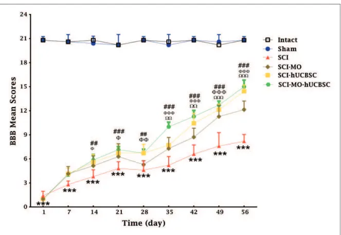
Figure 3. Effect of hUCBSC-MO treatment on motor function after SCI. Intraperitoneal injection of MO (150 mg/kg) was started 1 day after injury and continued once a day for 14 days after injury. Intraspinal grafting of hUCBSCs was started 24 hr after injury. Data are represented as mean of BBB scoreSEM, (n¼5–7) and analyzed by two-way ANOVA followed by post hoc Bonferroni’s multiple comparison test. ***p<.001 shows significant different between SCI versus intact., andshow significant difference between
SCI-MO and SCI.,, andshow significant difference between SCI-hUCBSC and SCI. ##, and ### show significant difference
between SCI-MO-hUCBSC and SCI (p<.05,p<.01, andp<.001, respectively).Note. hUCBSCs¼human umbilical cord blood stem cells;
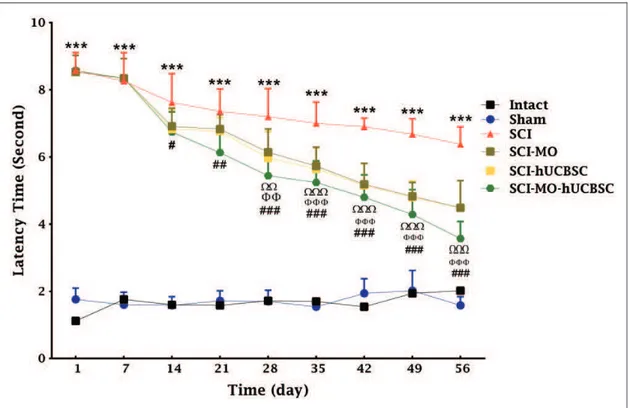
Figure 4. Effect of hUCBSC-MO treatment on sensory function after SCI. Intraperitoneal injection of MO (150 mg/kg) was started 1 day after injury and continued once a day for 14 days after injury. Intraspinal grafting of hUCBSCs was started 24 hr after injury. Data are represented as mean of latency timeSEM, (n¼5–7) and analyzed by two-way ANOVA followed by post hoc Bonferroni’s multiple comparison test. ***p<.001 significant difference between SCI versus intact., andshow significant difference between SCI-MO
and SCI., andshow significant difference between SCI-hUCBSC and SCI. #, ##, and ### show significant difference between
SCI-MO-hUCBSC and SCI (p<.05,p<.01, andp<.001, respectively).Note. hUCBSCs¼human umbilical cord blood stem cells;
MO¼Melissa officinalis; ANOVA¼analysis of variance; SCI¼spinal cord injury.
Figure 5. Effect of hUCBSC-MO on electromyographic activity after SCI. Intraperitoneal injection of MO (150 mg/kg) was started a day after injury and continued once a day for 14 days after injury. Intraspinal grafting of hUCBSCs was started 24 hr after injury. Data are represented as mean of recruitment indexSEM, (n¼5–7) and analyzed by two-way ANOVA followed by post-hoc Bonferroni’s multiple comparison test. ***p<.001 shows significant difference between SCI versus intact. ##p<.01 and ###p<.001 versus spinal cord injury.
F(40, 270)¼29.37, p<.001. Application of post hoc Bonferroni’s multiple comparison tests showed significant improvement in motor function following 150 mg/kg MO treatment on Day 35, 42 (p<.01), 49, and 56 (p<.001) and hUCBSCs therapy on Day 14, 21 (p<.05), 28 (p<.01), 35, 42, 49, and 56 (p<.001) in comparison with SCI group. Furthermore, the combination of MO and hUCBSCs significantly enhanced motor function on Day 14, 28 (p<.01), 21, 35, 42, 49, and 56 (p<.001) in com-parison with SCI group. There were no significant dierences between SCI-MO, SCI-hUCBSC, and SCI-MO-hUCBSC groups (Figure 3).
Statistical evaluations showed that the mean latency time of response to the painful stimulus decreased signifi-cantly in SCI-hUCBSC group when compared with SCI group. When intraperitoneal MO treatment (150 mg/kg) was added a day after the injury, it significantly enhanced
sensory recovery in rats when compared with SCI group. The application of two-way ANOVA showed significant interaction between variables including hUCBSCs, MO
treatment (150 mg/kg), and time, F(40, 270)¼12.39,
p<.001. Application of post hoc Bonferroni’s multiple comparison tests showed significant improvement in sen-sory function following 150 mg/kg MO treatment on Day 28 (p<.01), 35, 42, 49, and 56 (p<.001) and hUCBSCs therapy on Day 28 (p<.01), 35, 42, 49, and 56 (p<.001) in comparison with SCI group. Moreover, combination of MO and hUCBSCs significantly improved motor func-tion on Day 14 (p<.05), 21 (p<.001), 28, 35, 42, 49, and 56 (p<.001) in comparison with SCI group. There were
no significant differences between SCI-MO,
SCI-hUCBSC, and SCI-MO-hUCBSC groups (Figure 4).
Figure 6. Effect of hUCBSC-MO treatment on cavity formation after SCI. Intraperitoneal injection of MO (150 mg/kg) was started one day after injury and continued once a day for 14 days after injury. Intraspinal grafting of hUCBSCs was started 24 hr after injury. Data are represented as mean of the cavity areaSEM (n¼5–7) and analyzed by one-way ANOVA followed by post hoc
Bonferroni’s multiple comparison test. ***p<.001 shows significant
difference between SCI versus intact. ##p<.01, and ###p<.001
versus spinal cord injury.shows significant difference between
SCI-MO-hUCBSC and SCI-hUCBSC (p<.05). $ shows significant
difference between SCI-MO-hUCBSC and SCI-MO (p<.05,
p<.01, andp<.001, respectively).Note. hUCBSCs¼human
umbilical cord blood stem cells; MO¼Melissa officinalis; SCI¼spinal cord injury; SEM¼standard error of the mean; ANOVA¼analysis of variance.
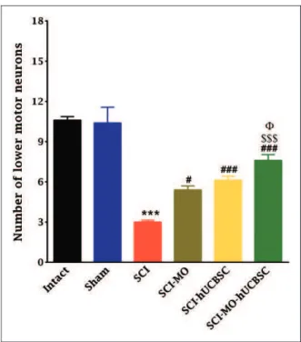
Figure 7. Effect of hUCBSC-MO treatment on cell loss in ventral horn of spinal cord after injury. Intraperitoneal injection of MO (150 mg/kg) was started one day after injury and continued once a day for 14 days after injury. Intraspinal grafting of hUCBSCs was started 24 hr after injury. Data are represented as mean number of ventral horn motor neuronsSEM (n¼5–7) and analyzed by one-way ANOVA followed by post hoc Bonferroni’s multiple compari-son test. ***p<.001 shows significant difference between SCI
versus intact. #p<.05 and ###p<.001 versus spinal cord injury.
show significant difference between MO-hUCBSC and SCI-hUCBSC (p<.05). $$$ shows significant difference between
SCI-MO-hUCBSC and SCI-MO (p<.05,p<.01, andp<.001,
respectively).Note. hUCBSCs¼human umbilical cord blood stem cells; MO¼Melissa officinalis; SCI¼spinal cord injury;
Electrophysiological Results
Combination of MO extract and hUCBSCs transplantation increased the recruitment pattern of hind limbs after SCI.
Although after the application of two-way ANOVA, there was no significant dierence between the right and left hind limb. The statistical analysis indicated that the means of recruitment index were significantly increased for left and right hind limbs in SCI-MO, SCI-hUCBSC, and SCI-MO-hUCBSC groups when compared with SCI group, F(5, 60)¼0.01, p<.001. Application of post-hoc Bonferroni’s
multiple comparisons test as well as Bartlett’s test for equal variances showed significant improvement in electro-physiological activity of left and right hind limbs following 150 mg/kg of MO extract administration (p<.01), hUCBSC therapy, and hUCBSC-MO treatment (p<0.001) in com-parison with SCI group. There were no significant dierences between SCI-MO, SCI-hUCBSC, and SCI-MO-hUCBSC groups (Figure 5).
Histological Results
Efficacy of MO-hUCBSCs treatment in reduction of cavity forma-tion after SCI. In the intact group, spinal cord segments were
not damaged in both white and gray matter. Application of one-way ANOVA demonstrated that the mean cavity size in terms of mm2was significantly reduced in treatment groups,
F(5, 30)¼27.95,p<.001. Moreover, post hoc Bonferroni’s multiple comparison test illustrated the significant reduction in the mean cavity area in SCI-MO, SCI-hUCBSC (p<.01), and SCI-MO-hUCBSC (p<.001) groups when compared with SCI group. Furthermore, application of one-way ANOVA showed that mean cavity area in SCI-MO-hUCBSC group was significantly reduced in comparison with SCI-MO and SCI-hUCBSC groups (p<.05; Figure 6).
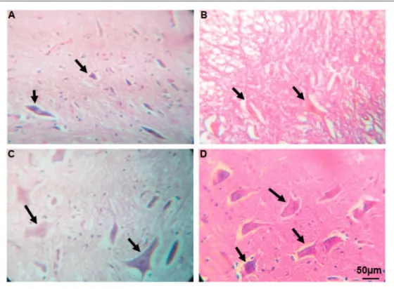
Combination of MO extract and hUCBSCs transplantation decreased the lesioning of lower motor neurons in ventral horn of spinal cord after its injury. Statistical evaluations showed the significant dierences between SCI-MO, SCI-hUCBSC, and SCI-MO-hUCBSC groups when compared with SCI group based on the number of ventral horn lower motor neurons, F(5, 30)¼30.86, p<.001. Application of post hoc Bonferroni’s multiple comparisons test as well as Bartlett’s test for equal variances revealed significant increase in the number of ventral horn motor neurons in SCI-MO (p<.05), SCI-hUCBSC, and SCI-MO-hUCBSC
(p<.001) treatment groups when compared with SCI group. Moreover, application of one-way ANOVA showed signifi-cant dierence in SCI-MO-hUCBSC group when compared with SCI-MO (p<.001) and SCI-hUCBSC (p<.05) groups (Figures 7 and 8).
Immunohistochemistry and TEM Results
Effects of MO extract administration along with hUCBSCs trans-plantation on GFAP expression after SCI. Statistical evaluations revealed that the number of GFAPþastrocytes was signifi-cantly increased in SCI group. However, this activation was significantly attenuated in the treatment groups, F(5, 30)¼45.49,p<.001. Application of post hoc Bonferroni’s multiple comparisons test as well as Bartlett’s test for equal variances showed a significant reduction in the GFAP
expression in SCI-MO, SCI-hUCBSC (p<.01), and SCI-MO-hUCBSC (p<.001) treatment groups when compared with SCI group. Statistically significant dierence was found in GFAP expression between SCI-MO-hUCBSC group in comparison with SCI-MO (p<.01) and SCI-hUCBSC (p<.05) groups (Figures 9 and 10).
Data obtained revealed that the density of astrogliosis in the ventral horn of spinal cord was significantly reduced in treatment groups in comparison with SCI
group, F(5, 30)¼17.66, p<.001. Moreover, post hoc
Bonferroni’s multiple comparison test showed that the density of gliosis was significantly reduced in
SCI-hUCBSC (p<.01) and SCI-MO-hUCBSC (p<.001)
groups when compared with SCI group. Statistical evalu-ations showed significant differences between
SCI-MO-hUCBSC, SCI-MO (p<.001), and SCI-hUCBSC
(p<.05) groups (Figure 11).
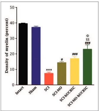
Effects of MO extract administration along with hUCBSCs trans-plantation on myelination after SCI. Application of one-way ANOVA demonstrated that density of myelin in the dorsal white matter of spinal cord was significantly increased in treatment groups when compared with SCI group, F(5, 30)¼62.11, p<.001. Furthermore, post hoc Bonferroni’s multiple comparison test revealed that the density of myelin was significantly increased in SCI-MO (p<.05), SCI-hUCBSC, and SCI-MO-hUCBSC (p<.001) groups compared with SCI group. Furthermore, statistical evalu-ations showed significant dierences between SCI-MO-hUCBSC, SCI-MO (p<.001), and SCI-hUCBSC (p<.05) groups (Figures 12 and 13).
Conversely, evaluation of electron microscopic pic-tures from all groups by application of one-way ANOVA revealed that MI was reduced in treatment
groups, F(5, 6)¼102.1, p<.001. Moreover, post hoc
Bonferroni’s multiple comparison test showed that MI was significantly reduced in SCI-MO, SCI-hUCBSC (p<.01), and SCI-MO-hUCBSC (p<.001) groups than in SCI group. Statistical evaluations showed significant differences between SCI-MO-hUCBSC, SCI-MO, and SCI-hUCBSC (p<.05) groups (Figures 13 and 14).
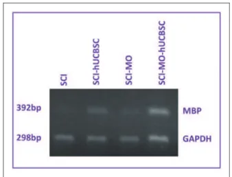
RT-PCR Results
Combination of MO extract and hUCBSCs transplantation enhanced expression of MBP after SCI. For further confirm-ation of myelinconfirm-ation process and the synthesis of MBP by MO-hUCBSC treatment after SCI, RT-PCR analysis was used. There was a change in the mRNA levels after SCI was determined utilizing standardized RT-PCR analysis. Qualitative analysis of RT-PCR findings in all groups revealed considerable upregulation of mRNA gene of MBP in SCI-MO-hUCBSC treated group when compared with SCI group. Application of one-way ANOVA revealed that the density of RT-PCR bands was increased in treatment
Figure 9. Effect of hUCBSC-MO treatment on astrogliosis for-mation in ventral horn of spinal cord after injury. Intraperitoneal injection of MO (150 mg/kg) was started one day after injury and continued once a day for 14 days after injury. Intraspinal grafting of hUCBSCs was started 24 hr after injury. Data are represented as mean of GFAP-positive astrocytesSEM (n¼5–7) and analyzed by one-way ANOVA followed by post hoc Bonferroni’s multiple comparison test. ***p<.001 shows significant difference between
SCI versus intact. ##p<.01, and ###p<.001 versus spinal cord
injury.shows significant difference between SCI-MO-hUCBSC
and SCI-hUCBSC (p<.05). $$ shows significant difference between
SCI-MO-hUCBSC and SCI-MO (p<.05,p<.01, andp<.001,
respectively).Note. hUCBSCs¼human umbilical cord blood stem
groups, F(5, 6)¼1077, p<.001. Moreover, post hoc Bonferroni’s multiple comparison test revealed that the density of RT-PCR bands was significantly increased in SCI-MO (p<.01), SCI-hUCBSC. and SCI-MO-hUCBSC (p<.001) groups than in SCI group. Application of one-way ANOVA showed a significant dierence between SCI-MO-hUCBSC, SCI-MO, and SCI-hUCBSC groups (p<.05; Figures 15 and 16).
Discussion
Although some research work have indicated that stem cell transplantation for treatment of SCI is unsuccessful (Ru˚zˇicˇka et al., 2013), many investigations have demon-strated that stem cells are effective in SCI (Veeravalli et al., 2009; Caron et al., 2016; Satti et al., 2016). As a result, there has been controversy about this issue.
This research was hinged on the promotion of the therapeutic properties of hUCBSCs in combination with a neuroprotective agent to induce curative effects to SCI in rats. The present study demonstrated that com-bination of MO extract and hUCBSCs transplantation has neuroprotective properties in treatment of SCI. Although this combination promoted the motor, sensory, and EMG functions, there were no significant differences
between SCI-MO, SCI-hUCBSCs, and
SCI-MO-hUCBSC groups. A number of previous investigations have revealed functional improvement after transplant-ation of hUCBSCs in SCI (Saporta et al., 2003; Kuh et al., 2005; Dasari, Spomar, et al., 2008; Rodrigues et al., 2012). Previous research has shown that adminis-tration of MO extract itself improved neurological and cellular outcomes in rat SCI (Hosseini et al., 2015). MO has neuroprotective and neurotrophic effects, including
promotion of functional recovery, thereby suggesting that it has therapeutic effect on neurodegenerative dis-eases (Bayat et al., 2012; Sepand et al., 2013). MO has acetylcholinesterase inhibitory properties (Dastmalchi et al., 2009). Anticholinesterases increase the residence time of acetylcholine in the synapse. This allows rebind-ing of the transmitter to nicotinic receptors. It thus gives acetylcholine the competitive advantage over the neuro-muscular blocking agent (Nair and Hunter, 2004). Our results revealed that the combination of hUCBSCs and MO prevented cell loss, formation of cavity, and astro-gliosis in ventral horn and also enhances the myelination in the dorsal white matter of spinal cord after injury. The effect of hUCBSCs and MO on enhancing functional recovery, myelination, and reducing astrogliosis and cavity formation is likely attributable to the inhibition of pro-inflammatory cytokines. Inflammatory processes have fundamental roles in the pathophysiology of SCI.
Figure 11. Effect of hUCBSC-MO treatment on density of astrogliosis in ventral horn of spinal cord after injury.
Intraperitoneal injection of MO (150 mg/kg) was started one day after injury and continued once a day for 14 days after injury. Intraspinal grafting of hUCBSCs was started 24 hr after injury. Data are represented as mean of gliosis densitySEM (n¼5–7) and analyzed by one-way ANOVA followed by post hoc Bonferroni’s multiple comparison test. ***p<.001 shows significant difference
between SCI versus intact. #p<.05, ##p<.01, and ###p<.001
versus spinal cord injury.shows significant difference between
SCI-MO-hUCBSC and SCI-hUCBSC (p<.05). $$ shows significant
difference between SCI-MO-hUCBSC and SCI-MO (p<.05,
p<.01, andp<.001, respectively).Note. hUCBSCs¼human
umbilical cord blood stem cells; MO¼Melissa officinalis; SCI¼spinal cord injury; SEM¼standard error of the mean; ANOVA¼analysis of variance.
Figure 12. Effect of hUCBSC-MO treatment on density of myelin in dorsal white matter of spinal cord after injury. Intraperitoneal injection of MO (150 mg/kg) was started one day after injury and continued once a day for 14 days after injury. Intraspinal grafting of hUCBSCs was started 24 hr after injury. Data are represented as mean of myelin densitySEM (n¼5–7) and analyzed by one-way ANOVA followed by post hoc Bonferroni’s multiple comparison test. ***p<.001 shows significant difference between SCI versus
intact. #p<.05 and ###p<.001 versus spinal cord injury.shows
significant difference between SCI-MO-hUCBSC and SCI-hUCBSC (p<.05). $$ shows significant difference between
SCI-MO-hUCBSC and SCI-MO (p<.05,p<.01, andp<.001, respectively).
Pro-inflammatory cytokines, including IL-1 and TNF-a, are released by activation of neurons, astrocytes, microglia, and endothelial cells after injury. Thereafter, the secondary inflammatory response and activation of IL-6 and IL-8 are induced by those cytokines (Zhu et al., 2014).
It has been shown that, transplantation of human umbilical cord blood mesenchymal stem cells (hUCB-MSCs) decreases the number of activated microglia and inhibits the permeation of immune cells and cellular apoptosis in the brain after ischemic brain injury (Yang et al., 2013; Zhu et al., 2014; Wang et al., 2016). Researchers have shown that the injection of hUCB-MSCs throughout the early stage of ischemic brain
injury decreased the IL-1b, IL-6, and TNF-aexpression
levels in the serum and increased IL-10 expression levels. The significant increase of pro-apoptotic genes such as Bad, Bax, p53, AFAP1, caspase 3, and caspase 9 has
been observed after the SCI (Sabapathy et al., 2015). IL-10 can decrease the expression of those genes, oxygen free radicals, and cytokines. Upregulation of
IL-1b, IL-6, and TNF-a can initiate neuronal death and
improve the synthesis of nitric oxide after injury. IL-1b
can improve the intracellular calcium concentration and release neurotropic factors, which can induce neuronal
apoptosis. Moreover, TNF-a can induce arachidonic
acid metabolite release, which enhance extracellular accu-mulation of glutamate and generate neurodegenerative toxicity. Glutamate-induced cell death in the CNS con-tains upregulation of caspase 3 and its activation via a caspase-dependent pathway involves mitochondrial sig-naling. hUCBSCs decline caspase-3 and -7 activities and are responsible for activation of the Akt pathway
and regulation of N-methyl-D-aspartic acid receptors,
thereby giving neuroprotection to cortical neurons (Dasari, Veeravalli, et al., 2008). Therefore, hUCBSCs
Figure 13. Ultra structural characteristics of myelination in dorsal white matter of spinal cord at the level of T12-L1 of all groups which were evaluated in this study on Day 56. (b) Low power view reveals the distribution of myelinated axons. Three representative high power photographs show the typical appearance of myelinated axons with extensive myelin sheath wrapped around an axon (a, d, and e). Densitometry of MBP in dorsal white matter of spinal cord at the level of T12-L1 is shown in the left part of any electron microscopy pictures. a¼Intact, b¼SCI, c¼SCI-MO, d¼SCI-hUCBSC, e¼SCI-MO-hUCBSC.Note. MBP¼myelin basic protein; hUCBSC¼human
transplantation could provide neuroprotection by regulating the balance of pro- and anti-inflammatory cytokines.
Conversely, it has been shown that MO extracts have anti-inflammatory properties (Bounihi et al., 2013; Mu¨zell et al., 2013). Its anti-inflammatory effects are due to rosmarinic acid, flavonoids, and terpenoids present in the extract. Probably, flavonoids have a more effective role by facilitating the synthesis of prostaglandin. MO administration can suppress the pro-inflammatory
cytokines such as IL-1b, IL-6, and TNF-a (Bounihi
et al., 2013).
Our results have demonstrated that the combination of MO extract administration and hUCBSCs transplant-ation prevented cell loss and enhanced myelintransplant-ation. A possible explanation to this is that, the transplantation of hUCBSCs can increase the length of neurofilament-positive fibers and increase the numbers of growth
cone-like structures at the lesion site. Grafted
hUCBSCs can survive, move over short distances, and produce large amounts of glial cell line-derived neuro-trophic factors and neurotrophin-3 (NT3) in the host spinal cord (Rodrigues et al., 2012). It has also been con-firmed that the hUCBSCs can form morphologically myelin sheaths in the spinal cord. It has been revealed that oligodendrocytes derived from human umbilical cord blood secrete NT3 and brain-derived neurotrophic factor. Cord blood stem cells promote the synthesis of MBP and proteolipid protein in the injured areas, thereby facilitating the process of remyelination (Dasari et al., 2009). Conversely, a large number of experimental evi-dences support oxidative stress as important mediators of secondary cell death after SCI (Cuzzocrea and Genovese, 2008; Jia et al., 2012; Fatima et al., 2015). It has been shown that administration of MO extract has powerful antioxidant effects which are probably exerted through
Figure 14. The effect of hUCBSC-MO treatment in reducing of myelin index in dorsal white matter of the spinal cord after injury. Intraperitoneal injection of MO (150 mg/kg) was started one day after injury and continued once a day for 14 days after injury. Intraspinal grafting of hUCBSCs was started 24 hr after injury. Data are represented as mean of myelin indexSEM, (n¼2) and
ana-lyzed by one-way ANOVA followed by post-hoc Bonferroni’s mul-tiple comparison test. ***p<.001 shows significant difference
between SCI versus intact. ##p<.01, and ###p<.001 versus
spinal cord injury.shows significant difference between
SCI-MO-hUCBSC and SCI-SCI-MO-hUCBSC (p<.05). $ shows significant difference
between SCI-MO-hUCBSC and SCI-MO (p<.05,p<.01, and
p<.001, respectively).Note. hUCBSCs¼human umbilical cord
blood stem cells; MO¼Melissa officinalis; SCI¼spinal cord injury; SEM¼standard error of the mean; ANOVA¼analysis of variance.
Figure 15. The effect of hUCBSC-MO treatment in upregulation of myelin basic protein in the spinal cord after injury. Intraperitoneal injection of MO (150 mg/kg) was started one day after injury and continued once a day for 14 days after injury. Intraspinal grafting of hUCBSCs was started 24 hr after injury. Data are represented as mean of RT-PCR bands densitySEM (n¼2) and analyzed by one-way ANOVA followed by post hoc Bonferroni’s multiple compari-son test. ***p<.001 shows significant difference between SCI
versus intact. #p<.05, ##p<.01, and ###p<.001 versus spinal
cord injury.shows significant difference between
SCI-MO-hUCBSC and SCI-SCI-MO-hUCBSC (p<.05). $, $$, and $$$ show
signifi-cant difference between SCI-MO-hUCBSC and SCI-MO (p<.05,
the rosmarinic acid and the benzodioxole present in the extract. Moreover, compounds such as linoleic acid, car-nosic acid, and ursolic acid are also present in the extracts, all of which have antioxidant properties.
The present study demonstrated that, the combination of MO and hUCBSCs inhibited astrogliosis. This issue has shown that the matrix metalloproteinase (MMP) as a proteolytic enzyme advances functional recovery after SCI by directing the development of a glial scar. Treatment with hUCBSCs after SCI altered the expression of different MMPs in rats. hUCBSCs transplantation in SCI causes upregulation of MMP-2 and therefore reduced the development of the glial scar at the lesion site (Veeravalli et al., 2009). In addition, as mentioned earlier, MO extract can inhibit the pro-inflammatory cytokines and reactive oxygen species. These two factors are key medi-ators of reactive astrogliosis in SCI (Bharne et al., 2013).
The limitation of this study is the nonmeasurement of the inflammatory factors and immunostaining for macro-phage markers (F4/80 or Iba-1). Further investigations on differentiation of hUCBSCs along with the use of MO extracts will provide further evidence regarding the therapeutic effectiveness of hUCBSCs after SCI.
Conclusions
SCI causes motor and sensory dysfunction, tissue deformity, and cell death, the formation of astrogliosis, and degeneration of axons. In conclusion, hUCBSCs enhanced motor and sensory dysfunction as well as pro-moting morphological improvement in SCI contusion
model in comparison with SCI. Our results showed that MO extract can promote the neuroprotective effects of hUCBSCs. Further studies are needed to clarify the underlying mechanisms of these results.
Author Notes
The authors alone are responsible for the content and writing of this paper.
Acknowledgments
The authors would like to thank the Neuroscience Research Center of Baqiyatallah, University of Medical Sciences for supporting this research. This research is dedicated to MAHAK (The Society to Support Children Suffering from Cancer) and to the National Cancer Institute (NCI) for helping little children.
Declaration of Conflicting Interests
The authors declared no potential conflicts of interest with respect to the research, authorship, and/or publication of this article.
Funding
The authors received no financial support for the research, author-ship, and/or publication of this article.
References
Agrawal, G., Kerr, C., Thakor, N. V., & All, A. H. (2010). Characterization of graded MASCIS contusion spinal cord injury using somatosensory evoked potentials.Spine(Phila Pa 1976),35, 1122–1127.
Barros Filho, T. E. P. D, & Molina, A. E. I. S. (2008). Analysis of the sensitivity and reproducibility of the Basso, Beattie, Bresnahan (BBB) scale in Wistar rats. Clinics (Sao Paulo), 63, 103–108.
Bayat, M., Tameh, A. A., Ghahremani, M. H., Akbari, M., Mehr, S. E., Khanavi, M., & Hassanzadeh, G. (2012). Neuroprotective properties ofMelissa officinalis after hypoxic-ischemic injury both in vitro and in vivo.Daru,20, 42–50.
Bharne, A. P., Upadhya, M. A., Shelkar, G. P., Singru, P. S., Subhedar, N. K., & Kokare, D. M. (2013). Neuroprotective effect of cocaine-and amphetamine-regulated transcript peptide in spinal cord injury in mice.Neuropharmacology,67, 126–135. Bounihi, A., Hajjaj, G., Alnamer, R., Cherrah, Y., & Zellou, A. (2013). In vivo potential anti-inflammatory activity ofMelissa officinalis L. essential oil. Advances in Pharmacological Sciences,2013, 101759.
Byrnes, K. R., Fricke, S. T., & Faden, A. I. (2010). Neuropathological differences between rats and mice after spinal cord injury. Journal of Magnetic Resonance Imaging, 32, 836–846.
Caron, I., Rossi, F., Papa, S., Aloe, R., Sculco, M., Mauri, E.,. . .Parazzi, V. (2016). A new three dimensional biomimetic hydrogel to deliver factors secreted by human mesenchymal stem cells in spinal cord injury.Biomaterials,75, 135–147. Chhabra, H. S., & Sarda, K. (2015). Stem cell therapy in spinal
trauma: Does it have scientific validity? Indian Journal of Orthopaedics,49, 56–71.
Coutts, M., & Keirstead, H. S. (2008). Stem cells for the treatment of spinal cord injury.Experimental Neurology,209, 368–377. Cuzzocrea, S., & Genovese, T. (2008). Role of free radicals and
poly (ADP-ribose) polymerase-1 in the development of spinal cord injury: New potential therapeutic targets. Current Medicinal Chemistry,15, 477–487.
Dasari, V. R., Spomar, D. G., Gondi, C. S., Sloffer, C. A., Saving, K. L., Gujrati, M.,. . .Dinh, D. H. (2007). Axonal remyelination by cord blood stem cells after spinal cord injury. Journal of Neurotrauma,24, 391–410.
Dasari, V. R., Spomar, D. G., Li, L., Gujrati, M., Rao, J. S., & Dinh, D. H. (2008). Umbilical cord blood stem cell mediated down-regulation of Fas improves functional recovery of rats after spinal cord injury.Neurochemical Research,33, 134–149. Dasari, V. R., Veeravalli, K. K., & Dinh, D. H. (2014).
Mesenchymal stem cells in the treatment of spinal cord injuries: A review.World Journal of Stem Cells,6, 120–133.
Dasari, V. R., Veeravalli, K. K., Saving, K. L., Gujrati, M., Fassett, D., Klopfenstein, J. D.,. . .Rao, J. S. (2008). Neuroprotection by cord blood stem cells against glutamate-induced apoptosis is mediated by Akt pathway. Neurobiology of Disease, 32, 486–498.
Dasari, V. R., Veeravalli, K. K., Tsung, A. J., Gondi, C. S., Gujrati, M., Dinh, D. H., & Rao, J. S. (2009). Neuronal apoptosis is inhibited by cord blood stem cells after spinal cord injury. Journal of Neurotrauma,26, 2057–2069.
Dastmalchi, K., Ollilainen, V., Lackman, P., Boije af Genna¨s, G., Dorman, H. J., Ja¨rvinen, P. P.,. . .Hiltunen, R. (2009). Acetylcholinesterase inhibitory guided fractionation ofMelissa officinalisL.Bioorganic & Medicinal Chemistry,17, 867–871. Deng, L.-X., Hu, J., Liu, N., Wang, X., Smith, G. M., Wen, X., & Xu, X.-M. (2011). GDNF modifies reactive astrogliosis allow-ing robust axonal regeneration through Schwann cell-seeded guidance channels after spinal cord injury. Experimental Neurology,229, 238–250.
Edalat, H., Hajebrahimi, Z., Pirhajati, V., Movahedin, M., Tavallaei, M., Soroush, M.-R., & Mowla, S. J. (2013). Transplanting p75-suppressed bone marrow stromal cells pro-motes functional behavior in a rat model of spinal cord injury. Iranian Biomedical Journal,17, 140–145.
Faramarzi, H., Mehrabani, D., Fard, M., Akhavan, M., Zare, S., Bakhshalizadeh, S.,. . .Shirazi, R. (2016). The potential of men-strual blood-derived stem cells in differentiation to epidermal lineage: A preliminary report.World Journal of Plastic Surgery, 5, 26–31.
Fatima, G., Sharma, V., Das, S., & Mahdi, A. (2015). Oxidative stress and antioxidative parameters in patients with spinal cord injury: Implications in the pathogenesis of disease.Spinal Cord, 53, 3–6.
Fleming, J. C., Norenberg, M. D., Ramsay, D. A., Dekaban, G. A., Marcillo, A. E., Saenz, A. D.,. . .Weaver, L. C. (2006). The cellular inflammatory response in human spinal cords after injury.Brain,129, 3249–3269.
Fouad, K., Bennett, D. J., Vavrek, R., & Blesch, A. (2013). Long-term viral brain-derived neurotrophic factor delivery promotes spasticity in rats with a cervical spinal cord hemisection. Frontiers in Neurology,4, 187.
Geoffroy, C. G., & Zheng, B. (2014). Myelin-associated inhibitors in axonal growth after CNS injury. Current Opinion in Neurobiology,27, 31–38.
Hosseini, R., Kaka, G., Joghataei, M., Hooshmandi, M., Sadraie, S., Mansouri, K.,. . .Mohammadi, A. (2015). Neuroprotective effect of Melissa officinalis in animal model of spinal cord injurya. Medicinal & Aromatic Plants. doi:10.4172/2167-0412.S1-004.
Jia, Z., Zhu, H., Li, J., Wang, X., Misra, H., & Li, Y. (2012). Oxidative stress in spinal cord injury and antioxidant-based intervention.Spinal Cord,50, 264–274.
Kamdem, J. P., Adeniran, A., Boligon, A. A., Klimaczewski, C. V., Elekofehinti, O. O., Hassan, W.,. . .Athayde, M. L. (2013). Antioxidant activity, genotoxicity and cytotoxicity evaluation of lemon balm (Melissa officinalis L.) ethanolic extract: Its potential role in neuroprotection. Industrial Crops and Products,51, 26–34.
Kim, J. Y., Oh, C. H., Huang, X., Kim, M. H., Yoon, S. H., Kim, K. H.,. . .Choi, B. H. (2013). Improvement in sensory function via granulocyte-macrophage colony-stimulating factor in rat spinal cord injury models: Laboratory investigation. Journal of Neurosurgery: Spine,18, 69–75.
Kuh, S.-U., Cho, Y.-E., Yoon, D.-H., Kim, K.-N., & Ha, Y. (2005). Functional recovery after human umbilical cord blood cells transplantation with brain-derived neutrophic factor into the spinal cord injured rat. Acta Neurochirurgica (Wien), 147, 985–992.
Li, J., & Lepski, G. (2013). Cell transplantation for spinal cord injury: A systematic review. BioMed Research International, 2013, 786475.
Mu¨zell, D. P., Lunardelli, A., Leite, C. E., Fagundes, R. M., Saciura, V. C., Reichel, C. L.,. . .Astarita, L. V. (2013). Nephroprotective and anti-inflammatory effects of aqueous extract of Melissa officinalis L, on acetaminophen-induced and pleurisy-induced lesions in rats. Brazilian Archives of Biology and Technology,56, 383–392.
Nair, V. P., & Hunter, J. M. (2004). Anticholinesterases and anti-cholinergic drugs. Continuing Education in Anaesthesia, Critical Care, & Pain,4, 164–168.
Niapour, A., Karamali, F., Nemati, S., Taghipour, Z., Mardani, M., Nasr-Esfahani, M. H., & Baharvand, H. (2012). Cotransplantation of human embryonic stem cell-derived neural progenitors and Schwann cells in a rat spinal cord con-tusion injury model elicits a distinct neurogenesis and functional recovery.Cell Transplantation,21, 827–843.
Pang, Y., Zheng, B., Kimberly, S. L., Cai, Z., Rhodes, P. G., & Lin, R. (2012). Neuron-oligodendrocyte myelination co-culture derived from embryonic rat spinal cord and cerebral cortex. Brain and Behavior,2, 53–67.
Pereira, R. P., Fachinetto, R., de Souza Prestes, A., Puntel, R. L., da Silva, G. N. S., Heinzmann, B. M.,. . .Morel, A. F. (2009). Antioxidant effects of different extracts fromMelissa officina-lis, Matricaria recutita, and Cymbopogon citratus. Neurochemical Research,34, 973–983.
Popovich, P. G., & Jones, T. B. (2003). Manipulating neuroinflam-matory reactions in the injured spinal cord: Back to basics. Trends in Pharmacological Sciences,24, 13–17.
Ru˚zˇicˇka, J., Romanyuk, N., Hejcˇl, A., Vetrik, M., Hruby´, M., Cocks, G.,. . .Jendelova´, P. (2013). Treating spinal cord injury in rats with a combination of human fetal neural stem cells and hydrogels modified with serotonin. Acta Neurobiologiae Experimentalis(Wars),73, 102–115.
Sabapathy, V., Tharion, G., & Kumar, S. (2015). Cell therapy aug-ments functional recovery subsequent to spinal cord injury under experimental conditions.Stem Cells International,2015, 132172.
Saporta, S., Kim, J.-J., Willing, A. E., Fu, E. S., Davis, C. D., & Sanberg, P. R. (2003). Human umbilical cord blood stem cells infusion in spinal cord injury: Engraftment and beneficial influ-ence on behavior. Journal of Hematotherapy & Stem Cell Research,12, 271–278.
Satti, H. S., Waheed, A., Ahmed, P., Ahmed, K., Akram, Z., Aziz, T.,. . .Malik, S. A. (2016). Autologous mesenchymal stromal cell transplantation for spinal cord injury: A phase I pilot study.Cytotherapy,18, 518–522.
Sepand, M. R., Soodi, M., Hajimehdipoor, H., Soleimani, M., & Sahraei, E. (2013). Comparison of neuroprotective effects of Melissa officinalis total extract and its acidic and non-acidic fractions against a b-induced toxicity. Iranian Journal of Pharmaceutical Research: IJPR,12, 415–423.
Sta˚lberg, E., Falck, B., Gilai, A., Jabre, J., Sonoo, M., & Todnem, K. (1998). Standards for quantification of EMG and neurogra-phy. The international federation of clinical neurophysiology. Electroencephalography and Clinical Neurophysiology Supplement,52, 213–220.
Veeravalli, K. K., Dasari, V. R., Tsung, A. J., Dinh, D. H., Gujrati, M., Fassett, D., & Rao, J. S. (2009). Human umbilical cord blood stem cells upregulate matrix metalloproteinase-2 in rats after spinal cord injury.Neurobiology of Disease,36, 200–212. Wang, G.-H., Liu, Y., Wu, X.-B., Lu, Y., Liu, J., Qin, Y.-R.,. . .Duan, H.-F. (2016). Neuroprotective effects of human umbilical cord–derived mesenchymal stromal cells com-bined with nimodipine against radiation-induced brain injury through inhibition of apoptosis.Cytotherapy,18, 53–64. Webb, A. A., Ngan, S., & Fowler, D. (2010). Spinal cord injury II:
Prognostic indicators, standards of care, and clinical trials. Canadian Veterinary Journal,51, 598–604.
Wrathall, J. R., Li, W., & Hudson, L. D. (1998). Myelin gene expression after experimental contusive spinal cord injury. The Journal of Neuroscience,18, 8780–8793.
Yang, Z., Chen, P., Yu, H., Luo, W., Pi, M., Wu, Y.,. . .Gou, Y. (2013). Combinatorial effects of conception and governor vessel electroacupuncture and human umbilical cord blood-derived mesenchymal stem cells on pathomorphologic lesion and cellu-lar apoptosis in rats with cerebral ischemia/reperfusion.Journal of Traditional Chinese Medicine,33, 779–786.