Universidade Federal de São Paulo, S. Paulo Brasil (UNIFESP):1P rofessor Adjunto e Chefe da Disciplina de Neuro c i ru rgia - Universidade Federal São Paulo - São Paulo, Brasil UNIFESP;2Laboratório de Técnicas Neuro c i r ú rgicas - Disciplina de Neuro c i ru rgia - UNIFESP; 3P rofessor Titular Departamento de Farmacologia Celular - UNIFESP;4P rofessor Adjunto de Neuropatologia Departamento de Patologia -UNIFESP.
Received 3 January 2005, received in final form 17 May 2005. Accepted 21 June 2005.
Dr. Mirto N. Prandini - Rua dos Crisântemos 117 - 040049-020 São Paulo SP - Brasil. E-mail: [email protected]
MILD HYPOTHERMIA REDUCES
POLYMORPHONUCLEAR LEUKOCYTES
INFILTRATION IN INDUCED BRAIN INFLAMMATION
Mirto N. Prandini
1, Antonio Neves Filho
2, Antonio J. Lapa
3, João N. Stavale
4ABSTRACT - Over the last 50 years deep hypothermia (230C) has demonstrated to be an excellent
neuro-p rotective agent in cerebral ischemic injury. Mild hyneuro-pothermia (31-330C) has proven to have the same
neu-roprotective properties without the detrimental effects of deep hypothermia. Mechanisms of injury that are exaggerated by moderate hyperthermia and ameliorated by hypothermia include, reduction of oxygen radical production, with peroxidase damage to lipids, proteins and DNA, microglial activation and ischemic depolarization, decrease in cerebral metabolic demand for oxygen and reduction of glycerin and excitatory amino acid (EAA) release. Studies have demonstrated that inflammation potentiates cerebral ischemic in-jury and that hypothermia can reduce neutrophil infiltration in ischemic regions. To further elucidate the mechanisms by which mild hypothermia produces neuro p rotection in ischemia by attenuating the inflamma-tory response, we provoked inflammainflamma-tory reaction, in brains of rats, dropping a substance that provokes a heavy inflammatory reaction. Two groups of ten animals underwent the same surgical pro c e d u re: the s k u l l bone was partially removed, the duramater was opened and an inflammatory substance (5% carr a g e e n i n ) was topically dropped. The scalp was sutured and, for the group that underwent neuroprotection, an ice bag was placed covering the entire skull surface, in order to maintain the brain temperature between 29,5-3 10C during 120 minutes. After three days the animals were sacrificed and their brains were examined. The
g roup protected by hypothermia demonstrated a remarkable reduction of polymorphonuclear leukocytes (PMNL) infiltration, indicating that mild hypothermia can have neuro p rotective effects by reducing the in-flammatory reaction.
KEY WORDS: cerebral hypothermia, brain protection, brain inflammation, hypothermia.
A hipotermia moderada reduz a infiltração leucocitária na inflamação encefálica induzida
RESUMO - Nos últimos 50 anos, a hipotermia tem demonstrado ser um excelente agente neuro p ro t e t o r nas lesões isquêmicas encefálicas. A hipotermia moderada (310C - 330C) provou também apresentar as m e
s-mas propriedades protetoras, sem os efeitos deletérios da hipotermia profunda. Dentre alguns mecanis-mos de lesão que são melhorados pela hipotermia e piorados pela hipertermia moderada, podemecanis-mos citar a diminuição da demanda de oxigênio pelo encéfalo e a redução da glicina e aminoácidos excitatórios, e v i-tando a produção de radicais de oxigênio, com aumento da peroxidase e conseqüente lesão aos lípides, p ro-teínas e DNA, assim como pela ativação microglial e despolarização isquêmica. Alguns estudos demonstra-ram que a inflamação potencializa a lesão isquêmica e que a hipotermia pode reduzir a infiltração leu-cocitária nas áreas isquêmicas. Para melhor elucidar os mecanismos pelos quais a hipotermia apre s e n t a efeito neuro p rotetor através da redução da inflamação, no processo isquêmico, escolhemos o método utili-zando a indução de uma reação inflamatória com a utilização de uma substância com capacidade pro m o-ver intensa reação inflamatória em encéfalos de ratos. Dois grupos de dez animais foram submetidos a um mesmo procedimento cirúrgico: a calota craniana foi parcialmente removida, a duramáter aberta e uma substância com potente efeito inflamatório (carragenina a 5%) foi gotejada. A pele foi suturada e, para o g rupo com neuro p roteção, uma bolsa de gelo foi colocada, cobrindo toda a superfície craniana, de modo a manter a temperatura encefálica entre 29,50C e 310C durante 120 minutos. Três dias após, os animais
foram sacrificados e os encéfalos examinados. O grupo protegido pela hipotermia apresentou considerável redução na infiltração leucocitária, demonstrando que a hipotermia pode apresentar função neuro p ro t e-tora por meio de uma redução no processo inflamatório.
experimental occlusion of the middle cerebral ar-tery in dogs. Deep hypothermia became an adju-vant method for neuro p rotection in cases where c i rc u l a t o ry arrest in complex aneurysm surg e ry was n e c e s s a ry. However the detrimental effects of pro-longed deep hypothermia including delayed re c o v-e ry from anv-esthv-esia, acidosis, hv-emodynamic comp ro-mise, blood hyperc o a g u l a b i l i t y, hypotension and m y o c a rdial arrhythmia have limited the use the t e c h n i q u e2 - 4.Over the last 20 years a large num-ber of studies have demonstrated that mild hypo-t h e rmia (31-330C) can have the same neuro p ro-tective effect provided by deep hypothermia in ischemic brain3 , 5 - 1 1. The mechanisms underlying this n e u ro p rotection have been attributed to decre a s e in cerebral metabolic demand to oxygen and re-duction of glycine and excitatory aminoacid (EAA) release. Glutamate release occurs 1-5 h after is-chemic onset and mild hypothermia can be pro t e c-tive even if delayed by 2 hours1 2. Corbett et al.1 0 demonstrated that delayed hypothermia re d u c e s focal ischemic injury. There f o re, although re d u c-ing EAA re l e a s e5and glycine and glutamate re-l e a s e1 2, mild hypothermia can have other neuro-p rotective effects other than EEA, glutamate and glycine release. Even more, while neuro p ro t e c t i o n by deep hypothermia can be explained by a dec re a-se in cerebral blood flow and metabolic demand f o r oxygen, this by itself cannot fully explain the equal-ly protection that has been shown when the tem-p e r a t u re is lowered by only a few degre e s1 3 , 1 4. A h i g h d e g ree of neuro p rotection was conferred by pos-tischemic cooling (2h) to 320C which is virt u a l l y equivalent to that observed with intraischemic co-oling at the same level in focal cerebral ischemia1 4. It has been documented that inflammation con-tributes significantly to cerebral injury following i s c h e m i a7 , 1 5. Inflammatory cells presumably pro m o-te ischemic cell damage by microvascular occlus i o n . This may prolong and intensify the ischemic e v e n t1 6. Cytotoxic inflammatory reactions caused by micro-glial activation and blood-borne neutrophils have been implicated in the pathogenesis of ischemia/re-p e rfusion brain injury2 , 3 , 1 4 , 1 7 - 1 9. Neutrophils began to infiltrate into an infarct area soon after ische-mia. Cytokine expression may be the earliest sign of the inflammatory response. Cytokines activate m i c roglia and stimulate expression of endothelial adhesion leading to leukocyte infiltration. PMNL
mechanisms by which hypothermia reduces ische-mic neuronal injury13,19,20.
Since it has been demonstrated that carr a g e-enin is a highly reactive substance with inflamma-t o ry pro p e rinflamma-t i e s2 1and brain inflammation can be induced with its subarachnoidal injection in brains of mice2 2, in this study we aimed to demonstrate that mild hypothermia can have neuroprotective e ffects in brains of rats submitted to inflammatory injury by means of the use of carrageenin.
METHOD
Animal protocols were approved by the Federal Uni-versity of S. Paulo animal’s ethic board. Institutional gui-delines were followed in all protocols. All animal exper-iments were conducted in accordance with the NIH gui-de for the care and use of laboratory animals (NIH publi-cation 80-23). All eff o rts were made to minimize ani-mal suffering, and only the sani-mallest number of aniani-mals were used to generate reliable scientific data.
Fig 1. Line of incision of the skin.
Fig 3. Brain of rat. Haematoxylin-eosin 200X. Slices obtained three days after 5% Carrageenin was dropped. No neuro p rotection was performed. Small necro t -ic area and marked inflammatory infiltration can be seen.
Fig 4. Brain of rat. Haematoxylin-eosin 200X. Slices obtained three days after 5% Carrageenin was dropped. Neuro p rotection with hypothermia (30oC) for
120 minutes. Macrophage infiltration with capillary proliferation can be seen.
T h ree groups of ten Wistar E.PM rats weighing bet-ween 290g and 330 g were studied.
G roup 1 - control group, Group 2 - received no pro-tection by hypothermia and; Group 3 - received pro t e c-tion by hypothermia.
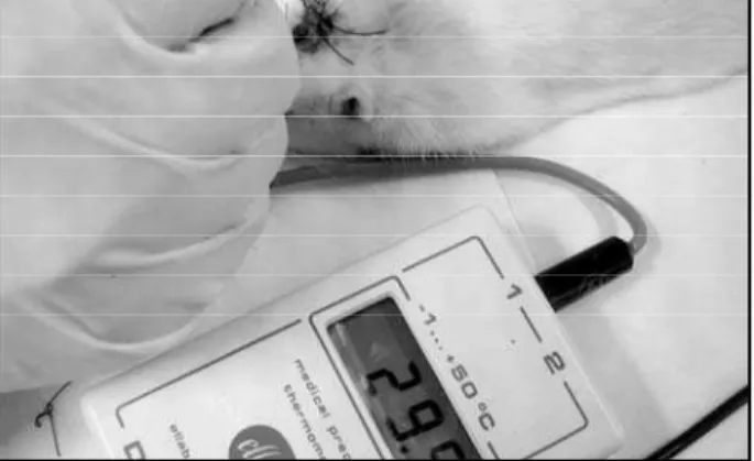
General anesthesia was given by means of IM injec-tion of tiletamin chloridrate + zolazepan chloridrate in the pro p o rtion of 100mg/kg .The animals were placed i n a specially designed table covered by an homeotherm i c blanket control unit (Harv a rd Apparatus Limited Cat 50-7079 Edenbridge Kent). The core temperature was main-tained in 370 C and was measured with a flexible fibber with the sensor tip placed into each animal’s rectum (El-lab medical precision thermometer DM 852). The scalp incision measuring 18mm was C shaped 4mm from the midline (Fig 1). The muscles and fascia were dissected f rom the cranium. Under an operating microscope a 10-12 mm burr hole was drilled and the dura was exposed. After the duramater had been incised, 2-3 drops of solu-tion of 5% beta-carrageenin was topically dropped. The scalp was sutured with mononylon 3-0. In the group 3, (the one protected by hypothermia), an ice bag was
pla-low 29,5 C and replaced when the temperature re a c h e d 3 10C. Brain temperature was measured with a needle
p robe placed subcutaneously inserted 5mm into the brain parenchyma (Fig 2) (Ellab medical precision ther-mometer DM 852 ). The hypothermia was maintained for 120-130 minutes. The control group underwent the same surgical procedure but no inflammatory solution was dropped, nor was hypothermia performed.
After 3 days the animals were anesthetized and sacri-ficed. All bone of the superior part of the skull was with-drawn in order to permit the removal of the whole brain that was immediately fixed in formalin 10,0%. The speci-mens were allowed to fix for 24 hours, and then embed-ded in paraffin.
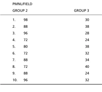
Histopatological examination –Brains were sliced into 18-µm-thick coronal sections and stained with he-matoxylin and eosin with magnification X40, X100, X200. High microscopy examination aimed to demonstrate the number of PMNL per field. Four fields were exami-ned. Total number of cells was counted using original magnification X 200. Histopatological analysis re v e a l e d acute lymphocytic and macrocytic cell pre d o m i n a n c e ; small areas of necrosis were also seen (Fig 3) in some ca-ses without neuro p rotection. Inflammatory infiltration was only seen in the specimens where carrageenin was d ropped (Fig 4). In brain hemisphere that has not re-ceived carrageenin (Group 1) no significant sign of i n f l a m-m a t o ry reaction was identified. Norm-mal and pathologi-cal areas can be seen in Figure 5. In group 2 and 3, the total number of PLNL is demonstrated in the Table 1.
Statisical analysis– Data are presented as means ± s t a n d a rd deviation. They were compared by analysis of variance followed by Mann Whitney Test. Data were considered different at p<0.5 (Fig 6).
DISCUSSION
R o s o m m o ff1first re p o rted that deep hypother-mia reduced ischemic damage after experimental occlusion of the MCA in dogs. The use of deep hy-p o t h e rmia has been limited because of several un-desirable side effects including acidosis, blood hyper-c o a g u l a b i l i t y, delayed re hyper-c o v e ry from anesthesia, hemodynamic compromise, myocardial arrh y t h m i a and hypotension2 - 4. More recently several studies have shown that mild hypothermia can have the same neuro p rotective effect in ischemic models using rodents, cats and dogs and the severe limitations as-sociated with deep hypothermia can be avoided5 - 1 0. Prandini et al.11have demonstrated that mild hypo-t h e rmia, locally produced in rabbihypo-t’s brains, can re d
uFig 6. Scatter plot of PMNL accumulated after topical Carr a g e -enin application.
GROUP 2 GROUP 3
1. 98 30
2. 88 38
3. 96 28
4. 72 24
5. 80 38
6. 72 32
7. 88 34
8. 72 40
9. 88 24
ce infarct size in cases of ischemia produced by coag-ulation of the middle cerebral art e ry.
It has been a matter of debate the mechanisms implicated in the neuro p rotective effects of hypo-t h e rmia. A large number of shypo-tudies have shown hypo-t h a hypo-t the mechanisms of neuro p rotection conferred b y moderate hypothermia are multifactorial5 , 1 4 - 1 6. M e-chanisms of injury that are ameliorated by hypo-t h e rmia and exaggerahypo-ted by moderahypo-te hyperhypo-t h e r-mia include, oxygen radical production, with pexidase damage to lipids, proteins and DNA, m i c ro-glial activation and ischemic depolarizat i o n5 , 1 4 - 1 6, d e c rease in cerebral metabolic demand for oxy-gen and reduction of EEA and glycerin re l e a s e1 2a l s o cannot fully account for the neuro p rotective ef-fect of hypothermia. Recent studies have demon-strated that inflammation is a major determ i n a n t of neural death following ischemia1 5 , 1 7 - 1 9 , 2 4. Focal i s-chemia and re p e rfusion of the neocortex elicit a substantial cell-mediated inflammatory re s p o n s e and produced cell infarc t i o n2 3 - 2 5. Cellular inflamma-tion is initiated by ischemia at the blood micro v a s-cular endothelial cell interface14.
Polymorphonuclear leukocytes are early part i c-ipants in the cerebral microvascular response to fo-cal ischemia1 6 , 1 8. The presence of PMNL in occluded m i c rovessels within 60 minutes after MCA occlusion in baboons has been demonstrated1 5. Developing i n f a rction was accompanied by accumulation of i n f l a m m a t o ry cells of both intrinsic (microglia) and extrinsic (macroglia) origin2 5. Treatment with anti-p y retic and antiinflammatory drug dianti-pyrone delays n e u ronal damage in the rat hippocampus, cort e x and striatum2 6. Anti-adhesion molecule antibodies selectively reduces apoptosis in transient middle ce-rebral art e ry in rat brain2 7. Toyoda et al.2 4d e m o n-strated that intraischemic hypothermia attenuates n e u t rophil infiltration in the rat neocortex after fo-cal ischemia-re p e rf u s i o n - i n j u ry. Kawai et al.1 3h a v e demonstrated that ischemic brain damage can be reduced with delayed hypothermia and pro l o n g e d postischemic hypothermia in a focal model of tran-sient cerebral ischemia in rats. One of the mecha-nisms by which hypothermia reduces ischemic neu-ronal injury is by attenuating the inflammatory re s-p o n s e1 3 , 1 9. Intraischemic hypothermia reduced the volume of infarction by 59% compared with the n o rm o t h e rmic animals. Since the accumulation of PMNL is maximal at 48-72h after ischemic insult, our specimens were obtained only 72 h after the inflam-m a t o ry reaction had begun.
While several studies have begun to elucidate the contribution of the inflammatory response to ce-rebral ischemic injury, the effect of hypothermia on the inflammatory response is still unclear7 , 1 0 , 1 5 , 1 7 - 1 9.
C a rrageenin was used in our experiments be-cause for more than 50 years it has been conside-red as one of the best drugs to produce inflamma-t o ry acinflamma-tiviinflamma-ty2 1and its inflammatory pro p e rties w h e n subarachnoidally injected have been demonstrat-e d2 2. Our results indicate that mild hypothermia e f-fectively reduced the leukocyte infiltration on b r a i n s of rats subjected to this potent inflammatory sub-stance. This could explain one additional mecha-nism of protection provided by mild hypotherm i a .
REFERENCES
1. R o s o m o ff HL. Hypothermia and cerebral vascular lesions 1; experi-mental interruption of the middle cerebral artery during hypothermia. J Neurosurg 1956;13:244-255.
2. Guegan Y, Scarabin JM, LeGilcher C, Guillion I. Extracorporeal circu-lation with deep hypothermia and circulatory arrest in the tre a t m e n t of aneurysm. Surg Neurol 1985;24:441-448.
3. Goto Y, Kassell NF, Hiramatsu K, Soleau SW, Lee KS. Effects of intrais-chemic hypothermia on cerebral damage in a model of reversible focal ischemia. Neurosurgery 1993;32:980-984.
4. Steen PA, Midle JH, Michenfelder JD. The detrimental effects of pro-longed hypothermia and rewarming in the dog. Anesthesiology 1980; 52:224-230.
5. Busto R, Dietrich WD, Globus MY, Ginsberg MD. The importance of brain temperature in cerebral ischemic injury. Stroke 1989;20:111 3 - 111 4 . 6. BakerCJ, Onesti ST, Barth KN, Prestigiacomo CJ, Solomon RA. Hy-pothermic protection following middle cerebral artery occlusion in the rat. Surg Neurol 1991;36:175-180.
7. Connolly ES, Wi n f ree CJ, Springer TA, et al. Cerebral protection in ho-mozygous null ICAM-I mice after middle cerebral artery occlusion: ro-le of neutrophil adhesion in the pathogenesis of stroke. J Clin In-vest1996;97:209-216.
8. Karibe H, Zarow GJ, Graham SH, Weinstein PR. Mild intraischemic hypothermia reduces postischemic hyperperfusion, delayed postische-mic hypoperfusion, blood-brain barrier disruption, brain edema, and n e u ronal damage volume after temporary focal cerebral ischemia in rats. J Cer Blood Flow Metab 1994;14:620-627.
9. Makarian GZ, Lee JH, Stein DJ, Hong SC. Mild hypothermia: thera-peutic window after experimental cerebral ischemia. Neuro s u rg e r y 1996;38:542-551.
10. Corbett D, Hamilton M, Colbourne F.Persistent neuroprotection with p rolonged postischemic hypothermia in adult rats subjected to tran-sient middle cerebral artery occlusion. Exp Neurol 2000;163:200-206. 11. Prandini MN, Lacanna SN, Valente PR, Stavale JN. Regional mild
hy-pothermia in the protection of the ischemic brain Acta Cir Bras 2002; 17:232-235.
12. Huang F, Zhou LF, Yang GY. Effects of mild hypothermia on the re l e a s e of regional glutamate and glycine during extended transient focal cere-bral ischemia in rats. Neurochem Res 1998;23:991-996.
13. Kawai N, Okauchi M, Morisaki K, Nagao S. Effects of delayed intrais-chemic and postisintrais-chemic hypothermia on a focal model of transient cerebral ischemia in rats Stroke 2000;31:1982-1989.
14. Colbourne F, Li H, Buchan AM. Indefatigable CAI sector neuro p ro t e c-tion with mild hypothermia induced 6 hours after severe fore b r a i n ischemia in rats. J Cer Blood Flow Metab1999;19:742-749.
17. Huh PW, Belayev L, Zhao W, Koch S, Busto R, Ginsberg MD. Com-parative neuro p rotective efficacy of prolonged moderate intraischemic and postischemic hypothermia in focal cerebral ischemia. J Neuro s u rg 2000;92:91-99.
18. Hernandez LA, Gnsham MB, Twohlg B, Arfors KE, Harlan JM, Granger DN. Role of neutrophils in ischemia-reperfusion induced micro v a s c u-lar injury. Am J Physiol. 1987;253:699-703.
19. Kochanek PM, Hallenbeck JM. Polymorphonuclear leukocytes and mo-n o c y t e s / m a c rophages imo-n the pathogemo-nesis of cerebral ischemia amo-nd stroke. Stroke 1992;23:1367-1379.
20. Wang GJ, Deng CM, Maier GH, Sun GH, Yenaryy MA. Mild hypother-mia reduces ICAM-expression, neutrophil infiltration and micro-glia/monocyte accumulation following experimental stroke. Neuros-cience 2002;114:1081-1090.
21. Smith DB, Cook WH, Neal JL. Physical studies on carrageenin and car-rageenin fractions. Arch Biochem 1954;53:192-204.
1995;274:95-99.
23. Matsuo Y, Onodera H, Shozuhara H, et al. Role of cell adhesion mole-cules in brain injury after transient middle cerebral artery occlusiont h e rat. Brain Res 1994;656:344-352.
24. Toyoda T, Suzuki S, Kassel NF, Lee KS. Intraischemic hypothermia attenuates neutrophil infiltration in the rat neocortex after focal ischemia-reperfusion injury. Neurosurgery. 1996;39:1200-1204. 25. Kato H, Kogure K, Liu XH, et al. Pro g ressive expression of
immunomol-ecules on activated microglia and invading leukocytes following focal cerebral ischemia in the rat. Brain Res 1996;734:203-212.
26. Coimbra C, Drake M, Boris-Möller F, Wieloch T. Long-lasting neuro-p rotective effect of neuro-postischemic hyneuro-pothermia and treatment with an anti-inflammatory/antipyretic drug. Stroke 1996;27:1578-1585. 27. Chopp M, Li Y, Jiang N, Zhang R, Prostak J. Antibodies against