SELECTIVE HYPOTHERMIA
An experimental study on traumatic brain injury in rats
Amylcar Edemilson Dvilevicius
1, Mirto Nelso Prandini
2Abstract – Objective: To evaluate the efficiency of selective hypothermia in the treatment of the traumatic brain injury in rats. Method: After the trauma produced for the model of cortical impact, a small craniectomy in the right frontoparietal region was carried through; after the procedure the animals had been divided in two groups of 15 each. Group A, without treatment with hypothermia (control group) and group B, treated with selective hypothermia for a period to 5 to 6 hours. After this time all the animals were sacrificed, their brains had been removed and histopathological analysis was carried through. Results: Comparison between both groups was done using the counting of neurons injured for field. Counting in the control group n=15 had an average of 70.80 neurons injured for field against an average of 21.33 neurons injured for field in group B (submitted to the treatment with hypothermia), with n=15 also. The difference was statiscally significant. Conclusion:
Based in the quantification of the neurons injured for field, the effectiveness of the treatment with selective hypothermia was demonstrated.
Key Words: selective hypothermia, hypothermia, neuroprotection, traumatic brain injury.
Hipotermia seletiva: estudo experimental de traumatismo crânio-encefálico em ratos
Resumo – Objetivo: Avaliar a eficiência da hipotermia seletiva no tratamento do traumatismo crânio-encefálico (TCe) em ratos. o trauma foi produzido por um modelo de impacto cortical desenvolvido exclusivamente para o estudo. Método: Após o TCe produzido pelo modelo de impacto cortical, foi realizada pequena craniectomia na região fronto-parietal direita; após o procedimento os animais foram divididos em dois grupos de 15 cada um. o grupo A, sem tratamento com hipotermia (grupo controle) e grupo B, tratado com hipotermia seletiva por período de 5 a 6 horas. depois deste tempo todos os animais foram sacrificados, seus encéfalos foram removidos e realizada a análise anatomopatológica. Resultados: Na comparação entre o grupo tratado com hipotermia e o grupo controle utilizou-se a contagem de neurônios lesados por campo. Tal contagem no grupo A (controle/sem tratamento) com n=15 teve media de 70,80 neurônios lesados por campo contra a media de 21,33 nerônios lesados por campo no grupo B (submetido ao tratamento com hipotermia), com n=15 também. diferença estatísticamente significativa pôde ser demonstrada. Conclusão: A análise anatomopatológica dos encéfalos dos animais estudados, baseada na quantificação dos neurônios lesados por campo demonstrou efetividade do tratamento com hipotermia seletiva com diferença estatística significativa.
PAlAvrAs-ChAve: hipotermia seletiva, hipotermia, neuroproteção, traumatismo crânio-encefálico.
1Neurosurgeon, Pos-graduate student at Universidade Federal de são Paulo, Brazil (UNIFesP/ePM); 2Neurosurgeon UNIFesP/ePM. received 5 december 2007, received in inal form 27 March 2008. Accepted 14 April 2008.
Dr. Amylcar Edemilson Dvilevicius – Avenida República do Líbano 370 - 82520-500 Curitiba PR - Brasil. E-mail: [email protected] The use of hypothermia in clinical practice has, since
1940, been supported by numerous reports from the 50s and 60s1-4. however, the side effects occurred due to its
use such as coagulopathy, cardiac arrhythmias, severe pneumonia and metabolic disturbances, led to a decrease in the number of hypothermia procedures as tic practice and a decline in the conventional therapeu-tic arsenal5,6. In the 1980’s and 1990’s, several authors
pub-lished studies on the use of mild and moderate hypother-mia offering new guidelines for its use and
demonstrat-ing once more its eficacy. In their studies, they claimed that lower degrees in whole body cooling presented few-er complications7-9.
Inspired by the positive findings of that time, new studies10-20 investigated the use of head cooling, also
known as selective hypothermia. These studies demon-strated encouraging results, and so did the studies pub-lished by Prandini et al. on ischemic brain lesions21 and
hypo-thermia gave rise to this study. The use of mild and mod-erate systemic hypothermia and its benefits for brain protection have been extensively reported. on the oth-er hand, fewoth-er publications on selective hypothoth-ermia are found. Consequently, a larger number of studies are re-quired to show its effectiveness.
This assertion led to the development of an experi-mental animal model to demonstrate the hypothermia effectiveness, in a selective way, in traumatic brain inju-ries23 as well as to develop a technique which could
po-tentially be adapted for use in humans.
METHOD
For the present study, 30 (thirty) Wistar/ePM male rats were used, their weight ranging between 300 and 400 grams. They were kept in the bioterium of the Federal University of são Paulo under ideal lighting, feeding and hygiene conditions. This study was analyzed and approved by the ethics Commission of UNIFesP. The procedures were developed in the Neuromicro-surgery laboratory of UNIFesP.
Controlled cortical impact model
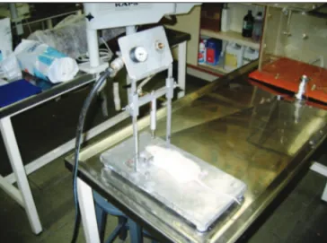
In order to induce the encephalic cranial trauma on the rats, we developed a controlled cortical impact device. The device consists of a pneumatic piston, built with adequate dimensions for the model, supported by two height-regulating metallic rods; metallic platform for the rats’ head likely to be adapted and ad-justed for either foam or rubber platforms; control panel with a pressure gauge to calibrate and measure the impact intensity; and a trigger switch.
Trauma inducement and surgical procedures
After anesthesia with Zoletil 50® 0.1 mg/Kg (combination of tiletamine/zolazepam - virbac s/A - France), the rats
under-went a 150 psi impact on the right frontal parietal region. After trauma inducement, they were observed and anesthetized again. A small linear incision was performed on the right frontal pa-rietal region, followed by a 3 X 3 mm craniectomy, without in-juring the dura mater. The usual surgical care procedures about hemostasis were performed, and a small cotton wound dress-ing was left on the incision.
Selective hypothermia
After craniectomy, the rats were divided into two random-ly selected groups of 15 rats each: Group A, without treatment (control group) and Group B, the group which underwent hypo-thermic treatment. The rats treated with hypothermia received latex ice bags, specially manufactured for this study, set on the frontal parietal region. As the ice melted, the bags were replaced by new ones. Throughout the analysis period, which lasted from 5 to 6 hours, all rats of both groups were kept anesthetized with Zoletil 50®, lying on a thermic mattress, their body temperature
Fig 1. Anesthetized animal on the cortical impact model.
ranging from 33 to 36ºC in both groups. Group B (hypothermia) had their brain surface temperature between 29-31ºC measured with the precision thermometer ellab® dM 852.
After 5 to 6 hours, the rats received an additional anesthe-sia dosage and were killed by decapitation.
Brain removal and storage
After decapitation, the brains were carefully removed, with-out incurring lesions, and the whole brains were stored in glass containers with a 20% formol solution. In order to prevent the brains from becoming deformed, a 3-0 cotton thread was in-serted through the cerebral trunk and the rats were left hang-ing without touchhang-ing the bottom of the container.
Histopathologic analysis
The removed brains of the 30 rats, duly numbered and iden-tiied, were submitted to an histopathologic analysis. In order to quantify the generated lesions, the author followed the pro-cedures established by medical practice18,24, and selected the
percentage count of damaged neurons. The brains were cut at coronal sections in right frontoparietal region with 5µ-thick sec-tions, stained with hematoxiline-eosine, and counted by means of an optical microscope, enlarged 400. other alterations were observed in the analysis including hemorrhagic petechias, brain concussions and edemas; however, they were not included in this study (Fig 2).
RESULTS
Variables and data
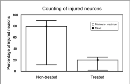
The variable of concern in this study was the counting of the injured neurons. The data obtained for each group of rats are in Table 1.
There was signiicant difference between the number of neurons counted in brain of the animals of both exper-Table 1. Results of control group and treated with selective
hipothermia group (number of injured neuron).
Group A = Control group
Group B = Treated with selective hypothermia Animal 1A = 12 Animal 1B = 2 Animal 2A = 30 Animal 2B = 5 Animal 3A = 50 Animal 3B = 12 Animal 4A = 30 Animal 4B = 20 Animal 5A = 90 Animal 5B = 25 Animal 6A = 90 Animal 6B = 60 Animal 7A = 80 Animal 7B = 2 Animal 8A = 90 Animal 8B = 80 Animal 9A = 80 Animal 9B = 2 Animal 10A = 80 Animal 10B = 10 Animal 11A = 90 Animal 11B = 25 Animal 12A = 90 Animal 12B = 20 Animal 13A = 80 Animal 13B = 12 Animal 14A = 90 Animal 14B = 20 Animal 15A = 80 Animal 15B = 25
Table 2. Comparative results.
Group N Average Mean Minimum Maximum standard deviation
A (non-treated) 15 70.80 80.00 12.00 90.00 26.53
B (treated) 15 21.33 20.00 2.00 80.00 21.83
imental groups in favor of the group treated with selec-tive hypothermia (p<0.00001, Mann-Whitney). see Table 2 and Figure 3.
DISCUSSION
hypothermia, regardless of the type used (systemic or selective), aims at preventing secondary brain injuries, and, ultimately, providing brain protection. The brain tissue of the primary brain injury hardly ever survives. on the other hand, the mechanisms that generate secondary brain inju-ry, such as brain edema, ischemia and decrease in cerebral perfusion pressure (CPP) caused by the increase in intra-cranial pressure (ICP) are considered the possible targets in the acute phase treatment. The factors that promote these physiological changes such as free radicals, blood-brain barrier dysfunction, excitatory amino acid levels and high intracellular calcium concentrations are taken into consideration at the time of the treatment25.
The primary brain injury involves the destruction of brain tissue, vascular lesion and release of potassium and vasoactive substances such as endogenous opioids, catecolamines, serotonin and excitatory amino acids such as aspartate and glutamate. These changes are developed immediately following brain ischemia, which follows syn-aptic paralysis and temporary cardiopulmonary dysfunc-tion. In animal studies as the ones published by Baker et al.26, the initial release of the excitatory amino acids
glu-tamate and aspartate, controlled by the constitutive nitric oxide (No) radicals, is reported in the rise of intracel-lular calcium ions in the injured brain tissue. The release of glutamate, high levels of intracellular calcium and the presence of constitutive free radicals cause severe lesion in the intracellular homeostasis and membrane dysfunc-tion as well.
Wang et al.27 claim that the brain reactions that happen
to protect the injured tissue, such as the neutralization of neuronal acidosis, the normalization of the neurotoxic glutamate concentrations, the removal of free radicals, the restoration of neurotrophic factors and the ph con-trol of glial cells, are not suficient in most of the cases. The brain tissue rich in protrombine, when severely in-jured, generates microcirculation disturbances with rapid progression into brain ischemia followed by brain edema and ICP rise. Brain ischemia, brain edema and ICP rise are the most signiicant pathophysiological alterations at ear-ly acute stages of severe brain injuries. due to the patho-physiological alteration, the ICP rise leads to the decrease in cerebral perfusion pressure (CPP) and brain ischemia with compartmental syndromes27.
When ICP is above 20-25 mmhg, the cerebral perfusion pressure (CPP) decreases, as these values are higher than
the ones for brain capillary pressure. Consequently, in the intensive care unit (ICU) it is essential that the patients with brain trauma have their CPP kept above 80mmhg and the ICP below 20mmhg. Macintosh28 showed that
tis-sue destruction, vascular ingurgitation, brain edema and inlammatory cytokines with blood-brain barrier dysfunc-tion were considered the major causes of brain edema and ICP rise. Before ICP rise and progression of brain ede-ma, functional changes in the vascular permeability begin between 1 and 15 hours. Before the progression of diffuse edema, selective and speciic neuronal lesions in the basal ganglia were reported. The release mechanism of excit-atory amino acids (glutamate) has been said to be the ma-jor responsible for the neuronal death. Consequently, it is extremely important to prevent neural excitement when handling severe brain traumas.
The brain edema and the intracranial hypertension start to progress after 24 hours, and are followed by re-actions involving free radicals. At this point, intravascular release of thromboplastine, activation of intravascular co-agulation and consumption of tromboplastine ibrinoly-sis agents lead to changes in vascular permeability of the microcirculation. vascular ingurgitation, microcirculation disturbances, ICP rise and reactions involving free radi-cals generate a malignant ischemic cycle, leading to brain edema and intracranial hypertension. As the lesions take place in the injured tissue, iniltrate of immunoprotective cells is observed, with neutrophils and macrophages pro-ducing cytokines, nitric oxide (No) and superoxide radi-cals. The release of cytokines increases intravascular co-agulation by activating adhesion molecules to the intima of the vascular wall and activates alterations on vascular permeability. The complications due to systemic infec-tions promote these alterainfec-tions more easily and more se-verely, and free radicals cause larger brain lesions25.
administration of metabolic substrates and management of the secondary lesion are required25,28.
The effects of hypothermia described in medical lit-erature include its effects on excitatory amino acids, free radicals, decline in vascular permeability, nitric oxide, cerebral blood low, (due to the decline in the regional oxygen low and oxygen consumption), decline of the in-lammatory reaction and decrease in the genic expression and apoptosis21,22,25-28.
The current use of systemic hypothermia and prospective use for selective hypothermia – Mild (34-36ºC) and moderate (28-33.5ºC) systemic hypothermia have been reported to present better results obtained in the treatment of severe brain injury with fewer systemic complications such as arrhythmia and blood dyscrasia. Nevertheless, a study by harris et al.29 claims that more controlled, multi-center
and random studies are necessary in order to permanently deine the indications for use and the procedures of this therapeutic practice.
recent published studies have proposed the use of systemic hypothermic treatments25,27 with the brain
tem-perature between 32ºC and 34ºC for severe brain injury, without compromising the systemic circulation and me-tabolism, and performing neuro-hormonal management and stabilization of the immune function. The initial “tar-gets” of this therapeutic procedure for severe traumat-ic brain injury with hypothermia are not the decline of the brain metabolism to prevent brain ischemia and the decrease of ICP. Currently, in the attempt to restore the nerve tissue injured due to trauma, researchers advocate the use of hypothermia to attenuate the neuro-hormonal reactions of the hypothalamic-pituitary-adrenal axis, com-plemented with the adequate administration of oxygen and substrates in order to stabilize the brain metabolism25.
other applications of hypothermia as well as its use during complex brain aneurysm surgeries, brain strokes and post cardiac arrest have yielded promising results.
several studies, which describe the mild and moder-ate systemic hypothermia side effects, include indings about the decline of the enzymatic activity, changes in pharmacokinetics and in pharmacodynamics, and chang-es in the neuromuscular rchang-esponse. Complications include postoperative tremors and myocardial ischemia, operative wound infections and coagulopathies concomitant with peroperatory blood loss20,25,28.
In the last 50 years, several attempts to develop tech-niques of hypothermia exclusively targeted at the brain have been described, and more common used term in literature was selective hipothermia. These techniques include perfusion, injections of cold substances or
di-rect cooling of the brain by means of several methods and helmet for head cooling. however, since senning and olsson’s pioneer study30, few clinical studies on selective
hypothermia have been carried out with adequate meth-odology that would promote the adoption of the therapy on a regular basis. larger, controlled and random studies are necessary.
The objective to propose a new therapy to treat se-vere brain trauma with selective hypothermia (cooling of the brain, keeping the corporal temperature constant) was researched and developed following the literature param-eters which report the new indings about traumatic brain injury and hypothermia. The original technique discussed in this study proposes the bone removal of a brain region (craniectomy), with the preservation of the dura mater, the animal anesthetized, and the selective hypothermia treatment with an ice bag, gently set on the exposed re-gion. The aim is to propose a viable procedure, easily rep-licated in future clinical trials.
For the generation of the brain trauma in rats, a con-trolled cortical impact model was developed, speciically built for the trial, based on available data. The need to show its eficacy also led the author to determine the parameter with which measure the extent of the gener-ated brain lesion. The counting of injured neurons was the criterion selected as it would be dificult to analyze the data obtained by means of determining the extent of the lesion due to brain edema or the hemorrhagic petechias. This option was done because brain edema and hemor-rhagic petechias have dificult quantiication.
The results obtained have shown that the group treat-ed with selective hypothermia presenttreat-ed a lower percent-age of injured neurons when compared to the control group. It was possible to determine the eficacy of the selective hypothermia despite the short treatment time (5 to 6 hours). other pathological conditions, such as brain edema and hemorrhagic brain patechias were described by the pathologist, who reports a lower incidence of edema in the group treated with hypothermia. however, these data were not used in the analysis of this study.
REFERENCES
1. Fay T. Observations on prolonged human refrigeration. NY State J Med 1940;40:1351-1354.
2. Rosomoff HL, Shulman K, Raynor R, Grainger W. Experimental brain injury and delayed hypothermia. Surg Gynecol Obstet 1960;110:27-32. 3. Drake CG, Barr WK, Coles JC, Gergely NF. The use of extracorporeal circulation and profound hypothermia in the treatment of ruptured in-tracranial aneurysm. J Neurosurg 1964;21:575-581.
4. Covino BG, D’Amato HE. Mechanism of ventricular ibrillation in hy -pothermia. Circ Res 1962;148-155.
5. Steen PA, Mide JH, Michenfelder JD. The detrimental effects of prolonged hypothermia and rewarming in the dog. Anesthesiology 1980; 52:224-230. 6. Sands MP, Mohri H, Sato S. Hematorheology during deep
hypother-mia. Cryobiology 1979;16:229-239.
7. Busto R, Dietrich WD, Globus MY, Ginsberg MD. The importance of brain temperature in cerebral ischemic injury. Stroke 1989;20:1113-1114. 8. Marion DW, Obrist WD, Carlier PM, Penrod LE, Darby JM. The use of moderate therapeutic hypothermia for patients with severe head inju-ries: a preliminary report. J Neurosurg 1993;79:354-362.
9. Clifton GL, Jiang JY, Lyeth BG, et al. Marked protection by moderate hypothermia after experimental traumatic brain injury. J Cereb Blood Flow Metab 1991;11:114-121.
10. Huh PW, Belayev L, Zhao W, et al. Comparative neuroprotective efi -cacy of prolonged moderate intraischemic and postischemic hypother-mia in focal cerebral ischehypother-mia. J Neurosurg 2000;92:91-99.
11. Okubo K, Itoh S, Isobe K, et al. Cerebral metabolism and regional
cere-bral blood low during moderate systemic cooling in newborn piglets.
Pediatrics Internat 2001;43:496-501.
12. Inderbitzen B, Yon S, Lasheras J, et al. Safety and performance of a nov-el intravascular catheter for induction and reversal of hypothermia in a porcine model. Neurosurgery 2002;50:364-370.
13. Horn M, Schlote W, Henrich HA. Global cerebral ischemia and subse-quent selective hypothermia. A neuropathological and morphomet-rical study on ischemic neuronal damage in cat. Acta Neuropathol 1991;81:443-449.
14. Kuluz JW, Gregory GA, Yu ACH, et al. Selective brain cooling during and after prolonged global ischemia reduces cortical damage in rats. Stroke 1992;23:1792-1797.
15. Gelman B, Schleien CL,Lohe A, Kuluz JW. Selective brain cooling in infant piglets after cardiac arrest and resuscitation. Crit Care Med 1996;24:1009-1017.
16. Ohta T, Kuroiwa T, Sakaguchi I, et al. Selective hypothermic perfusion of canine brain. Neurosurgery 1996;38:1211-1215.
17. Gunn AJ, Gunn TR, Haan HH, Williams CE, Gluckman PD. Dramatic neuronal rescue with prolonged selective head cooling after ischemia in fetal lambs. J Clin Invest 1997;99:248-256.
18. Thoresen M, Simmonds M, Satas S, et al. Effective selective head cool-ing durcool-ing posthypoxic hypothermia in newborn piglets. Pediatric Res 2001;49:594-599.
19. Laptook AR, Shalak L,Corbett RJT. Differences in brain temperature and
cerebral blood low during selective head versus whole-body cooling.
Pediatrics 2001;108:1103-1110.
20. Wang H, Olivero W, Lanzino G, et al. Rapid and selective cerebral hypo-thermia achieved using a cooling helmet. J Neurosurg 2004;100:272-277. 21. Prandini MN, Laccana SN, Vallente PR, Stavale JN. Regional mild hy-pothermia in the protection of ischemic brain. Acta Cir Bras 2002;17: 232-235.
22. Prandini MN, Neves A,Lapa AJ, Stavale JN. Mild hypothermia reduced
polymorphonuclear leukocytes iniltration in induced brain inlamma -tion. Arq Neuropsiquiatr 2005;63:779-784.
23. Nilsson B, Ponten V, Voigt G. Experimental head injury in the rat: Part I. Mechanics, pathophysiology and morphology in an impact trauma model. J Neurosurg 1977;47:241-251.
24. Foda MAAE, Marmarou A. A new model of diffuse brain injury in rats: Part II. Morphological characterization. J Neurosurg 1999;80:301-313. 25. Hayashi N, Dietrich DW. Brain hypothermia treatment. Berlin: Springer
2004;23-89.
26. Baker AJ, Zornow MH, Scheller MS, et al. Changes in extracellular con-centrations of glutamate, aspartate, glycine, dopamine, serotonin and dopamine metabolites after transient global ischemia in the rat. J Neu-rochem 1991;57:1370-1379.
27. Wang Y, Chang CF, Morales M, Chian YH, Hoffer J. Protective effects of glial cells line derived neurotrophic factor in ischemic brain injury. Ann NY Acad Sci 2002;962:423-237.
28. Macintosh TK. Neurological sequele of traumatic brain injury: thera-peutic implications. Cerebrovasc Brain Metab Rev 1994;6:109-162. 29. Harris OA, Colford JM Jr, Good MC, Matz PG. The role of hypothermia
in the management of severe brain injury: a meta-analysis. Acta Neu-rol 2002;59:1077-1083.