Cop
yright
© ABE&M t
odos os dir
eit
os r
eser
vados
.
Novel
DMRT1
3’UTR+11insT
mutation associated to XY
partial gonadal dysgenesis
Nova mutação 3’UTR+11insT no gene DMRT1
associada à disgenesia gonadal parcial XY
Maricilda Palandi de Mello1, Fernanda Borchers Coeli1,2, Juliana Godoy Assumpção1,3, Tammy Mazeo Castro1,
Andréa Trevas Maciel-Guerra4, Antônia Paula Marques-de-Faria4, Maria Tereza Matias Baptista5, Gil Guerra-Júnior6
SUMMARY
The Y-chromosome-located SRY gene encodes a small testis-specific protein containing a
DNA--binding motif known as the HMG (high mobility group) box. However, mutations in SRY are
not frequent especially in cases of 46,XY partial gonadal dysgenesis. Several sex-determining genes direct the fate of the bipotential gonad to either testis or ovary. In addition, heterozygous small deletions in 9p can cause complete and partial XY gonadal dysgenesis without other symptoms. Human DMRT1 gene, which is located at 9p24.3, is expressed in testis and ovary
and has been considered, among others, a candidate autosomal gene responsible for gonadal dysgenesis. In this report we describe a nucleotide insertion in DMRT1 3’UTR in a patient of XY
partial gonadal dygenesis. The 3’UTR+11insT is located within a conserved motif important for mRNA stabilization. Arq Bras Endocrinol Metab. 2010;54(8):749-53
SUMÁRIO
O gene SRY, localizado no cromossomo Y, codifica uma proteína testículo-específica conten-do um conten-domínio HMG (grupo de alta mobilidade) de ligação ao DNA. No entanto, mutações no gene SRY não são frequentes, especialmente nos casos de disgenesia gonadal parcial em indivíduos 46,XY. São atualmente conhecidos vários genes que participam do processo de di-ferenciação gonadal, tanto para o desenvolvimento testicular quanto para o ovariano. Além disso, pequenas deleções heterozigotas em 9p podem causar disgenesia gonadal XY completa ou parcial, sem outros sintomas associados. O gene DMRT1 humano, que está localizado em
9p24.3, é expresso no testículo e ovário no período fetal e tem sido considerado um dos genes autossômicos envolvido na etiologia das disgenesias gonadais. Neste trabalho, descrevemos a inserção de um nucleotídeo em 3’UTR do gene DMRT1 em um paciente 46,XY com disgenesia
gonadal parcial. A mutação 3’UTR+11insT está localizada dentro de um motivo conservado importante para a estabilização do mRNA. Arq Bras Endocrinol Metab. 2010;54(8):749-53
1 Center for Molecular Biology and
Genetic Engineering (CBMEG), Universidade Estadual de Campinas (Unicamp), Campinas, SP, Brazil
2 Department of Clinical Medicine,
Faculdade de Medicina de Ribeirão Preto, Universidade de São Paulo (FMRP-USP), Ribeirão Preto, SP, Brazil
3 Pediatric Hematology Services,
Hospital das Clínicas, Universidade Federal de Minas Gerais (UFMG), Belo Horizonte, MG, Brazil
4 Department of Medical
Genetics, Faculdade de Ciências Médicas (FCM), Unicamp, Campinas, SP, Brazil
5 Department of Clinical Medicine,
FCM, Unicamp, Campinas, SP, Brazil
6 Department of Pediatrics,
Unicamp, Campinas, SP, Brazil
Correspondence to:
Maricilda Palandi de Mello Laboratório de Genética Molecular Humana, Centro de Biologia Molecular e Engenharia Genética, Unicamp
13083-875 − Campinas, SP, Brazil Caixa Postal 6010
Received on Jul/30/2010 Accepted on Nov/17/2010
INTRODUCTION
D
isorders of sex development (DSD) are deined as congenital conditions in which development of chromosomal, gonadal, or anatomical sex is atypi-cal (1). XY gonadal dysgenesis (OMIM ID #400044) is a DSD in which the embryonic gonadal development is defective (2). Clinically, XY gonadal dysgenesis may manifest as complete or partial forms. CompleteCop
yright
© ABE&M t
odos os dir
eit
os r
eser
vados
.
tations in are not frequent especially in cases of 46,XY partial gonadal dysgenesis (6,7).
Several sex-determining genes direct the fate of the bipotential gonad to either testis or ovary (8). It is well known that heterozygous small deletions in 9p can cause complete or partial XY gonadal dysgen-esis (OMIM ID #154230) without other symptoms (9,10). A human gene located at 9p24.3 with sequence similarities to genes that regulate sexual development in insects and nematodes has been described as respon-sible for XY gonadal dysgenesis. This gene is called
DMRT1 (doublesex and mab-3 related transcription
factor 1) and in adults it is expressed in the testis and ovary (11,12). The gene has ive exons and is predicted to encode a protein of 373 amino acids with a DM do-main near its N-terminal portion (13). There are four
DMRT1 mutations in codons 45, 221, 281, and 295,
but their association to a characteristic 46,XY gonadal dysgenesis phenotype is not well deined (13).
We report here a DMRT1 nucleotide insertion in
3’UTR found by sequence analysis in a case of XY par-tial gonadal dygenesis. The 3’UTR+11insT is located within a conserved motif important for directing alter-native splicing and/or promoting mRNA stabilization.
SUBJECT AND METHODS
Blood specimens and clinical data of the patient and relatives were collected with approval by the appropri-ate Institutional Review Board; signed informed con-sent was obtained.
Clinical data
A 7-month-old male infant was referred to us due to sex ambiguity. The child was delivered by cesarean section after an uneventful 39-week pregnancy. Birth weight was 3,100 g and length 48 cm. He was the second child of young healthy unrelated parents. When irst exam-ined by us, weight was 6,820 g and length 65.1 cm.
and anti-müllerian hormone (114 pMol/L, NR = 265-679 pMol/L), but also normal values of ACTH (21 pg/mL, NR < 46 pg/mL), cortisol (12 μg/dL, NR = 5-25 μg/dL), progesterone (0.7 ng/mL, NR = 0.1-1.4 ng/mL), 17-OH progesterone (0.8 ng/mL, NR = 0.2-1.5 ng/mL), androstenedione (1.0 ng/mL, NR = 0.7-3.6 ng/mL) and DHEA (3.7 ng/mL, NR = 3.0-6.1 ng/mL). G-banding karyotype in 32 cells revealed a 46,XY karyotype. After three injections of testoster-one enanthate (50 mg over successive months), phallus size increased 2 cm. The diagnosis of 46,XY DSD due to partial gonadal dysgenesis was conirmed by biopsy of both gonads, which revealed bilateral prepubertal testis with marked tubular hypoplasia, severe germinal hypoplasia and Sertoli cells hyperplasia. This patient is patient number 5 of the paper published by Ribeiro-Scolfaro and cols. (4).
Molecular analysis
Genomic DNA was extracted from peripheral blood leukocytes by the standard phenol/chloroform method.
For the microdeletion investigation, the following polymorphic markers located in the distal part of 9p: D9S143 (14); D9S1779, D9S1858, D9S1813, and D9S54 (http://www.ensembl.org/Homo_sapiens/ index.html) were used. Each microsatellite was ampli-ied in an independent radioactive PCR. PCR was per-formed in a inal volume of 12.5 µL containing 50-100
ηg of genomic DNA, 20 ρmol of each primer, 200 M
of each dNTPs (0.2 mM dATP + 0.2 mM dTTP + 0.2 mM dGTP + 0.1 mM dCTP + 1 μCi [α-32P] dCTP),
0.25 U Taq DNA polymerase (Invitrogen, CA, USA) reaction buffer for the enzyme, 1.0 mM to 1.5 mM MgCl2. PCR cycles were: 94oC for 5 minutes, 94oC for
30 seconds, 57oC for 30 seconds, 72oC for 30 seconds
(25 cycles) and a inal step of 72oC for 5 min.
Cop
yright
© ABE&M t
odos os dir
eit
os r
eser
vados
.
denatured for 5 minutes at 94oC and applied to
dena-turing polyacrylamide gel 6%. Electrophoresis was per-formed at 1500 V, 50 W, 50 mA, for 2-4 hours. Gels were placed in a gel dryer for an hour and then exposed to X-ray ilms (Hyperilm MP − Amersham-Pharmacia Biotech) at -70°C for 2-8 hours.
DMRT1 gene was ampliied by PCR
ampliica-tion of the entire coding region including exon-intron junctions and both 5’UTR and 3’UTR regions us-ing synthetic oligonucleotides (Invitrogen) as primers (Table 1) which were designed using Primer 3 open access software (http://primer3.sourceforge.net). The ampliied fragments were directly sequenced using Big Dye TM Terminator Cycle Sequencing Kit V3.1 Ready Reaction (ABI PRISM/PE Biosystems, Foster City, CA, USA). The sequences obtained in an ABI3700 Automated Sequencer (ABI PRISM/PE Biosystems) were compared to the normal sequence of the gene (ENSG00000137090).
RESULTS
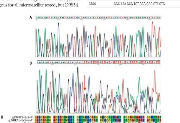
To investigate the possibility of a 9p deletion in the pa-tient, a study using DNA microsatellite in the critical in-terval 9p24.3 was carried out. There was no evidence of microdeletion in the DMRT1 region because the patient
was heterozygous for all microsatellite tested, but D9S54.
DMRT1 sequence analysis on the patient’s DNA
re-vealed the heterozygous insertion of a thymine located 12 nucleotides beyond the stop codon in the 3’UTR (Figure 1). Sequencing also revealed heterozygosity for a nucleotide change T>A in exon 1 that caused p.S45T protein variation (data not shown), considered to play no biological effect on protein function, therefore a polymorphism (rs16925431-dbSNP125). Unfortu-nately, parents were not available to evaluate the segre-gation of both allelic variants.
Table 1.Primers for DMRT1 gene amplification and sequencing
Primer Sequence (5 → 3’) Tm (°C)
E1FA CTC CGG AGC GTC GCT GTC CGT CGG 63
EIRA GAG CCA AGA TCG CGC CAC TAC ACT GC 59
E1FS TCC CTG GCA GCA GTC TCC AGG CGA G 61
E1RS AGC CTG GCA ACC GAG CGA GAC TC 57
E2FS GTG TTT TGG CAA AGC TGA TTC TGG 51
E2RS TGC AAC CTT CGG CCT CCC TCA TGC 57
E3FS AGA AGT AAA GTT TCG TGG ACT AAC 47
E3RS TGC ACA TGC ATG TGG CTT TCA CAC 52
E4F CAC TGT GCC CAG CCT GTT ACC TTG 56
E4R AAG CCA TTA GAC ACA GCT AAT GAC 49
E5F GAA TAA TGA ATG TAT AAA GAC CAG CCA C 50
CR64 CAG CCT CTG GAC TAA ACT CTA AGG 52
CR76 GGC AAA GCG TCT GGG GCG CTA GTG 59
Figure 1. Eletropherograms showing the end of exon 5 and part of 3’UTR of DMRT1 gene. (A) normal sequence. (B) Heterozygosis for a T insertion in the patient’s
sample; the red arrow indicates the insertion site. (C) Sequence alignment: sequence on the top shows the normal allele, in the middle, 3’UTR+11insT, and at the bottom sequencing reading of the heterozygous patient; the stop codon TGA is underlined. (K) = G or T; (M) = A or C; (S) = G or C; (Y) = C or T; (W) = A or T.
A
B
Cop
yright
© ABE&M t
odos os dir
eit
os r
eser
vados
.
humans (15,16). Cheng and cols. (15) have character-ized multiple transcript isoforms in human testis. These transcripts code for different putative proteins, two of them generated by alternative splicing in the 3’UTR. Other transcripts are results of the intronic exonization including Alu elements that are not randomly distribut-ed in the non-coding regions. In mice, alternative splic-ing processes were detected in adult testis and in undif-ferentiated gonads. All forms are produced with a similar pattern of expression peaking at 13.5 days after fertiliza-tion and maintaining a baseline pattern of expression in male gonads (16). These experiments indicate an impor-tant role of this gene in the testicular determination and differentiation in mammals. The genomic region where the 3’UTR+11insT mutation occurred is recognized as an Exonic Splicing Enhancer by PSEX (Putative Exonic Splicing Enhancers/Silencers) algorithm (17,18). A pre-liminary in silico analysis using ESEinder 3.0 to identify exonic splicing enhancers (19,20) indicates the abolish-ment of a SC35 protein recognition site in the presence of the 3’UTR+11insT mutation.
Herpin and cols. (21) studied mechanisms that
reg-ulate the expression of speciic genes in the gonads dur-ing embryonic development. They found the protein-binding conserved motif CUGCUGCCGAU located in the DMRT1 3’UTR that participates in the
gonad-speciic stabilization of mRNA during development. The presence of this sequence indicates the importance of cis and trans elements forming mRNA-protein com-plexes in the control of speciicity and selectivity of gene expression. The insertion described here is located exactly in the middle of the highly conserved region changing this sequence to CUGCUUGCCGAU.
In conclusion, the mutation identiied in the patient with partial gonadal disgenesis may cause the pheno-type either by modiications in the alternative splicing process or by preventing mRNA stabilization by pro-tein binding and, consequently, leading to mRNA deg-radation.
REFERENCES
1. Hughes IA. Disorders of sex development: a new definition and classification. Best Pract Res Clin Endocrinol Metab. 2008;22:119-34. 2. Andrade JG, Guerra-Júnior G, Maciel-Guerra AT. 46,XY and
45,X/46,XY testicular dysgenesis: similar gonadal and genital phenotype, different prognosis. Arq Bras Endocrinol Metabol. 2010;54:331-4.
3. Michala L, Goswami D, Creighton SM, Conway GS. Swyer syn-drome: presentation and outcomes. BJOG. 2008;115:737-41. 4. Ribeiro-Scolfaro M, Cardinalli IA, Stuchi-Perez EG, de Mello MP,
Assumpção JG, Baptista MTM, et al. Morphometry and histology of gonads from 13 children with dysgenetic male pseudoherma-phroditism. Arch Pathol Lab Med. 2001;125 652-6.
5. Berta P, Hawkins JR, Sinclair AH, Taylor A, Griffiths BL, Goo-dfellow PN, et al. Genetic evidence equating SRY and the testis--determining factor. Nature. 1990;348:448-50.
6. Knower KC, Kelly S, Harley VR. Turning on the male-SRY, SOX9 and sex determination in mammals. Cytogenet Genome Res. 2003;101:185-98.
7. Tagliarini EB, Assumpção JG, Scolfaro MR, Mello MP, Maciel--Guerra AT, Guerra Jr G, et al. Mutations in SRY and WT1 genes required for gonadal development are not responsible for XY partial gonadal dysgenesis. Braz J Med Biol Res. 2005;38:17-25. 8. Biason-Lauber A. Control of sex development. Best Pract Res Clin
Endocrinol Metab. 2010;24(2):163-86.
9. Veitia RA, Nunes M, Quintana-Murci L, Rappaport R, Thibaud E, Jaubert F, et al. Swyer syndrome and 46,XY partial gonadal dysgenesis associated with 9p deletions in the absence of mono-somy-9p syndrome. Am J Hum Genet. 1998;63:901-5.
10. Calvari V, Bertini V, De Grandi A, Peverali G, Zuffardi O, Ferguson--Smith M, et al. A new submicroscopic deletion that refines the 9p region of sex reversal. Genomics. 2000; 65:203-12.
11. Raymond CS, Shamu CE, Shen MM, Seifert KJ, Hisch B, Hodgkin J, et al. Evidence for evolutionary conservation of sex-determining genes. Nature. 1998;391:691-4.
12. Pask AJ, Behringer RR, Renfree MB. Expression of DMRT1 in the mammalian ovary and testis--from marsupials to mice. Cytoge-net Genome Res. 2003;101:229-36.
13. Raymond CS, Parker ED, Kettlewell JR, Brown LG, Page DC, Kusz K, et al. A region of human chromosome 9p required for testis development contains two genes related to known sexual regula-tors. Hum Mol Genet. 1999;8:989-96.
14. Furlong RA, Lyall JE, Lush MJ, Affara NA, Ferguson-Smith MA. Four dinucleotide repeat polymorphisms on chromosome 9 (D9S143-146).Hum Mol Genet. 1992;1:447.
Cop
yright
© ABE&M t
odos os dir
eit
os r
eser
vados
.
16. Lu H, Xiao H, Liao Z, Yiqing G, Hanhua C, Rongjia Z. Multiple al-ternative splicing of mouse Dmrt1 during gonadal differentiation. Biochem Biophys Res Commun. 2007;352: 630-4.
17. Smith PJ, Zhang C, Wang J, Chew SL, Zhang MQ, Krainer AR. An increased specificity score matrix for the prediction of SF2/ASF-specific exonic splicing enhancers. Hum Mol Genet. 2006;15:2490-508.
18. Cartegni L, Wang J, Zhu Z, Zhang MQ, Krainer AR. ESEfinder: a web resource to identify exonic splicing enhancers. Nucleic Acid Res. 2003;31:3568-71.
19. Zhang XH, Chasin LA. Computational definition of sequen-ce motifs governing constitutive exon splicing. Genes Dev. 2004;18:1241-50.
20. Zhang XH, Kangsamaksin T, Chao MS, Banerjee JK, Chasin LA. Exon inclusion is dependent on predictable exonic splicing enhancers. Mol Cell Biol. 2005;25:7323-32.