Role of
β
-lactamase operon on mecA
expression in Staphylococcus aureus
Joana Henriques Ministro
November, 2011
Role of
β
-lactamase operon on mecA
expression in Staphylococcus aureus
Joana Henriques Ministro
November, 2011
Dissertation for the Master Degree in Medical
Microbiology
Dissertation supervised by Dr. Duarte Carvalho Oliveira
CREM - Centro de Recursos Microbiológicos
Departamento de Ciências da Vida
ACKNOWLEDGMENTS
First of all, I would like to express my gratitude to Dr. Duarte Oliveira, Assistant Researcher at Centro de Recursos Microbiológicos, my supervisor, for welcome me into his laboratory. Above all, I would like to thank him for all the knowledge he share with me, for the several fruitful discussions and for his constant availability to help me in every question I had.
To my colleague Pedro Arêde, for all his priceless help, support, knowledge share and also for the good times spending inside and outside the laboratory.
To Centro de Recursos Microbiológicos (CREM), for providing me the excellent facilities to perform my practical work.
To my colleagues from CREM, Isabel Correia, Lia Godinho, Mário Ferreira, Renato Pires, Márcia Rato, Pedro Almeida, Carla Gonçalves, Márcia Palma and Marco Coelho for the helpful work environment.
To Fundação para a Ciência e Tecnologia for the financial support.
To my master colleagues and friends João Bota, Cátia Piedade and Susana Silvestre, for the constant sharing of ideas about science and for the excellent moments we spent together.
To my family, for being there every moment, for supporting, listening and for giving me the strength to go on.
ABSTRACT
Methicillin-resistant Staphylococcus aureus (MRSA) is an important nosocomial pathogen and is also emerging in the community. MRSA is cross-resistant to virtually
all β-lactam antibiotics and has acquired two main resistance mechanisms: production
of β-lactamase (bla), coded by blaZ, and production of penicillin binding protein 2a (PBP2a), coded by mecA. Both genes are regulated by homologous sensor-transducers (BlaR1 and MecR1) and repressors (BlaI and MecI), and coregulation of mecA and blaZ
by both systems has been demonstrated, although with remarkable different efficiencies. In fact, induction of mecA by mecI-mecR1 is so slow that it is believed it is not functional in most MRSA strains.
However, recent data from our laboratory has unexpectedly demonstrated that not only there is no correlation between the presence of mecI gene and the resistance level in epidemic MRSA strains, but also that for most strains there were no significant changes on the resistance phenotype upon the mecI overexpression in trans. Interestingly, the two strains in which mecI overexpression affected the resistance expression were negative for the bla locus, suggesting that this locus may interfere directly with the MecI-mediated repression of mecA and account for those puzzling observations.
RESUMO
Os Staphylococcus aureus resistentes à meticilina (MRSA, do inglês
“methicillin-resistant Staphylococcus aureus”) são um dos principais agentes
responsáveis por infeções hospitalares. Os MRSA são resistentes a praticamente todos os antibióticos β-lactâmicos devido a dois mecanismos principais: produção de β -lactamase (bla), codificada pelo gene blaZ, e produção de uma proteína de ligação à
penicilina (PBP2a, do inglês “penicillin binding protein 2”), codificada pelo gene mecA. Estes dois genes são regulados por sistemas homólogos, constituídos por um sensor-transdutor (BlaR1 e MecR1) e um repressor (BlaI e MecI), de tal modo que ambos os sistemas são capazes de co-regular os genes mecA e blaZ, embora com eficiências de indução muito diferentes. De facto, a indução mediada pelo sistema mecI-mecR1 é tão lenta que se acredita que este sistema não está funcional na maioria das estirpes MRSA.
No entanto, dados recentes do nosso laboratório, demonstram a ausência de relação entre a presença do gene mecI e o nível de resistência à meticilina em estirpes MRSA epidémicas, e também que, o fenótipo de resistência da grande maioria das estirpes não é perturbado pela sobre-expressão em trans do repressor mecI. Curiosamente, as duas estirpes em que a expressão da resistência foi afetada pela sobre-expressão do mecI são negativas para o locus da β-lactamase, o que sugere que este
TABLE OF CONTENTS
ACKNOWLEDGMENTS ... ii
ABSTRACT ... iii
RESUMO ... iv
ABBREVIATIONS ... vii
LIST OF TABLES ... ix
LIST OF FIGURES ... xi
INTRODUCTION ... 1
1. The Staphylococcus genus ... 1
2. Staphylococcus aureus as a human pathogen ... 1
2.1 Clinical relevance ... 1
2.2 Virulence factors ... 2
2.3 Antibiotic resistance ... 3
2.4 Epidemiology of antibiotic resistance ... 3
2.5 Treatment and prevention ... 5
3. β-lactam resistance mechanisms... 6
3.1 Cell wall: the β-lactams target... 6
3.2 Penicillin resistance ... 7
3.2.1 β-lactamase ... 7
3.2.2 β-lactamase operon ... 8
3.3 Methicillin-Resistant Staphylococcus aureus (MRSA) ... 10
3.3.1 mecA gene ... 10
3.3.2 SCCmec ... 10
3.3.3 Heterogeneous and homogeneous resistance ... 11
3.3.4 Origin and evolution ... 12
3.3.5 Molecular epidemiology of MRSA ... 13
4. Regulation of β-lactam resistance ... 14
5. Role of β-lactamase operon in the stabilization and expression of methicillin resistance in S. aureus ... 17
1. Bacterial strains and plasmids ... 20
2. Molecular methods ... 23
2.1 DNA isolation ... 23
2.2 DNA purification and manipulation ... 24
2.3 Electrophoresis analysis of PCR and DNA restriction reactions ... 24
2.4 Electroporation of recombinant plasmids into S. aureus ... 25
3. Overexpression of β-lactamase regulatory genes ... 26
4. Genetic knock-out of β-lactamase regulatory genes ... 27
5. Phenotypic analysis ... 29
RESULTS ... 31
1. Introduction of native β-lactamaseplasmid into prototype strains ... 31
1.1 Introduction of native β-lactamaseplasmid into strain COL-I ... 31
1.2 Introduction of native β-lactamase plasmid into VNG17 and VNG17-I strains ………...33
1.3 Introduction of native β-lactamaseplasmid into RJP17 and RJP17-I strains .. 35
1.4 Introduction of native β-lactamaseplasmid into HT0350 strain ... 36
2. Phenotypic effect of clavulanic acid ... 38
3. Introduction of β-lactamase regulators into COL-I strain ... 40
3.1 Introduction of blaIblaR1 into COL-I strain ... 40
3.2 Introduction of blaR1 and blaI genes into COL-I strain ... 40
3.3 Introduction of blaR1 domains into COL-I strain ... 41
4. Deletion of blaregulators from native β-lactamase plasmid ... 43
DISCUSSION AND CONCLUSION ... 44
REFERENCES ... 52
ABBREVIATIONS
Amp: Ampicillin
ATc: Anidrotetracycline
bla: β-lactamase bp: Base pairs
CA: Clavulanic acid
CA-MRSA: Community acquired methicillin-resistant Staphylococcus aureus
CC: Clonal complex Cd: Cadmium
Cf: Final concentration
Cm: Chloramphenicol
dNTP’s: Desoxiribonucleotides
GlcNAc: N-acetylglucosamine
IPTG: Isopropyl β-D-1-thiogalactopyranoside IS: Insertion sequence
kb: Kilobase
MIC: Minimum inhibitory concentration MLST: Multilocus sequence typing
MRSA: Methicillin-resistant Staphylococcus aureus
MSSA: Methicillin-susceptible Staphylococcus aureus
MurNAc: N-acetylmuramic acid
Oxa: Oxacillin
PBP: Penicillin binding protein PCR: Polymerase chain reaction Pen: Penicillin
PFGE: Pulse field gel electrophoresis
SCCmec: Staphylococcal cassette chromosome mec spaA: Staphylococcus aureus protein A
ST: Sequence type
Tc: Tetracycline TSA: Triptic soy agar TSB: Triptic soy broth
Vf : Final Volume
VISA: Vancomycin-intermediate Staphylococcus aureus
LIST OF TABLES
Table 1– Strains and plasmids………... 20
Table 2– Oxacillin-resistance of parental strain COL and recombinant strains……... 32
Table 3– Oxacillin-resistance of parental strain VNG17 and recombinant strains…... 34
Table 4– Oxacillin-resistance of parental strain RJP17 and recombinant strains……. 36
Table 5 –Oxacillin-resistance of parental strain HT0350 and recombinant strains….. 37
Table 6 –Clavulanic acid effect on oxacillin and penicillin-resistance phenotype…... 39
Table 7 – Oxacillin-resistance of strain COL transformed with β-lactamase regulators ………. 42
Table 8 –Culture media….……… 64
Table 9 –Buffer solutions………. 64
Table 10 –Media and solutions for transduction experiments……….. 65
Table 11 –Cell-wall lysis enzymes……… 65
Table 12 – Antibiotic solutions……….. 66
Table 13 –Restriction endonucleases……… 66
Table 15– Routine PCR reaction mix……….……….. 68
Table 16– High-fidelity PCR mix………. 68
Table 17– Thermocycling conditions……… 69
LIST OF FIGURES
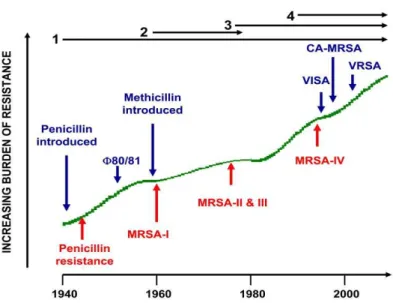
Figure 1– Timeline of the four resistance waves in S. aureus……….. 4
Figure 2– Structure of Penicillin and Methicillin………..7
Figure 3–β-lactamase operon………... 9
Figure 4– Regulation of β-lactam resistance………. 16
Figure 5– Map of pKOR1………. 28
Figure 6 – Phenotypic expression of oxacillin resistance in COL and recombinant strains……….. 32
Figure 7 – Phenotypic expression of oxacillin resistance in VNG17 and recombinant strains……….. 34
Figure 8 – Phenotypic expression of oxacillin resistance in RJP17 and recombinant strains……….. 35
Figure 9 – Phenotypic expression of oxacillin resistance in HT0350 and recombinant strains……….. 37
Figure 10–Membrane topology of the penicillin-sensory transducer BlaR1 ………...49
INTRODUCTION
1. The Staphylococcus genus
Staphylococci are gram positive cocci of about 0,8-1,0 µm in diameter that divide in perpendicular planes to form irregular clumps. They are facultative anaerobes, nonsporulating, but are resistant to drying and are readily dispersed in dust particles through the air and surfaces (77, 105).
Staphylococcus genus belongs to Staphylococcaceae family and so far 45 species and 24 subspecies have been identified (38). The genus contains common pathogens of humans and animals and commonly infects the skin and wounds, occasionally causing life-threatening diseases (92). In humans two major species are recognized, Staphylococcus epidermidis, a nonpigmented, comensal, nonpathogenic organism usually found on the skin or mucous membranes and Staphylococcus aureus, a yellow pigmented species that is often associated with pathological conditions (96).
2. Staphylococcus aureus as a human pathogen
2.1Clinical relevance
asymptomatically (148). The nasal carriage can be a risk factor for endogenous infection as these commensal bacteria have the ability to disseminate and invade the host organism (111) but, normally, these individuals do not acquire disease, presumably because the other resident microorganisms compete successfully for resources and limit pathogen growth. In addition, in healthy individuals the innate immune system is particularly active at mucosal surfaces and may inhibit the microbial growth. Most infections result from a colonized individual that transmits to a weakened individual or infects a damaged tissue (25). The most common infections, impetigo, cellulitis and abscesses are the result of invasion and laceration of the skin or cellular tissues. The dissemination to adjacent tissues can originate bacteremia, endocarditis, osteomyelitis, arthritis and pneumonia. S. aureus infections are also associated with the presence of medical devices in the organism (78).
2.2Virulence factors
The virulence of S. aureus is due to a combination of many virulent factors such as toxins, enzymes, cell wall components and antigens (92). S. aureus secrete several toxins responsible for different symptomatologies as food poisoning (enterotoxins A, B, C, D, E, G and H), scalded skin syndrome (exfoliating toxin A and B) and toxic shock syndrome (enterotoxin TSST-1). These toxins can work as superantigens stimulating large numbers of immune response cells, resulting in extended inflammatory reactions (91).
Along with toxins this bacteria secretes hemolisins (α, β, γ and δ), a leucocidin and a few enzymes (coagulase, hialurodinase, fibrinolisine, catalase, lipase and nucleases) that contribute to damage the host cell or stimulate a large number of lymphocytes and cause systemic inflammatory responses (32). The fibrin matrix produced as a result of coagulase activity protects the bacteria from attack by host cells and probably accounts for the extremely localized nature of many S. aureus infections as in boils and pimples (148, 150). This enzyme enables differentiation of S. aureus
2.3Antibiotic resistance
Antibiotic resistance is associated with the permanent change of the highly flexible bacterial genome under pressure (43). In fact, although Staphylococcus aureus
is naturally susceptible to virtually every antibiotic developed so far (18), it is one of the pathogens of greatest concern because of its incredible facility to acquire antibiotic resistance traits along with the ability to cause life-threatening infections and to adapt to different conditions (89). Although chromosomal mutations are also important, resistance is often a consequence of horizontal gene transfer, mostly occurring in hospitals and healthcare institutions, where the selective pressures for resistance are greatest (27, 94).
As new antibiotics have emerged, such as, quinolones, aminoglycosides, oxalidiones, S. aureus has developed efficient mechanisms to neutralize them (88). But the increasing overall burden of staphylococcal disease in many countries in both healthcare and community settings is mainly caused by methicillin-resistant S. aureus
strains (MRSA), which are virtually resistant to all classes of β-lactams (44). Infections caused by antibiotic-resistant strains of S. aureus have reached epidemic proportions in many parts of the world and resistant strains that are contained within hospitals temporarily, can eventually arise within the community (52).
2.4Epidemiology of antibiotic resistance
worldwide pandemic of MRSA in hospitals, although the prevalence of infections may vary significantly in different countries (49, 136) (Fig. 1).
In response to β-lactams, S. aureus has acquired two main resistance
mechanisms: production of β-lactamase, that hydrolyze the β-lactam ring of penicillin, and production of PBP2a, an extra penicillin binding protein (PBP) with low affinity to virtually all β-lactams (60, 95). This latter mechanism, characteristic of MRSA, together with the former, confers resistance to all β-lactams, including penicillins,
cephalosporins and carbapenems. The β-lactam resistance genes were spread over time duo to horizontal transfer and clonal expansion in several waves (18).
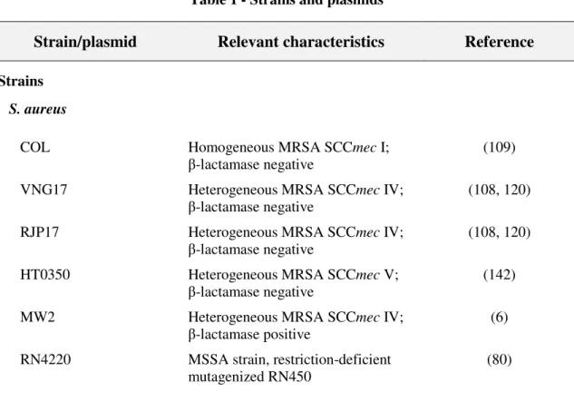
Figure 1 - Timeline of the four resistance waves in S. aureus
(18). Wave 1 began after the introduction of penicillin into clinical
practice and continues till today. Wave 2 had arisen after the
introduction of methicillin into clinical practice (first MRSA
strains). Wave 3 began with emergence of new MRSA strains,
marking the on-going worldwide pandemic of MRSA. Wave 4
Traditionally, MRSA strains have been recognized mainly as nosocomial pathogens. However, in recent years, its epidemiology has radically changed, being increasingly isolated in the community and affecting people without known risk factors (52). The first cases of community-acquired MRSA (CA-MRSA) infections were reported in indigenous populations in Australia, in the early 1990’s (144), and soon after in the United States (113), which were followed by several reports worldwide (15, 123). Unlike hospital clones, CA-MRSA’s were susceptible to most antibiotics but contained several virulence factors (22, 61, 106).
Most contemporary MRSA strains are resistant to many classes of antimicrobial agents leaving physicians with few therapeutic options. Glycopeptides are considered the last resort therapy against MRSA (76). However, in 1997, the first case of reduced susceptibility to vancomycin, designated VISA from vancomycin-intermediate
Staphylococcus aureus, was described (63). Since then, several MRSA strains with reduced susceptibility to vancomycin have been found throughout the world (93, 133, 134). In 2002, for the first time, a strain fully resistant to vancomycin was identified, designated VRSA from vancomycin-resistant Staphylococcus aureus (33). In contrast to the chromosomally mediated resistance for VISA strains that result in a thickened cell wall (54, 55), the VRSA strains acquired the vanA operon from Enterococcus faecalis, which allows synthesis of the terminal peptide ended in Ala-Lac, rather than D-Ala-D-Ala. This new terminus has a remarkable reduced affinity for vancomycin (48, 129). Together with CA-MRSA, VISA and VRSA are the most recent waves of antimicrobial resistance in S. aureus (18).
2.5Treatment and prevention
Extensive use of antibiotics has promoted the selection of resistant
surgery wards and nurseries the carriers of known pathogenic strains must be isolated or treated to eradicate the carrier state (35, 101). Nowadays, the treatment of choice for S. aureus infection in most countries is a penicillin-resistant β-lactam antibiotic (for example, oxacillin or cloxacillin) or a lipopeptide (daptomycin) (9, 127). Combination therapy with gentamicin may be used to treat serious infections like endocarditis, but its use is controversial because of the high risk of damage to the kidneys (23).
3.
β
-lactam resistance mechanisms
3.1 Cell wall: the β-lactams target
In gram positive bacteria the peptidoglycan is the main constituent of cell wall. It is a polymer with a complex organization that confers mechanic resistance to the cell (125). The basic unit of the peptidoglycan is a disaccharide-pentapeptide composed of the amino sugars N-acetylglucosamine (GlcNAc) and N-acetylmuramic acid (MurNAc),
which are linked together by β-1,4 glycosidic bonds. In S. aureus each MurNAc is attached to a short amino acid chain that can be cross-linked to an amino acid chain of another strandthrough a pentaglycine, allowing the formation of peptide cross bridges (126). The polymerization of the newly synthesized disaccharide-peptide and incorporation into the growing peptidoglycan are achieved through the action of four penicillin-binding proteins (PBP1, PBP2, PBP3 and PBP4), which catalyze the transpeptidation and transglycosylation reactions responsible for the formation of the peptidic and glycosidic bonds, respectively (47, 124, 132).
stands up in clinical practice due to the low toxicity in eukaryotic organisms, since they act in an exclusively bacterial structure (145).
3.2 Penicillin resistance
3.2.1 β-lactamase
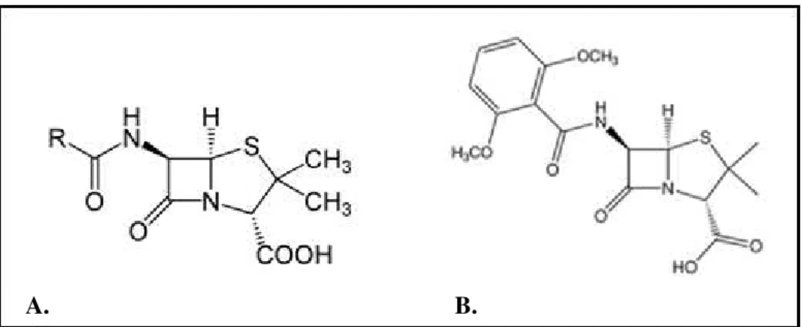
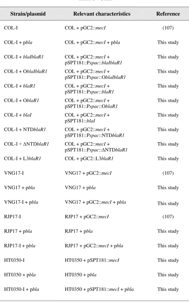
In bacteria, four types of β-lactamases (A, B, C, D) have been distinguished by serotyping and differences in hydrolysis rates of selected β-lactam substrates (74). Types A, C and D are usually located on plasmids and are active-site serine enzymes, whereas type B enzymes typically reside in the chromosome and are zinc-dependent (97, 147). Structural evidences support the proposal that β-lactamases descended from the cell wall PBP’s (97). The action of this group of enzymes consists on the interaction with the β-lactam antibiotic and subsequent disruption of the amide bond in the four-membered β-lactam ring, rendering the antibiotic inactive in an irreversibly manner (19, 86, 153).
Figure 2 - Structure of Penicillin (A) and Methicillin (B).
Staphylococcal β-lactamases are from type A (115) and are large surface attached molecules that reduce the external level of active drug. Penicillin-resistant strains have acquired an exogenous plasmid coding for penicillinase, which confers resistance only to penicillin (86). When penicillin was introduced into clinical practice, only about 5% of S. aureus isolates acquired the plasmid but, since then, through horizontal transfer of the plasmid and strain selection, 80 to 90% of isolates carry the -lactamase gene (82, 85, 86).
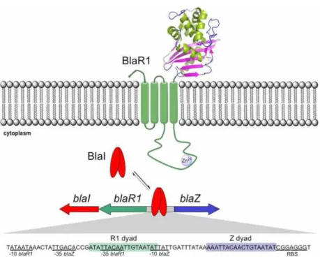
3.2.2 β-lactamase operon
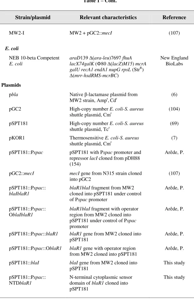
In S. aureus the operon responsible for synthesis of β-lactamase is located in transposon Tn552 (117) and contains the blaZ gene, which encodes for β-lactamase, and the regulatory genes blaR1 and blaI (4, 139). These regulatory genes are divergently transcribed from blaZ (20) (Fig. 3).
BlaR1 is a high molecular weight sensor-transducer transmembrane protein and consists of two domains (97). One is a carboxyl-terminal domain of approximately 27-kDa, the sensor domain, extending to the extracellular medium and containing penicillin binding motifs, that have been shown to bind to β-lactam compounds, and an active site serine, which is involved in activation of the signaling cascade (73, 151). The other domain is an amino-terminal domain of approximately 38 kDa, the transducer, that is intracellular and contains four transmembrane α-helices (TM1, TM2, TM3, TMA4) (57). These transmembrane segments are interconnected by three loops (L1, L2, L3), where L1 and L3 connect to cytoplasm and L2 is exposed on the outside of the cell. The L3 segment has a zinc metalloprotease domain, defined by a histidine sequence and a glutamic acid, which is believed to interact in an unknown way with promoter-bound BlaI dimers (56, 155).
repressor has two functional domains. The amino-terminal domain of approximately 11 kDa is responsible for operator recognition, and the carboxyl-terminal domain of approximately 3 kDa for subunit dimerization (50). DNA-binding experiments demonstrate that formation of BlaI dimer, as well as intact amino and carboxyl termini, are absolutely required for the binding activity of the protein (20, 152). Proteolytic cleavage disrupts the dimer interface, causing its dissociation and releasing from the operator (155). In 1968, Cohen and Sweeney have speculated on the existence of another regulator, the chromosomal gene blaR2, involved in the induction of β -lactamase (21).
3.3 Methicillin-Resistant Staphylococcus aureus (MRSA)
3.3.1 mecA gene
The characteristic element of all methicillin-resistant S. aureus (MRSA) strains is a specific gene, mecA, which codes for PBP2a, an inducible 76 kDa PBP that is absent in susceptible strains (14, 60). In MRSA, PBP2a, which has low binding affinity
to β-lactam antibiotics, can substitute for essential functions of high affinity PBP’s and enables staphylococci to survive under exposure to high concentrations of these agents (60, 112). The organization of the mec operon is similar to the bla operon, containing the mecA gene and the respective regulators, mecI and mecR1 (62). In fact, bla and mec
regulatory genes have been shown to be interchangeable in vivo (83, 99).
Although clearly necessary, there are some evidences suggesting that mecA may not be sufficient to assure high-level resistance to methicillin. As a matter of fact, it has been shown that MRSA strains with virtually identical amounts of PBP2a, showed methicillin inhibitory concentrations (MIC) values spread over a range of several hundred fold (59, 119). Later it was demonstrated the importance of many chromosomal genes in defining resistance levels, namely the fem genes (for factor essential for methicillin resistance), that do not interfere with the transcription and transduction of
mecA (10, 11). De Lencastre et al. proposed that the survival and growth of these
bacteria in the presence of β-lactams require the cooperative functioning of a large number of genes, a process similar to the bacterial stress response mechanism (28).
3.3.2 SCCmec
contains the mec gene complex, which includes mecA and the regulatory genes mecI
and mecR1, and the ccr gene complex, which encodes for recombinases that allow mobility of SCCmec (68). Besides these gene complexes, there are also three so-called J-regions (J1, J2 and J3), which constitute non-essential components of the cassette and may carry additional antimicrobial resistance determinants (65, 66).
SCCmec elements are highly diverse in their structural organization and genetic content (37) and have been classified into types and subtypes. Types are defined by the combination of the ccr gene complex allotype and the class of mec gene complex. Variations in the J regions within the same mec-ccr complex are used for defining subtypes. To date, eight major SCCmec types, designated I to VIII, have been recognized along with numerous subtypes (29, 68) and three new types, IX to XI, have been recently described (84, 131).
There are three classes for the mec gene complex in S. aureus. The class A mec
gene complex, the prototype complex, contains intact mecA, mecR1 and mecI, a hypervariable region (HVR) and insertion sequence (IS) IS431. The class B mec gene complex is composed of intact mecA, a truncated mecR1 (N-terminal inducer domain only) that resulted from insertion of IS1272, a HVR and the same IS as class A. The class C mec is similar to class B but mecR1 gene is truncated by IS431 (first 111 bp
only) and is subdivided in two sub classes depending on the orientation of the IS’s. The
ccr complex consists of two adjacent genes, ccrA and ccrB, in SCCmec I-IV, VI and VIII, and ccrC in V and VII (65, 68).
3.3.3 Heterogeneous and homogeneous resistance
The phenotypic expression of methicillin resistance is highly dependent on growth conditions such as, temperature, medium salt concentration, growth phase and other external factors (128). In addition, many MRSA strains exhibit a heterogeneous expression profile, in which the majority of cells are susceptible to low concentrations
homogeneous population of highly resistant cells that can grow at high concentration of antibiotic (59). In laboratory, with repeated subculture in antibiotic-free medium, the proportion of highly resistant cells gradually diminishes and the original heterogeneous pattern reemerges in most strains (141). There are some rare clinical isolates that consistently are homogeneous despite repeated subculture, with the COL strain of S. aureus being one of these (16, 59).
The phenomenon of heterogeneous and homogeneous resistance in wild-type strains is still unexplained. Heterogeneous strains may be deficient in a factor or lack a critical modification in a biochemical pathway, possibly for cell wall synthesis, that is important for functions of PBP2a. Homogeneous strains then arise from heterogeneous strains by antibiotic selective pressure favoring clones whose genetic background is well adapted for a fully functional PBP2a (16, 138).
In the clinical setting, the heterogeneous phenotypic expression of oxacillin resistance is a major problem, since it may originate false negatives in the phenotypic detection assays of resistance. Because these strains are in fact positive for mecA,
prescription of β-lactam antibiotics may select for high-level β-lactam resistant MRSA subpopulations, causing treatment failures (42, 64).
Another type of methicillin resistance is the borderline (or low-level) resistance, exhibited by strains with a minimum inhibitory concentration (MIC) at or just above the susceptibility breakpoint (17, 102). Borderline strains can be divided in two types: strains with mecA gene and production of PBP2a, presenting a heterogeneous profile, and strains without mecA gene, which do not contain highly resistant subpopulations. These mecA-negative strains can result from modification of normal PBP genes or overproduction of staphylococcal β-lactamase (140). As this later mechanism led to low-level resistance it is not as clinically relevant as production of PBP2a (140).
3.3.4 Origin and evolution
In the search for the possible origin of mecA, some authors identified a genetic element closely related to the S. aureus mecA gene in the animal commensal species
determinant of penicillin binding protein 4 (PBP4) of S. sciuri that was shown to share several properties with S. aureus PBP2a (1). However, this homologue was not identified as part of the mec gene complex or of the SCCmec element. In 2010 Tsubakishita et al. observed a potential mechanism of the generation of a new SCCmec -like element in Macrococcus caseolyticus (143), and, more recently, a divergent mecA
homologue was discovered in human and bovine populations located in a novel staphylococcal cassette chromosome mec element (SCCmec type-XI) (45).
3.3.5 Molecular epidemiology of MRSA
Currently, the characterization of methicillin-susceptible Staphylococcus aureus
(MSSA) and MRSA clones is mostly based on three molecular methods.
Pulse field gel electrophoresis (PFGE) is a technique based on the resolution of large restriction fragments in an agarose gel, resulting from the digestion of total DNA with a rare cutter enzyme (e.g. SmaI) (98). The groups defined by PFGE are clustered into types, with a similarity coefficient of 80%, and subtypes, with a similarity coefficient of 95% (39).
Multilocus sequence typing (MLST) is a genotyping method based on sequence analysis of approximately 450 base pairs (bp) of seven housekeeping genes. Isolates with identical sequences at the seven genetic loci are grouped in the same sequence type (ST). S. aureus isolates that differ in less than three loci are assigned into the same clonal complex (CC) (36, 40).
Spa typing is a method designed for investigation of S. aureus outbreaks and relies upon analysis of variable numbers of tandem repeats in spa gene, which codes for protein A, a constituent of the cell wall. This method takes into account the number of repeats as well as point mutations (130).
with the hypothesis that MRSA derived recently from a limited number of MSSA lineages by acquisition of SCCmec (39, 66).
4.
Regulation of β
-lactam resistance
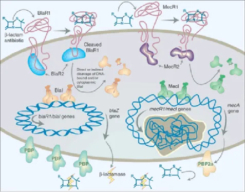
The signaling pathway that regulates β-lactam resistance has been studied for
production of β-lactamase and the respective regulators (50). When BlaR1 is cleaved it can no longer be functional (12). Thus, this protein must be continually produced in
order to sense the β-lactam and keep the signal-transduction active. This explains why
BlaR1 production is linked with β-lactamase production (155). When the extracellular antibiotic concentration decreases, BlaR1 is no longer auto-activated, and in consequence the BlaI proteolytic cleavage stops. The repressors gain conditions to dimerize again and to bind DNA, suppressing blaZ, blaR1 and blaI expression (99, 155).
This regulatory system is unique among bacteria. In fact, a signal transmitted by proteolytic events had not been described among these microorganisms so far (3, 155). Among the unknown questions concerning this signaling transduction pathway are the mechanism of BlaR1 acylation, the proteolytic cleavage events and the repressor mechanisms once the threat of the β-lactams has passed (43).
The main difference between these two systems is the kinetics of the signal-transduction. BlaR1 takes only a few minutes to induce blaZ/mecA expression, whereas MecR1 takes several hours (53, 99, 118). This might happen because MecI is a much stronger repressor than BlaI or because the poor response of MecR1 to some penicillins, such as methicillin and oxacillin (10). Moreover, the putative chromosomal encoded factor, BlaR2 may have a role in BlaI cleavage (21, 121). Actually, due to this slow induction most contemporary MRSA strains had lost or have mutations in the mec
regulatory genes (138). Others, called pre-MRSA, that carry mecA and fully functional
mecI and mecR1 genes are, in clinical terms, phenotypically susceptible to methicillin, precisely due to the strong repression of MecI (81). Some studies that corroborate this observation showed that, the in vitro deletion of mecI caused the increase in the resistance levels of β-lactams (31, 81). In fact, in 1992, Hiramatsu et al stated that the high level of resistance to β-lactam antibiotics acquired by MRSA was explained by some genetic alterations in the regulatory genes mecR1 and mecI (81, 138).
Interestingly, the great majority of clinical MRSA strains is positive for the β-lactamase
locus (86). Therefore, in clinical isolates, the regulation of PBP2a is accomplished mainly by bla regulatory genes because of deletions and mutations in the mec regulatory genes (72). All these facts lead to the proposal of the current model for the transcriptional expression of mecA in contemporary MRSA strains: high-level resistance
to β-lactams, implies non-functional mecI-mecR1 regulatory system, and, strains
possessing wild type regulatory genes present low resistance to β-lactams (81). Despite this fact, other studies have shown no correlation between the presence of MecI and the MIC level, as some strains negative for mec regulatory genes have a low resistance level, while other strains with intact locus are highly resistant (61, 103). Furthermore, two major pandemic nosocomial MRSA clones have a complete mecI-mecR1 locus, suggesting no correlation between the epidemicity of MRSA and regulators functionality (37, 109). Based on these contradictory observations, it has been postulated the existence of other unknown determinants controlling the mecA
expression (79, 149).
5.
Role of β
-lactamase operon in the stabilization and expression of
methicillin resistance in
S. aureus
The restricted distribution of the SCCmec element within S. aureus population may be partly determined by strain properties that contribute to transformation efficiency and the stability of PBP2a, suggesting that some genetic backgrounds are better adapted than others to SCCmec and mecA acquisition (72). Chambers et al.
observed that the major MRSA lineages might be favored recipients while MSSA strains can only tolerate mecA to some extent. This factor could account for the relatively limited clonal distribution of mecA in nature in addition to the fitness cost associated to SCCmec and the low antibiotic selective pressure (71).
the ability of mec or bla regulatory genes to stabilize mecA, a finding that suggests a role of these elements in facilitating the dissemination of this gene. As mecR1-mecI
genes strongly repress mecA expression, which is a survival disadvantage in the presence of a β-lactam antibiotic, it is likely that bla regulatory genes have played this role (72). In 1980 Stewart and Rosenblum observed that β-lactamase plasmid is a critical determinant for transduction of the methicillin resistance and reported that methicillin resistance tends to be unstable in clinical isolates when this plasmid is absent (137). These observations are understandable in view of the ability of the β-lactamase operon to stabilize mecA in some genetic backgrounds. The maintenance of a functional
blaZ gene might be also useful for bacteria as a “first line defense” against first generation β-lactams (i.e. penicillins) or because it may be linked to other positively selected genes, as the cadmium resistance genes present in some β-lactamase plasmids. Moreover, β-lactamase likely causes little fitness cost as it is a secreted enzyme and much smaller than PBP2a, which is a transpeptidase with poor cross-link activity that has to be integrated into the cell-wall machinery. Therefore, one can speculate that there is a major advantage for MRSA strains to keep the β-lactamase locus (100), and in fact more than 95% of MRSA strains are still positive for bla genes (86), despite the fact that mecA can provide resistance to virtually all β-lactams. In short, β-lactamase regulatory genes seem to provide a compromise solution to the need for some control over PBP2a production to minimize the cost of maintaining mecA while also being able to express the protein in the presence of an antibiotic.
A recent study by Oliveira and Lencastre has challenged the current model for the transcriptional control of mecA in clinical MRSA strains (107). The authors overexpressed in trans the wild-type mecI gene in a collection of prototype MRSA clinical strains. These strains came from different clonal types and some have wild-type
mecA regulatory genes, while others have mutations in these genes. According to the current model, it was expected a significant decrease in the oxacillin resistance phenotype, particularly for those strains with SCCmec types I and IV-VII, which do not have mecI gene. However, for virtually all strains, there was no significant decrease in the resistance phenotype, suggesting the presence of other yet unidentified elements that
the β-lactamase operon. This observation suggests that the other strains containing the
β-lactamase operon are, in some way, protected by the negative effect of the overexpressed MecI repressor in terms of resistance expression. Previous studies showing that the bla regulatory genes can efficiently control the mecA gene along with the mec regulators favor this hypothesis (53, 99), although the disruption of MecI-mediated repression directly by bla regulators has never been described.
MATERIALS AND METHODS
1.
Bacterial strains and plasmids
Culture media, reagents, buffer solutions and antibiotics are listed in ANNEX. The strains and plasmids used in this study are listed in Table 1. S. aureus cultures stored at -80ºC were routinely grown on TSB or TSA (Difco) with aeration at 37ºC. E. coli strains were grown on LB or LA (Roth) with aeration at 37 ºC. Culture media were supplemented with antibiotics, when appropriate, at the following concentrations: chloramphenicol at 10 µg/mL, ampicillin at 100 µg/mL, tetracycline at 10 µg/mL, CdCl2 at 50 µM, anidrotetracycline at 1 µg/mL and 2 µg/mL, and Isopropyl β -D-1-thiogalactopyranoside (IPTG) ranging from 1 mM to 1000 mM.
Table 1 - Strains and plasmids
Strain/plasmid Relevant characteristics Reference
Strains
S. aureus
COL Homogeneous MRSA SCCmec I; β-lactamase negative
(109) VNG17 Heterogeneous MRSA SCCmec IV;
β-lactamase negative
(108, 120) RJP17 Heterogeneous MRSA SCCmec IV;
β-lactamase negative
(108, 120) HT0350 Heterogeneous MRSA SCCmec V;
β-lactamase negative
(142) MW2 Heterogeneous MRSA SCCmec IV;
β-lactamase positive
(6) RN4220 MSSA strain, restriction-deficient
mutagenized RN450
Table 1 – Cont.
Strain/plasmid Relevant characteristics Reference
COL-I COL + pGC2::mecI (107) COL-I + pbla COL + pGC2::mecI + pbla This study COL-I + blaIblaR1 COL + pGC2::mecI +
pSPT181::Pspac::blaIblaR1
This study COL-I + OblaIblaR1 COL + pGC2::mecI +
pSPT181::Pspac::OblaIblaR1
This study COL-I + blaR1 COL + pGC2::mecI +
pSPT181::Pspac::blaR1
This study COL-I + OblaR1 COL + pGC2::mecI +
pSPT181::Pspac::OblaR1
This study COL-I + blaI COL + pGC2::mecI +
pSPT181::blaI
This study COL-I + NTDblaR1
COL + pGC2::mecI + pSPT181::Pspac::NTDblaR1
This study COL-I + ∆NTDblaR1 COL + pGC2::mecI +
pSPT181::Pspac::∆NTDblaR1
This study COL-I + L3blaR1 COL + pGC2::L3blaR1 This study VNG17-I VNG17 + pGC2::mecI (107) VNG17 + pbla VNG17 + pbla This study VNG17-I + pbla VNG17 + pGC2::mecI + pbla This study
Table 1 – Cont.
Strain/plasmid Relevant characteristics Reference
MW2-I MW2 + pGC2::mecI (107)
E. coli
NEB 10-beta Competent
E. coli
araD139 ∆(ara-leu)7697 fhuA
lacX74galK (Ф80 ∆(lacZ)M15) mcrA galU recA1 endA1 nupG rpsL (StrR) ∆(mrr-hsdRMS-mcrBC)
New England BioLabs
Plasmids
pbla Native β-lactamase plasmid from MW2 strain, Ampr,Cdr
(6) pGC2 High-copy number E. coli-S. aureus
shuttle plasmid, Cmr
(104) pSPT181 High-copy number E. coli-S. aureus
shuttle plasmid, Tcr
(69) pKOR1 Thermosensitive E. coli-S. aureus
shuttle plasmid, Cmr
(7) pSPT181::Pspac pSPT181 with Pspac promoter and
repressor lacI cloned from pDH88 (154)
Arêde, P.
pGC2::mecI mecI gene from N315 strain cloned into pGC2
(107) pSPT181::Pspac::
blaIblaR1
blaR1blaI fragment from MW2 cloned into pSPT181 under control of Pspac promoter
Arêde, P.
pSPT181::Pspac:: OblaIblaR1
blaR1blaI fragment with operator region from MW2 cloned into pSPT181 under control of Pspac
promoter
Arêde, P.
pSPT181::Pspac::blaR1 blaR1 gene from MW2 cloned into pSPT181
Arêde, P. pSPT181::Pspac::OblaR1 blaR1 gene with operator region
from MW2 cloned into pSPT181
Arêde, P. pSPT181::blaI blaI gene from MW2 cloned into
pSPT181
This study pSPT181::Pspac::
NTDblaR1
N-terminal cytoplasmic sensor domain of blaR1 cloned into pSPT181
Table 1 – Cont.
Strain/plasmid Relevant characteristics Reference
pSPT181::Pspac:: ∆NTDblaR1
Truncated N-terminal domain of blaR1
gene (without loop 3 metalloprotease) cloned into pSPT181
This study
pGC2::L3blaR1 504 bp of blaR1 N-terminal domain containing the loop 3 metalloprotease cloned into pGC2
This study
pKOR1::blaR1 1.0 kb upstream and downstream
blaR1 vicinities cloned into pKOR1
This study
2.
Molecular methods
2.1DNA isolation
Total DNA from S. aureus was isolated from bacterial cultures with the Wizard Genomic DNA purification Kit (Promega) according to the manufacturer’s recommendation and using lysostaphin (10 µg/mL) and RNAse (10 µg/mL) in the lysis step (5). Alternatively, genomic DNA of S. aureus was isolated by a boiling prep with a lysis step at 37ºC for 30-60 minutes with 10 µg/mL of lysostaphin. Plasmid DNA was isolated from bacterial cultures with the High Pure Plasmid Isolation Kit (Roche). For S.
aureus plasmid DNA isolation the cultures were incubated at 37ºC for 30-60 minutes
2.2DNA purification and manipulation
Restriction endonuclease digestions (New England Biolabs) were performed according to the manufacturer’s directions. Dephosphorylation of vector arms and insert ligation was performed with Rapid DNA Dephos & Ligation kit (Roche) according to
the manufacturer’s recommendations. Routine PCR was performed with Go Taq Flexi
DNA polymerase (Promega). PCR primers and reagents are listed in ANNEX. PCR amplification of cloning inserts was obtained with the proof reading Pfu Turbo DNA Polymerase (Agilent). Recombination between PCR products (containing attB sites) and a donor vector (containing attP sites) were performed with Gateway BP Clonase II
enzyme (Invitrogen) according to the manufacturer’s directions.
DNA purification from PCR and digestion reactions was performed with High Pure PCR Product Purification kit (Roche). For ligation protocols, the inserts and linearized plasmids were resolved in a low melting agarose gel (1%) (Invitrogen) and DNA bands were purified with Gene Clean Turbo kit (MP Biomedicals), following the
manufacturer’s recommendations.
Transformation of recombinant plasmids into NEB 10-beta Competent E. coli
cells (New England Biolabs) was performed in accordance with the manufacturer’s recommendations. Selection of transformants was performed with ampicillin at 100 µg/mL.
2.3Electrophoresis analysis of PCR and DNA restriction reactions
(Invitrogen). DNA was quantified by U.V. spectroscopy with NanoDrop ND-1000 instrument (Thermo Scientific).
2.4Electroporation of recombinant plasmids into S. aureus
Recombinant plasmids were introduced into electrocompetent restriction minus
S. aureus strain RN4220 as previously described (122). Briefly, DNA and competent-cells were mixed in an electroporation cuvette with 0.2 cm electrode and submitted to
electroporation in a Gene Pulser (BioRad) at the following settings: resistance 200 Ω,
capacitance 25 µF, and voltage 2,5 kV. Immediately after the electric shock 1 mL of TSB was added to the cuvette. The mixture was transferred to an eppendorff tube and placed in a rotating device at 37ºC for one hour. Aliquots of 200 and 20 µL were spread onto TSA supplemented with antibiotic and incubated overnight at 37ºC.
2.5Preparation of transducing lysates and transduction
Recombinant plasmids were transferred from strain RN4220 to other S. aureus
strains by bacteriophage-mediated transduction as previously described (110). Briefly, for preparation of transducing lysates, donor strains were grown on BHI slants (Difco) overnight at 37ºC. Cells were collected with 1 mL of TSB and calcium chloride was
was centrifuged at 4500 rpm for 20 minutes at 4ºC. The supernatant was collected and filtered through a 0,45 µL sterile filter.
For transduction, recipient strains were grown overnight in a BHI slant. 100 µL of cell suspension supplemented with 5mM of CaCl2 were added to 10 µL and 100 µL of the phage lysate from donor strain and phage buffer was added to a final volume of 300 µL. The mixture was incubated for 20 minutes at 37ºC and 3 mL of 0,3GL top agar, equilibrated at 45ºC, were added. The mixture was then poured onto plates of 0,3GL agar with a gradient of selective antibiotic. Plates were incubated overnight at 37ºC.
3.
Overexpression of β
-lactamase regulatory genes
A DNA fragment containing the wild-type blaI coding region and the putative ribosomal binding site from the prototype strain MW2 was amplified by PCR with the high-fidelity Pfu Turbo DNA polymerase (Agilent) with primers blaF5 and blaR9 (ANNEX) containing the recognition sequences for endonucleases PstI and BamHI, respectively. After double digestion and purification, the inserts were directionally cloned into the multiple cloning site of pSPT181. pSPT181 is a high-copy number E. coli/ S. aureus shuttle plasmid with resistance determinants to ampicillin (E. coli) and tetracycline (S. aureus), with a T6 promoter upstream to the multiple cloning site. The integrity of the insert was verified by DNA restriction and PCR analysis. Two recombinant plasmids were then introduced in parallel into the restriction-deficient S.
chloramphenicol (S. aureus), in which the multiple cloning site is flanked by the strong SP6 and T7 bacteriophage promoters.
The N-terminal cytoplasmic domain of blaR1 with or without the metalloprotease L3 domain were also PCR amplified with the Pfu polymerase along with the putative ribosomal binding site, from the prototype strain MW2, with primers blaR1F2, blaR1F3 and blaR10 (ANNEX) containing recognition sequences for endonuclease XmaI. After digestion of PCR fragments, linearization of expression vector pSPT181::Pspac and dephosphorylation of vector arms, fragments were ligated to the multiple cloning site of the vector. The recombinant plasmids were then transformed and propagated in NEB 10-beta Competent E. coli cells. After the verification of insert integrity and orientation, the recombinant plasmids were electroporated into RN4220 and subsequently transduced to COL + pGC2::mecI in two experimental replicas.
4.
Genetic knock-out of
β
-lactamase regulatory genes
(Work in progress)
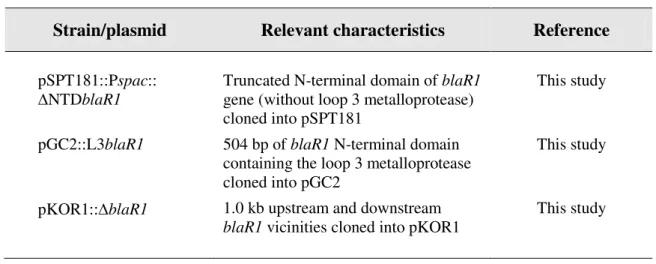
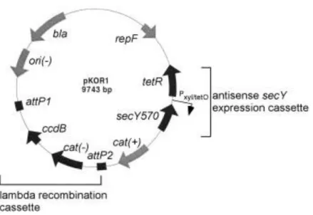
Genetic knock out’s were done by allelic replacement with inducible counter-selection using pKOR1 plasmid (7). This plasmid is an E. coli/ S. aureus shuttle vector with specific characteristics. It contains a lambda recombination cassette allowing efficient cloning without the use of restriction enzymes and ligases. This cassette encodes for ccdB, an E. coli gyrase inhibitor that suppresses the growth of cells containing pKOR1 without insert. pKOR1 has a thermosensitive origin of replication in
S. aureus which facilitates the chromosomal integration at non-permissive temperature (43ºC). Moreover, in S. aureus the plasmid allows selection for chromosomal excision and plasmid segregation via inducible antisense expression of the essential gene secY
For the genetic knock out of blaR1, DNA fragments of 1 kb were PCR amplified upstream and downstream of the regulator, using the primers attB1-blaF6 and
blaR6-BamHI, for the upstream fragment, and BamHI-blaF8 and attB2-blaR7, for the downstream fragment (ANNEX). The PCR products were digested with BamHI and ligated with T4 ligase. The ligation product was used for recombination with pKOR1 and the recombinant products were transferred to NEB 10-beta Competent E. coli cells.
The resulting plasmid, pKOR1::blaR1 was transferred via electroporation to the S. aureus restriction minus strain RN4220 and then transduced into the parental strain,
using phage 80α and selection with 10 µg/ mL of chloramphenicol.
Attempts to insert pKOR1::blaR1 in strains with bla locus integrated into the chromosome failed due to the resistance of those strains to bacteriophage infection. Therefore we set up a strategy to generate genetic knock-out’s in the -lactamase plasmid which is not single-copy. For the integration of recombinant pKOR1::blaR1
Figure 5 - Map of pKOR1. repF (Replication gene of pE194ts), secY570 (N-terminal 570 nucleotides of secY including ribosome binding site), cat (chloramphenicol acetyltransferase), attP (phage lambda attachment site), ori(-) (ColE1 plasmid replication origin), bla (β-lactamase). (+) or (-) indicates
into β-lactamase plasmid, COL strain transformed with both plasmids was grown at 43ºC on TSB supplemented with 10 µg/mL of chloramphenicol. A transducing-lysate of this culture was prepared and transduced back to strain COL with chloramphenicol and ampicillin selection, in order to select pbla-pKOR1::blaR1 co-integrates only. Next, one colony was picked and inoculated in TSB supplemented with 10 µg/mL of chloramphenicol at 30ºC, a permissive temperature, which enables co-integrate resolution. A transducing-lysate of this culture was prepared and transduced back to strain COL with ampicillin selection and anydrotetracyclin counter selection at 1 and 2 µg/mL. Control experiments were made without anydrotetracyclin. Plates were incubated at 30ºC for 2 days. Deletion of blaR1 was confirmed by PCR amplification with primers blaF9 and blaR8 (ANNEX).
5.
Phenotypic analysis
Susceptibility to oxacillin was routinely analyzed by disc diffusion method with 1 µg oxacillin discs prepared in-house. The cultures were homogeneously spread in a TSA plate with a swab and the antibiotic discs were carefully placed. The plate was incubated at 30ºC for 48 hours. The growth inhibition area was measured with a scale and compared with parental strains.
To infer the contribution of blaZ gene, TSA plates were supplemented with 2 µg/mL of clavulanic acid, a β-lactamase inhibitor, and susceptibility to oxacillin and penicillin was evaluated with 1 µg and 10 U diffusion discs, respectively. IPTG was added into TSA plates in a concentration range of 1 - 1000 mM when appropriate.
RESULTS
According to recent data from our laboratory, the oxacillin resistance phenotype of most MRSA strains is not affected by the overexpression in trans of the mecA
repressor (107). This surprising observation contradicts the current model and suggests the presence of other elements involved in the transcriptional control of mecA gene. The only two strains for which a decrease of the oxacillin-resistance phenotype was observed were negative for the -lactamase locus, suggesting that this locus might be involved in that phenomenon. In order to explore the putative effect of the β-lactamase operon in the protection against the repressive effect of mecI, several exploratory experiments were performed, as described below.
1.
Introduction of native
β
-lactamase
plasmid into prototype strains
1.1
Introduction of native
β
-lactamase
plasmid into strain COL-I
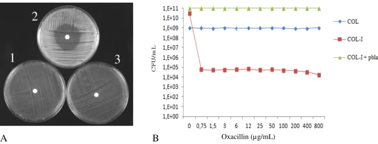
The prototype strain COL has a high and homogeneous level of oxacillin resistance, is negative for mecI and has a partially deleted mecR1, is naturally negative for the β-lactamase locus and has been used in many studies addressing the oxacillin-resistance mechanisms. Recombinant strain COL-I, overexpressing in trans the mecA
repressor, pGC2::mecI, is characterized by a massive decrease in the resistance level. In order to evaluate the role of β-lactamase operon in the observed “MecI-protection
effect”, we have first introduced the native β-lactamase plasmid into strain COL-I. Strain MW2, similarly to COL, has no mecI and a partially deleted mecR1, but is β -lactamase positive and its oxacillin resistance phenotype was not affected by the mecI
strain via 80α bacteriophage-mediated horizontal gene transfer with selection for ampicillin resistance. The strategy turned out successful with the introduction of the
large β-lactamase plasmid into strain COL first, followed by the introduction of the recombinant pGC2::mecI. The presence of both plasmids was confirmed by restriction
analysis and PCR detection of the β-lactamase operon and mecI gene. This experiment was done in two independent replicas.
When oxacillin susceptibility was tested, it was observed that the transformant COL-I in the presence of β-lactamase plasmid restored the resistance phenotype of strain COL in spite of the overexpression of mecI gene. These observations were also confirmed by population analysis profile (PAP) assays (Fig. 6 and Table 2).
Strain Relevant genotype Oxacillin-disc inhibition halo (mm)
COL mecI -∆mecR1 bla - 8
COL-I mecI ∆mecR1 bla - 32
COL-I + pbla mecI ∆mecR1 blaZ blaI blaR1 7
Table 2 - Oxacillin-resistance of parental strain COL and recombinant strains
1
1 2
3 4
2
3
Figure 6. Phenotypic expression of oxacillin resistance in COL and recombinant strains. A. Disc diffusion test 1: COL; 2: COL-I; 3: COL-I + pbla; B. Population analysis profile.
1
1 2
3 4
3
A B
2
3
1
3
1.2
Introduction of native
β
-lactamase
plasmid into VNG17 and
VNG17-I strains
Strain VNG17 and VNG17-I (overexpressing mecI) were transformed with the
β-lactamaseplasmid of strain MW2 (pbla). Given that strain COL-I showed a revertable phenotype in the presence of the β-lactamase plasmid we aimed to confirm these observations in other strains also negative for the β-lactamase locus. Strain VNG17 was the only other strain in which the overexpression of mecI caused a decrease in the oxacillin-resistance. Similarly to COL, strain VNG17 has no mecI and partially deleted
mecR1 but has a low-level resistance to oxacillin. The procedure was the same for introduction of the native β-lactamase plasmid into COL-I strain. The plasmid from prototype MW2 strain was introduced into VNG17 and VNG17-I strains via 80α bacteriophage-mediated horizontal gene transfer. For strain VNG17-I the protocol was also more successful with introduction of the β-lactamase plasmid into strain VNG17 first, following introduction of the pGC2::mecI. The presence of the plasmids was
confirmed by restriction analysis and PCR detection of the β-lactamaseoperon and mecI
gene.
As shown in Fig. 7 and Table 3, the introduction of the β-lactamase plasmid either in the parental or recombinant strain with mecI overexpression, caused a massive decrease in the susceptibility to oxacillin. In addition, the parental strain transformed with pbla showed a remarkable shift from low-level and heterogeneous to high-level and homogeneous expression of oxacillin resistance.
1
2
3
4
2
Strain Relevant genotype Oxacillin-disc inhibition halo (mm)
VNG17 mecI -∆mecR1 bla - 33/53a
VNG17 + pbla mecI -∆mecR1 blaZ blaI blaR1 13
VNG17-I mecI ∆mecR1 bla - 57
VNG17-I + pbla mecI ∆mecR1 blaZ blaI blaR1 33/38a a
Heterogeneous population
Table 3 - Oxacillin-resistance of parental strain VNG17 and recombinant strains
A B Oxacillin (µg/mL)
1
2
3
4
1.3
Introduction of native
β
-lactamase
plasmid into RJP17 and RJP17-I
strains
RJP17 strain is also a -lactamase negative strain, which belongs to the same clone of VNG17 and was isolated in the same country and time period. Similarly to VNG17, RJP17 is mecI negative, has a partial deleted mecR1 and expresses low level resistance to oxacillin. However, it was not detected any alteration in the oxacillin resistance phenotype upon the overexpression of mecI (strain RJP17-I) (107). Nevertheless, we have also evaluated the effect of the β-lactamase locus in this strain. Introduction of the β-lactamase plasmid in the recombinant strain overexpressing the
mecI gene, RJP17-I, was also tested.The procedure was exactly the same as performed for VNG17 and VNG17-I strains. Results are summarized in Fig. 8 and Table 4.
Since the resistant phenotype was not affected by the overexpression of MecI, the presence of pbla in strain RJP17-I did not cause any significant alterations. However, similarly to what was observed for strain VNG17, the parental strain transformed with pbla shifted to a high-level and homogeneous expression profile of oxacillin resistance.
Figure 8 - Phenotypic expression of oxacillin resistance in RJP17 and recombinant strains. A. Disc diffusion test 1: RJP17; 2: RJP17 + pbla; 3: RJP17-I; 4: RJP17-I + pbla; B. Population analysis profile.
A B Oxacillin (µg/mL)
1
2
1.4
Introductionof native
β
-lactamase
plasmid into HT0350 strain
HT0350 strain is other -lactamase negative strain but with a more extensive deletion of mecR1 due to the presence of IS431, a typical characteristic from SCCmec
type V strains. While strains COL, VNG17, RJP17 still have a complete N-terminal domain of mecR1 with the four-transmembrane segments (960 amino acids), strain HT0350 has only the first 36 amino acids of the MecR1. Similarly to VNG17 and RJP17, HT0350 is negative for mecI gene and expresses low-level resistance to oxacillin. The plasmid from prototype MW2 strain was introduced into this strain, via
80α bacteriophage-mediated horizontal gene transfer, in order to test, the effect of the β -lactamase locus. As we can see in Fig. 9 and Table 5 the β-lactamase plasmid promoted a significant increase in the oxacillin resistance phenotype.
Since strain HT0350 is intrinsically resistant to chloramphenicol, it was not included in previous mecI overexpression studies with plasmid pGC2, which carries a chloramphenicol resistance marker. Therefore, mecI was cloned in pSPT181, which carries a tetracycline resistance marker, and HT0350 phenotypic expression of oxacillin-resistance was evaluated in the presence of mecI overexpression in trans.
Strain Relevant genotype Oxacillin-disc inhibition halo (mm)
RJP17 mecI -∆mecR1 bla - 27/42a
RJP17 + pbla mecI -∆mecR1 blaZ blaI blaR1 15
RJP17-I mecI ∆mecR1 bla - 27/44a
RJP17-I + pbla mecI ∆mecR1 blaZ blaI blaR1 38/40a a
Heterogeneous population
Thereafter, theβ-lactamase plasmid was introduced in the HT0350-I strain, similarly to the above experiments. The results of the mecI overexpression and the later introduction of pbla are shown in Fig. 9 and Table 5. The presence of the β-lactamase plasmid in the parental strain promoted a shift from low-level and heterogeneous to high-level and homogeneous phenotypic expression of oxacillin resistance, and the recombinant strain HT0350-I restored the phenotype of the parental strain.
Strain Relevant genotype Oxacillin-disc inhibition halo (mm)
HT0350 mecI -∆mecR1 bla - 35
HT0350 + pbla mecI -∆mecR1 blaZ blaI blaR1 16
HT0350-I mecI ∆mecR1 bla - 56
HT0350-I + pbla mecI ∆mecR1 blaZ blaI blaR1 40
Table 5 - Oxacillin-resistance of parental strain HT0350 and recombinant strains Figure 9 - Oxacillin susceptibility test of parental strain HT0350 and recombinant strains.
A. Disc diffusion test. 1: HT0350; 2: HT0350 + pbla; 3: HT0350-I; 4: HT0350-I + pbla; B. Population analysis profile.
Oxacillin (µg/mL)
A B
1
2
Once we have confirmed the role of β-lactamase plasmid in the phenotypic expression of oxacillin-resistance and the interference with the mecI-mediated expression of mecA, we aimed to further experiments to investigate which element(s) of the β-lactamase operon would be, specifically, involved.
2.
Phenotypic effect of clavulanic acid
a
Heterogeneous population
Inhibition halo diameter (mm)
Oxa disc Pen disc
Clavulanic acid
(µg/mL) 0 2 0 2
MW2 30/39a 29/39a 8 11
MW2-I 30/40a 30/40a 7 11
COL 11 12 7 7
COL-I 15/40a 19/39a 28 27
COL-I + pbla 13 13 7 7
VNG17 34/52a 31/51a 15/45a 15/46a
VNG17 + pbla 17/20a 12/17a 7 10
VNG17-I 58 57 50 50
VNG17-I + pbla 35 35 7 10
RJP17 29/39a 27/38a 15/35a 14/34a
RJP17 + pbla 16 16 7 7
RJP17-I 29 28 16 15
RJP17-I + pbla 34 35 7 11
HT0350 35 35 7 11
HT0350 + pbla 16 16 7 22
HT0350-I 50 50 43 43
HT0350-I + pbla 38 37 7 7
3.
Introduction of
β
-lactamase regulators into COL-I strain
In order to explore the mechanisms by which the bla regulators interfere with
MecI repression and “boost” the phenotypic expression of oxacillin resistance, recombinant plasmids containing only theregulators were introduced into COL-I strain via bacteriophage-mediated horizontal gene transfer.
3.1
Introduction of
blaIblaR1
into COL-I strain
Previously, the blaIblaR1 coding region has been cloned in pSPT181 plasmid under the control of the inducible promoter Pspac (Arêde, P., unpublished data). At the same time, a similar construct was performed cloning this segment with the operator region, OblaIblaR1. These constructs were introduced into COL-I strain and oxacillin susceptibility was tested in the presence of several concentrations of IPTG. As shown in Table 7, the resistance phenotype of parental strain COL could not be restored in strain COL-I transformed with the β-lactamase regulators. Furthermore, the expression of these genes caused an increase in the susceptibility. The phenotype was not influenced by the presence or absence of the operator region, since it was not observed any significant difference between the two sets of experiments.
3.2
Introduction of
blaR1
and
blaI
genes into COL-I strain
Since introduction of both regulators simultaneously did not reproduce the effect