Marcelo Azevedo Costa , Ilka Afonso Reis , Claudio Antônio Bonjardim, Erna Geessien Kroon1, Jaquelline G. de Oliveira2☯‡
*, Paulo César Peregrino Ferreira1☯‡
1Departamento de Microbiologia, Instituto de Ciências Biológicas, Universidade Federal de Minas Gerais,
Belo Horizonte, Minas Gerais, Brasil,2Centro de Pesquisas René Rachou/FIOCRUZ, Belo Horizonte Minas Gerais, Brasil,3Departamento de Engenharia de Produção, Escola de Engenharia, Universidade Federal
de Minas Gerais, Belo Horizonte, Minas Gerais, Brasil,4Departamento de Estatística, Instituto de Ciências
Exatas, Universidade Federal de Minas Gerais, Belo Horizonte, Minas Gerais, Brasil
☯These authors contributed equally to this work.
¤ Current address: Departamento de Anatomia Patológica, Faculdade de Medicina, Universidade Federal
de Minas Gerais, Belo Horizonte, Minas Gerais, Brasil ‡These authors are senior authors on this work.
*jaque@cpqrr.fiocruz.br
Abstract
RAP1 (RAS proximate 1), a small GTP-binding protein of theRASsuperfamily, is a putative oncogene that is highly expressed in several malignant cell lines and types of cancers, in-cluding some types of squamous cell carcinoma. However, the participation of RAP1 in cer-vical carcinogenesis is unknown. We conducted a cross-sectional study of paraffin-embedded cervical biopsies to determine the association of RAP1 with cervical intraepithe-lial neoplasia (CIN). Standard and quantitative immunohistochemistry assessment of RAP1 expression in fixed tissue was performed on 183 paraffin-embedded cervical biopsies that were classified as normal or non-dysplastic mucosa (NDM) (n = 33); CIN grade 1 (n = 84) and CIN grade 2/3 (n = 66). A gradual increase in RAP1 expression in NDM<CIN 1<CIN 2/3 (p<0.001) specimens was observed and was in agreement with the histopathologic di-agnosis. A progressive increase in the RAP1 expression levels increased the risk of CIN 1 [odds ratio (OR) = 3.50; 95% confidence interval (CI) 1.30-10.64] 3.5 fold and the risk of CIN 2/3 (OR = 19.86, 95% CI 6.40-70.79) nearly 20 fold when compared to NDM. In addition, ste-reotype ordinal regression analysis showed that this progressive increase in RAP1 expres-sion more strongly impacted CIN 2/3 than CIN 1. Our findings suggest that RAP1 may be a useful biomarker for the diagnosis of CIN.
Introduction
Cervical cancer, which is caused by high-riskHuman papillomavirus(HR-HPV), is the second most frequent cancer in women worldwide [1]. Great efforts have been made during the last two decades to obtain useful markers that can be used in clinical practice for the proper
a11111
OPEN ACCESS
Citation:Pascoal-Xavier MA, Figueiredo ACC, Gomes LI, Peruhype-Magalhães V, Calzavara-Silva CE, Costa MA, et al. (2015) RAP1 GTPase Overexpression is Associated with Cervical Intraepithelial Neoplasia. PLoS ONE 10(4): e0123531. doi:10.1371/journal.pone.0123531
Academic Editor:Javier S Castresana, University of Navarra, SPAIN
Received:October 15, 2014
Accepted:February 19, 2015
Published:April 9, 2015
Copyright:© 2015 Pascoal-Xavier et al. This is an open access article distributed under the terms of the Creative Commons Attribution License, which permits unrestricted use, distribution, and reproduction in any medium, provided the original author and source are credited.
Data Availability Statement:All relevant data are within the paper and its Supporting Information files.
Funding:This research was supported by the Conselho Nacional de Desenvolvimento Científico e Tecnológico (CNPq-www.cnpq.br), Coordenação de Aperfeiçoamento de Pessoal de Nível Superior (CAPES—www.capes.gov.br), and Fundação de
identification and follow-up of precursor lesions of cervical cancer, also known as cervical intraepithelial neoplasia (CIN). Currently, the main biomarkers applied for CIN detection are proteins related to the cell cycle, such as p16INK4A, Ki-67, minichromosome maintenance 7 and 2 (MCM7 and 2), topoisomarase II alpha, and cyclin D1, all of which have altered expres-sion due to the effects of HPV on the cell cycle. The p16INK4Aprotein the best known surrogate biomarker of high grade lesions and also considered an indicator of the E6 and E7 overexpres-sion in cervical leoverexpres-sions [2–5]. Among the small GTPases, current research indicates the partici-pation of RAC1 and RHO in the progression of cervical neoplasia [6,7]. However, the
participation of RAP1 GTPase in cervical carcinogenesis remains unknown.
RAP1 (RAS proximate 1), a small GTPase of the RAS superfamily, and its regulatory pro-teins are involved in cell cycle progression, differentiation, survival and adhesion [8,9]. RAP1 is activated by a wide variety of external stimuli, and it functions as a signal transducer that switches between its inactive GDP-bound form and its active GTP-bound form. This switch is regulated by several guanine nucleotide exchanger factors (GEFs), which serve as activators, and GTPase-activating proteins (RAPGAPs), which act as inactivators [10,11]. RAP1 seems to play a role in cancer cell growth, invasion, and metastasis, and dysregulation of RAP1 activa-tion has been described in several malignant cell lines and cancers, such as oropharyngeal squa-mous cell carcinoma (SCC), papillary thyroid cancer, breast cancer, renal cell carcinoma, and melanoma [12–18].
Associations between HPV-mediated oncogenesis and alteration of the RAP1 signaling pathway have been described. The HR-HPV E6 oncoprotein targets the protein E6TP1, a known RAPGAP, for proteasome-dependent degradation and consequently enhances the GTP loading of RAP1 with subsequent activation of the MAPK signaling pathway and RAP1 over-expression [19,20]. Likewise, in HPV-infected epithelial cells, the interaction of the HPV E2 protein with the cellular bromodomain protein Brd4, a cell cycle progression regulator, en-hances the RAP GAP activity of SPA-1, which disrupts the proper balance of RAP1 activation [21].
Those findings led us to investigate the expression levels of RAP1 in low- and high-grade CIN lesions to address the potential use of this putative oncogene as a cervical neoplasia bio-marker. By comparing the immunostaining pattern of RAP1 in cervical specimens of CIN 1 and CIN 2/3 to that of non-dysplastic mucosa (NDM), we verified that RAP1 expression grad-ually increased with the severity of the cervical lesions. Moreover, we found a strong associa-tion of RAP1 overexpression in high grade lesions.
Materials and Methods
Ethics Statement
This cross-sectional study was approved by the institutional review board of the Universidade Federal de Minas Gerais (certificate number CAAE-0397.0.203.245–09). The need for written informed consent from the donor or the next of kin for the use of the paraffin-embedded sam-ples in this research was waived by the institutional review board of the Universidade Federal de Minas Gerais.
Study Participants
Two hundred and fifty-two paraffin-embedded cervical biopsies, normal or NDM (n = 65), CIN 1 (n = 102) and CIN 2/3 (n = 85), were collected from different individuals between May 2009 and March 2011 from the archives of the Hospital das Clínicas, Universidade Federal de Minas Gerais, Brazil. The majority of the patients were predominantly from the State of Minas Gerais, Brazil. Patients from other states, of the Southeast, North and Northeast regions of
Brazil were also included. The mean age of the patients was 38 years, ranging from 18 to 74 years old. Inclusion criteria were reviewed and classified as consensus diagnosis by two certi-fied pathologists (M.A.P.X. and L.P.F.) and HPV DNA detection. The study procedures are summarized in the flow diagram shown inFig 1.
HPV genotyping
DNA was extracted from five serial sections (10μm) of each paraffin-embedded sample using
the QIAamp DNA FFPE Tissue Kit (Qiagen, Düsseldorf, Germany) according to the manufac-turer's instructions. Nested PCR amplification was performed using the external primers MY11 and MY09 and the internal primers GP5+ and GP6+, which were designed to amplify a 150-bp product from the L1 gene of several HPV types, as described elsewhere [22,23]. For HPV genotyping, the gel-purified, 150-bp amplicons (QIAquick Gel Extraction Kit, QIAGEN Inc., CA, USA) were sequenced in both directions (Applied Biosystems 3730 DNA Sequencer; Life Technologies, USA), and a BLAST search was carried out to identify the HPV types. DNA extracted from HPV-18-infected HeLa cells was used as a positive control. To determine the
Fig 1. Study procedures and outcomes.The flow diagram shows the number of samples that were obtained from the laboratory file as well as those that were included in the immunohistochemistry and statistical analyses.
DNA integrity and identify the presence of PCR inhibitors, all samples were also tested by PCR using primers PC03 and PC04, which amplified the beta-actin gene [24].
Immunohistochemistry
RAP1 and p16INK4Aimmunostaining was performed on 4-μm sequential sections of
formalin-fixed, paraffin-embedded biopsy specimens using the Novolink Polymer Detection System (Novocastra, Leica Microsystems, USA). Deparaffinization with xylene and rehydration through a graded alcohol series was followed by antigen retrieval by heating to 90°C for 10 minutes in EDTA buffer, pH 9.0. After blocking endogenous peroxidases (DakoCytomation, Glostrup, Denmark), the deparaffinized sections were incubated for 1 hour at room tempera-ture with anti-RAP1 or anti-p16INK4Aantibodies (rabbit polyclonal to RAP1A+RAP1B; ab40814, Abcam, 1:120 dilution; mouse monoclonal to p16INK4A, 551154; clone G175-405, BD Pharmingen Bioscienses, dilution 1:110). The sections were then incubated at room tempera-ture for 30 minutes with the Novolink polymer DS and washed twice with Tris-borate saline for 5 minutes. Peroxidase activity was developed using a diaminobenzidine (DakoCytomation, Glostrup, Denmark) solution for 30 minutes at room temperature, counterstaining was per-formed using Harris hematoxylin, and then evaluated by light microscopy.
RAP1 and p16INK4Aexpression levels in the NDM and CIN groups were evaluated accord-ing to criteria previously described by Galgano and collaborators [25]. The scoring of RAP1 and p16INK4Awas graded as pattern 0 (no staining), pattern 1 (weak staining in 5%-25% of the cells distributed in the basal third of the epithelium), pattern 2 (moderate staining in 26%-50% of the cells distributed in the basal third of the epithelial thickness), and pattern 3 (strong stain-ing in>50% of the cells that were distributed in two-thirds of the entire epithelial thickness). RAP1 and p16INK4Aexpression levels were considered normal for pattern 0 or 1 and high for pattern 2 or 3.
To limit the potentially subjective visual assessment of the RAP1 and p16INK4A immunohis-tochemical score, we used the image-based uniplex method [16] to measure the relative intensi-ty (RI) of RAP1 and p16INK4Aprotein expression in a representative number of samples. The 16-bpp TIF images of the tissue were captured using an AxioCam MRc digital camera equipped with AxioVision software and attached to a Zeiss Axio microscope (Carl Zeiss Mi-croscopy, Cambridge, UK). Images spanning the full range of intensity levels were analyzed using ImageJ 1.37v processing software (National Institutes of Health), which provided area and pixel value statistics of the user-selected region of interest (ROI). Ten 1-mm2areas were se-lected as ROIs in the epithelium and stroma by a certified pathologist (M.A.P.X.). The RI of biomarker staining (relative protein expression) was calculated in RI units by dividing the mean pixel intensity of equal ROIs containing stroma (Ibackground) by the mean pixel intensi-ty of equal ROIs containing epithelial cells (Itest). This algorithm accounted for variations in background staining related to the immunohistochemistry routine.
Statistical analysis
Associations between categorical variables were evaluated using Fisher's exact test, and differ-ences between continuous variables were determined using the two-sample T-test. To estimate odds ratios (OR) and the corresponding 95% confidence intervals (CIs) for the association between CIN and change in RAP1 expression, we compared normal and high expression of NDM to that of CIN 1 and of CIN 2/3 and CIN 1 to that of CIN 2/3. To establish a relationship between CIN and the change in RAP1 expression, we fitted the stereotype ordinal logistic
re-gression model,log pj
pNDM
h i
classes, respectively, and x = 0,1,2,3 represents the RAP1 immunohistochemical expression, and we estimated the value ofβas proposed by Anderson [26]. The coefficient significance was tested using the Log-likelihood method. A result was considered statistically significant when p<0.05. The statistical analyses were performed using MINITAB 16 (Minitab Inc., State Col-lege, Pennsylvania) and R (Version 2.15.3;http://www.r-project.org) with the VGAM package statistical softwares.
Results
RAP1 expression and HPV detection
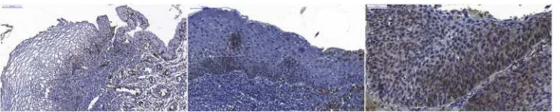
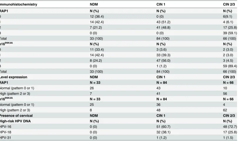
The RAP1 and p16INK4Aexpression levels in NDM, CIN 1, and CIN 2/3 lesions are shown in Fig 2, inS1 FigandTable 1, which also include the HPV-16, HPV-18 and HPV-31 types that were detected in the CIN groups. p16INK4Awas used as an immunohistochemical reference for our study. RAP1 was undetectable in 36.4% of the NDM samples (12/33), and once verified in the NDM group, RAP1 immunostaining was very weak and predominantly restricted to the basal layer of the epithelium in 42.4% of the samples (14/33) and was classified as pattern 1 (as described inMaterial and Methods). Only 7 of the 33 cases of NDM presented moderate stain-ing of RAP1 in 26%-50% of the cells that were distributed in the basal third of the epithelial thickness (pattern 2). Unlike what was observed in NDM, RAP1 expression was observed in 100% of the CIN 1 samples. About half of the CIN 1 samples (43/84) presented a moderate RAP1 immunostaining in dysplastic cells in the basal third of the epithelium (pattern 2), and the other half presented weak immunostaining of RAP1 expression, as described for pattern 1. In contrast, 85% (56/66) of the CIN 2/3 lesions presented moderate or strong immunostaining, indicating much higher RAP1 expression levels in high-grade lesions than those observed in NDM or CIN 1 biopsies (p<0.001). Indeed, a predominance of strong, diffuse RAP1 staining throughout the thickness of the epithelium was observed in 59.1% (39/66) of the CIN 2/3 le-sions. Notably, p16INK4Ashowed the same immunohistochemical patterns described by Gal-gano and colleagues in NDM, CIN1 and CIN2/3 samples [25]. Strong nuclear staining for RAP1 was observed in approximately 70% of the CIN 2/3 lesions (Fig 2C), while weak nuclear staining was only observed in some basal cells of the CIN 1 (Fig2Band2A). No cytoplasmic or nuclear staining was observed in intermediate or superficial cells of the NDM samples (Fig 2A). RAP1 was strongly expressed in neutrophils and endothelial cells and was weakly express-ed in lymphocytes and stromal mononuclear cells (Fig2Band2C).
A predominance of HPV-16 in 60.7% (51/84) and 72.7% (48/66) of the CIN 1 and CIN 2/3 samples, respectively, was observed. HPV-18 was detected in 38.1% (32/84) and 25.8% (17/66)
Fig 2. Immunohistochemical staining of RAP1.Left—Squamocolumnar junction of non-dysplastic cervical mucosa (NDM) showing very weak immunostaining for RAP1 in the cytoplasm of basal cells and no staining in the intermediate and superficial cells. Middle—CIN 1 showing moderate staining for RAP1 in cells distributed in the basal third of the epithelium and showing RAP1 expression in neutrophils. Right- CIN 2/3 showing strong staining for RAP1 in cells diffusely distributed throughout the entire epithelial thickness, with nuclear translocation of RAP1.
of the CIN 1 and CIN 2/3 samples, respectively. HPV-31 was detected in only one sample from each group (Table 1).
Morphometric evaluation of RAP1 expression
To limit the subjective visual assessment of immunohistochemical staining, we also performed a morphometric evaluation of RAP1 and p16INK4Aexpression levels. We measured the relative intensity (RI) of chromogen staining as a measure of the relative RAP1 and p16INK4Aprotein expression in NDM, CIN 1, and CIN 2/3 lesions (Fig 3). Our data showed higher RAP1 expres-sion in CIN 2/3 samples compared to NDM and CIN 1 samples (p<0.05) and in CIN 1 samples compared to NDM samples (p<0.05), similar to the data obtained by standard microscopy analyses. RAP1 and p16INK4Aimmunohistochemical morphometric patterns were similar ex-cept for the significant difference (p<0.05) between RAP1 and p16INK4Aexpression observed in the CIN2/3 group (Fig 3).
Overexpression of RAP1 and CIN progression
The association between RAP1 expression and risk of CIN progression was determined to per-form comparisons between all groups. Thetable 2presents the odds ratios (ORs) and 95%
Table 1. Scores of RAP1 and p16INK4Aimmunohistochemical expression in precursor lesions of cervical neoplasia.
Immunohistochemistry NDM CIN 1 CIN 2/3
RAP1 N (%) N (%) N (%)
0 12 (36.4) 0 (0) 6(9.1)
1 14 (42.4) 43 (51.2) 4 (6.1)
2 7 (21.2) 41 (48.8) 17 (25.8)
3 0 (0) 0 (0) 39 (59.1)
Total 33 (100) 84 (100) 66 (100)
p16INK4A N (%) N (%) N (%)
0 11 (33.4) 3 (3.6) 2 (3.0)
1 14 (42.4) 33 (39.3) 2 (3.0)
2 8 (24.2) 47 (56.0) 3 (4.5)
3 0 (0) 1 (1.2) 59 (89.4)
Total 33 (100) 84 (100) 66 (100)
Level expression NDM CIN 1 CIN 2/3
RAP1 N = 33 N = 84 N = 66
Normal (pattern 0 or 1) 26 43 10
High (pattern 2 or 3) 7 41 56
p16INK4A N = 33 N = 84 N = 66
Normal (pattern 0 or 1) 25 36 4
High (pattern 2 or 3) 8 48 62
Presence of cervical NDM CIN 1 CIN 2/3
High-risk HPV DNA N (%) N (%) N (%)
HPV-16 0 (0) 51 (60.7) 48 (72.7)
HPV-18 0 (0) 32 (38.1) 17 (25.8)
HPV-31 0 (0) 1 (1.2) 1 (1.5)
Abbreviations: NDM—non-dysplastic mucosa; CIN—cervical intraepithelial neoplasia.
confidence intervals (CIs) for the study of association between RAP1 expression levels (normal and high) in the NDM, CIN 1, and CIN 2/3 samples. We observed that high levels of RAP1 raised the risk of CIN 1 by 3.5 fold [(OR) = 3.50; 95% confidence interval (CI) 1.30–10.64] and the risk of CIN 2/3 by nearly 20 fold (OR = 19.86, 95% CI 6.40–70,80) compared to NDM and increased the risk of CIN 2/3 5.8 fold (OR = 5.80; 95% CI 2.51–14.52) compared to CIN 1.
The relationship between CIN grade and variation in RAP1 expression were determined using stereotype regression analysis. Thetable 3summarizes the results of the fitted model.
Ac-cording to the model,log pj
pNDM
h i
¼ ajþ ;jbx,j= 2,3 and x = 0,1,2,3 and with identifiability
constraints (;CIN 2/3= 1,;NDM= 0) as in Anderson and colleagues (26), we estimate;CIN 2/3= 0.36 andβ= 2.98. Therefore, we estimated the relation between risks ratio and RAP1
Fig 3. Morphometric analysis of RAP1 and p16INK4Aimmunohistochemical staining.Relative Intensity (RI) data of RAP1 anfd p16INK4Aexpression in NDM, CIN 1, and CIN 2/3 samples are expressed in box plot format, with boxes stretching from the 25thpercentile to the 75thpercentile and the line across the
box representing the median values. Statistically significant differences (p<0.05) are shown by dotted lines.
doi:10.1371/journal.pone.0123531.g003
Table 2. Associations between RAP1 expression and risk of CIN progression.
Ratio OR 95% CI p-value
CIN 1 / NDM 3.50 1.30,10.64 <0.001
CIN 2/3 / NDM 19.87 6.40,70.80 <0.001
CIN 2/3 / CIN 1 5.80 2.51,14.52 <0.001
Abbreviations: OR—Odds ratio; NDM—non-dysplastic mucosa; CIN—cervical intraepithelial neoplasia.
expression islog pCIN 2=3
pNDM
h i
¼ 4:56þ2:98xfor CIN2/3 andlog pCIN 1
pNDM
h i
¼ 0:30þ 0:36
ð Þ2:98xfor CIN1. Beta value is statistically different from 1, showing the influence of the
increasing in RAP1 in the risk of CIN compared to NDM.
However, since the estimate for the weight of the RAP1 expression influence in risks ratios for CIN1 class,;CIN 1, is statistically smaller than 1, stereotype ordinal regression analysis also showed that this progressive increase of RAP1 impacts more strongly in CIN2/3 than CIN1. Deviance residuals of the model indicated general goodness of fit of the model to the obtained data.
Discussion
In this cross-sectional study, we found a strong association of RAP1 with a significant increased risk of progression of CIN 1 and CIN 2/3 by comparing the RAP1 protein expression levels at different degrees of cervical intraepithelial neoplasia using standard and semi-quantitative ap-proaches. RAP1 expression levels were predominantly undetectable or basal in normal squa-mous mucosa, but RAP1 expression gradually increased according to the severity of the cervical lesions.
The two main limitations of this study were the quality of the DNA that was extracted from paraffin-embedded samples for HPV detection and typing and the subjective bias of the quali-tative assessment of immunohistochemistry. To circumvent those limitations, we used a large number of cervical lesions and performed morphometric analysis, respectively. Another limita-tion was the interference of nonepithelial cell immunostaining in the RAP1 morphometric analyses. The constitutive expression of RAP1 in neutrophils, lymphocytes and endothelial cells may have interfered with the calculation of the relative intensity (RI) index due to the in-creased number of inflammatory cells and angiogenesis that was frequently observed during CIN progression [27].
A very weak RAP1 cytoplasmic expression was observed in the basal cells of some NDM samples presenting inflammatory infiltrate in its the stromal tissue (Fig 2A). This may be due to cell proliferation induced by the inflammatory process of the cervical mucosa. Interestingly, a very discrete immunostaining of p16INK4Awas also observed in some NDM inflammatory cervical samples (S1 Fig) as also described elsewhere [28].
We observed a strong nuclear staining for RAP1 in CIN 2/3 samples, as previously reported in oropharyngeal SCC cell lines and human oral cancer specimens, and for other small GTPases, such as RHO and RAC1, in premalignant lesions and cervical cancer [6,7,12,29]. Be-cause only the active GTP-bound form of RAP1 translocates to the nucleus, we can infer that
Table 3. Estimated coefficients, standard errors, z-scores, and two-tailedp-values for the fitted stereotype regression model.
Coefficients Estimate (95% CI) SE z-score pvalue
;NDM 0* — — —
;CIN 1 0.36 (0.22,0.50) 0.07 4.73 <0.001
;CIN 2/3 1* — — —
αCIN 1 -0.30 (-1.04,0.44) 0.38 -0.79 0.2146
αCIN 2/3 -4.56 (-6.05,-3.07) 0.76 -5.97 <0.001
β 2.98 (2.16,3.80) 0.42 7.10 <0.001
Log-likelihood: -136.46
Abbreviations: SE—Standard error; NDM—non-dysplastic mucosa; CIN—cervical intraepithelial neoplasia.
*Identifiability constraints proposed proposed by Anderson [26].
influence theβvalue. However, the weight of the growing shift in the immunohistochemical
pattern of logitlog pCIN 2=3
pNDM
h i
is much more relevant than in the logitlog pCIN 1
pNDM
h i
. For such risk
es-timates, we use the parsimonious stereotype model, which is more flexible and better preserves the ordered outcomes by imposing a linear structure in the model’s logit [30,31]. In addition to its use as a diagnostic and potential risk progression biomarker, an advantage of RAP1 could be the manipulation of RAP1 signaling pathway components in the cervical microenvironment [32].
The very different expression patterns of RAP1, i.e., weak/moderate expression in CIN 1 lesions in contrast to the strong RAP1 expression observed in the vast majority of CIN 2/3 le-sions, suggest a distinct influence of HPV on RAP1 expression during the development of cer-vical carcinogenesis. CIN 1 and epithelial mucosa persistently infected with HPV are
characterized by high levels of cytoplasmic and nuclear HPV E2 protein, which binds to the Brd4 protein, a cellular protein that mediates several processes important for the viral life cycle, including viral genome maintenance and replication [33,34]. When bound to Brd4, E2 com-petes with SPA-1, a RAPGAP. Hence, it is possible that E2-Brd4 binding favors the dysfunction of SPA-1, creating a hyperactive state of RAP1 [35]. On the other hand, overexpression of HR-HPV E6 protein, especially after integration of the HPV genome into the host genome in high-grade dysplasia (CIN 2/3), may activate RAP1-mediated signaling pathways during cellu-lar transformation through the down-regulation of E6TP1 [36].
Our results suggest that RAP1 GTPase acts as a new biomarker with potential clinical value for severity assessment of cervical epithelial neoplasia. However, studies of RAP1 expression and its regulators in epithelial cells that are associated with HPV infection as well as prospec-tive observational studies with a long-term follow-up will be required to elucidate the role of RAP1 in cervical carcinogenesis and to identify the clinical relevance of RAP1 in cervical cancer progression.
Supporting Information
S1 Fig. Immunohistochemical staining of p16INK4A.Left—Squamocolumnar junction of non-dysplastic cervical mucosa (NDM) showing very weak immunostaining for p16INK4Ain the cytoplasm of basal cells and no staining in the intermediate and superficial cells. Middle—
CIN 1 showing moderate staining for p16INK4Ain cells distributed in the basal third of the epi-thelium. Right—CIN 2/3 showing very strong staining for p16INK4Ain cells diffusely distribut-ed throughout the entire epithelial thickness.
(TIF)
S1 Dataset. Data set forFig 3“Morphometric analysis of RAP1 and p16INK4A immunohis-tochemical staining.”
(XLSX)
S2 Dataset. Dataset forTable 1“Scores of RAP1 and p16INK4Aimmunohistochemical ex-pression in precursor lesions of cervical neoplasia”.
S3 Dataset. Dataset forTable 2“Associations between RAP1 expression and risk of CIN progression”.
(XLSX)
S4 Dataset. Dataset forTable 3“Estimated coefficients, standard errors, z-scores, and two-tailedp-values for the fitted stereotype regression model”.
(XLSX)
Acknowledgments
The authors are grateful to Dr. Lúcia Porto Fonseca (certified pathologist) for revising all cases included in this study and to João Rodrigues dos Santos, Clari Gandra and Luciana Lisboa from UFMG and CPqRR for their excellent technical support. The authors are also thankful for the financial support provided by Conselho Nacional de Desenvolvimento Científico e Tec-nológico (CNPq), Coordenação de Aperfeiçoamento de Pessoal de Nível Superior (CAPES), PDTIS-Fundação Oswaldo Cruz (Fiocruz) and Fundação de Amparo à Pesquisa do Estado de Minas Gerais (FAPEMIG).
Author Contributions
Conceived and designed the experiments: MAPX JGO PCPF. Performed the experiments: MAPX ACCF LIG VPM CECS. Analyzed the data: PCPF JGO MAPX MAC IAR. Contributed reagents/materials/analysis tools: CAB EGK. Wrote the paper: MAPX IAR JGO PCPF.
References
1. zur Hausen H. Papillomaviruses and cancer: from basic studies to clinical application. Nat Rev Cancer. 2002; 2:342–50. PMID:12044010
2. Portari EA, Russomano FB, de Camargo MJ, Machado Gayer CR, da Rocha Guillobel HC, Santos-Rebouças CB, et al. Immunohistochemical Expression of Cyclin D1, p16(INK4a), p21WAF1, and Ki-67 correlates with the severity of cervical neoplasia. Int J Gynecol Pathol. 2013; 32:501–8. doi:10.1097/ PGP.0b013e31826f5cf6PMID:23896712
3. Lobato S, Tafuri A, Fernandes PA, Caliari MV, Silva MX, Xavier MA, et al. Minichromosome mainte-nance 7 protein is a reliable biological marker for human cervical progressive disease. J Gynecol Oncol. 2012; 23:11–5. doi:10.3802/jgo.2012.23.1.11PMID:22355461
4. von Knebel Doeberitz M, Reuschenbach M, Schmidt D, Bergeron C. (2012) Biomarkers for cervical cancer screening: the role of p16(INK4a) to highlight transforming HPV infections. Expert Rev Proteo-mics. 2012; 9:149–63. doi:10.1586/epr.12.13PMID:22462787
5. Tsoumpou I, Arbyn M, Kyrgiou M, Wentzensen N, Koliopoulos G, Martin-Hirsch P, et al. p16(INK4a) immunostaining in cytological and histological specimens from the uterine cervix: a systematic review and meta-analysis. Cancer Treat Rev. 2009; 35:210–20. doi:10.1016/j.ctrv.2008.10.005PMID:
19261387
6. Mendoza-Catalán MA, Cristóbal-Mondragón GR, Adame-Gómez J, del Valle-Flores HN, Coppe JF, Si-erra-López L, et al. Nuclear expression of Rac1 in cervical premalignant lesions and cervical cancer cells. BMC Cancer. 2012; 12:116. doi:10.1186/1471-2407-12-116PMID:22443139
7. Tibúrcio MG, Pinheiro NM, Carboni Sde S, Rocha LP, Adad SJ, Maluf PJ, et al. GTPases Rho distribu-tion in intraepithelial and invasive neoplasias of the uterine cervix. Eur J Gynaecol Oncol. 2014; 35:284–8. PMID:24984542
8. Gloerich M, Bos JL. Regulating Rap small G-proteins in time and space. Trends Cell Biol. 2011; 21:615–23. doi:10.1016/j.tcb.2011.07.001PMID:21820312
9. Hattori M, Minato N. RAP1 GTPase: functions, regulation, and malignancy. J Biochem. 2003; 134:479– 84. PMID:14607972
13. Banerjee R, Henson BS, Russo N, Tsodikov A, D'Silva NJ. RAP1 mediates galanin receptor 2-induced proliferation and survival in squamous cell carcinoma. Cell Signal. 2011; 23:1110–8. doi:10.1016/j. cellsig.2011.02.002PMID:21345369
14. Banerjee R, Russo N, Liu M, Van Tubergen E, D'Silva NJ. RAP1 and its regulatory proteins: the tumor suppressor, oncogene, tumor suppressor gene axis in head and neck cancer. Small GTPases. 2012; 3:192–7. doi:10.4161/sgtp.20413PMID:22684501
15. Nellore A, Paziana K, Ma C, Tsygankova OM, Wang Y, Puttaswamy K, et al. Loss of RAP1GAP in pap-illary thyroid cancer. J Clin Endocrinol Metab. 2009; 94:1026–32. doi:10.1210/jc.2008-1042PMID:
19066305
16. Furstenau DK, Mitra N, Wan F, Lewis R, Feldman MD, Fraker DL. Ras-related protein 1 and the insulin-like growth factor type I receptor are associated with risk of progression in patients diagnosed with car-cinoma in situ. Breast Cancer Res Treat. 2011; 129:361–72. doi:10.1007/s10549-010-1227-yPMID:
20976540
17. Kim WJ, Gersey Z, Daaka Y. RAP1GAP regulates renal cell carcinoma invasion. Cancer Lett. 2012; 320:65–71. doi:10.1016/j.canlet.2012.01.022PMID:22266190
18. Gao L, Feng Y, Bowers R, Becker-Hapak M, Gardner J, Council L, et al. Ras-associated protein-1 regu-lates extracellular signal-regulated kinase activation and migration in melanoma cells: two processes important to melanoma tumorigenesis and metastasis. Cancer Res. 2006; 66:7880–8. PMID:
16912161
19. Gao Q, Srinivasan S, Boyer SN, Wazer DE, Band V. The E6 oncoproteins of high-risk papillomaviruses bind to a novel putative GAP protein, E6TP1, and target it for degradation. Mol Cell Biol. 1999; 19:733– 44. PMID:9858596
20. Singh L, Gao Q, Kumar A, Gotoh T, Wazer DE, Band H, et al. The high-risk human papillomavirus type 16 E6 counters the GAP function of E6TP1 toward small Rap G proteins. J Virol. 2003; 77:1614–20. PMID:12502878
21. Farina A, Hattori M, Qin J, Nakatani Y, Minato N, Ozato K. Bromodomain protein Brd4 binds to GTPase-activating SPA-1, modulating its activity and subcellular localization. Mol Cell Biol. 2004; 24:9059–69. PMID:15456879
22. Husnjak K, Grce M, MagdićL, PavelićK. Comparison of five different polymerase chain reaction meth-ods for detection of human papillomavirus in cervical cell specimens. J Virol Methmeth-ods. 2000; 88:125– 34. PMID:10960700
23. Fuessel Haws AL, He Q, Rady PL, Zhang L, Grady J, Hughes TK, et al. Nested PCR with the PGMY09/ 11 and GP5(+)/6(+) primer sets improves detection of HPV DNA in cervical samples. J Virol Methods, 2004; 122:87–93. PMID:15488625
24. Nonogaki S, Wakamatsu A, Filho AL, Roteli-Martins C, di Loreto C, Maeda MY, et al. Molecular strate-gies for identifying human papillomavirus infection in routinely processed samples: focus on paraffin sections. J Low Genit Tract Dis. 2005; 9:219–24. PMID:16205192
25. Galgano MT, Castle PE, Atkins KA, Brix WK, Nassau SR, Stoler MH. Using biomarkers as objective standards in the diagnosis of cervical biopsies. Am J Surg Pathol. 2010; 34:1077–87. doi:10.1097/ PAS.0b013e3181e8b2c4PMID:20661011
26. Anderson JA. Regression and ordered categorical variables. J R Stat Soc. 1984; 46:1–30.
27. Mazibrada J, RittàM, Mondini M, De Andrea M, Azzimonti B, Borgogna C, et al. Interaction between in-flammation and angiogenesis during different stages of cervical carcinogenesis. Gynecol Oncol. 2008; 108:112–20. PMID:17936343
28. Klaes R, Friedrich T, Spitkovsky D, Ridder R, Rudy W, Petry U, et al. Overexpression of p16(INK4A) as a specific marker for dysplastic and neoplastic epithelial cells of the cervix uteri. Int J Cancer. 2001; 92:276–84. PMID:11291057
29. Goto M, Mitra RS, Liu M, Lee J, Henson BS, Carey T, et al. RAP1 stabilizes beta-catenin and enhances beta-catenin-dependent transcription and invasion in squamous cell carcinoma of the head and neck. Clin Cancer Res. 2010; 16:65–76. doi:10.1158/1078-0432.CCR-09-1122PMID:20028760
31. Abreu MN, Siqueira AL, Cardoso CS, Caiaffa WT. Ordinal logistic regression models: application in quality of life studies. Cad Saude Publica. 2008; 24Suppl4:s581–91.
32. Schmid MC, Franco I, Kang SW, Hirsch E, Quilliam LA, Varner JA. PI3-kinaseγpromotes RAP1a-me-diated activation of myeloid cell integrinα4β1, leading to tumor inflammation and growth. PLoS One. 2013; 8(4):e60226. doi:10.1371/journal.pone.0060226PMID:23565202
33. McPhillips MG, Oliveira JG, Spindler JE, Mitra R, McBride AA. Brd4 is required for E2-mediated tran-scriptional activation but not genome partitioning of all papillomaviruses. J Virol. 2006; 80:9530–43. PMID:16973557
34. McBride AA, McPhillips MG, Oliveira JG. Brd4: tethering, segregation and beyond. Trends Microbiol. 2004; 12:527–9. PMID:15539109
35. Yan J, Li Q, Lievens S, Tavernier J, You J. Abrogation of the Brd4-positive transcription elongation fac-tor B complex by papillomavirus E2 protein contributes to viral oncogene repression. J Virol. 2010; 84:76–87. doi:10.1128/JVI.01647-09PMID:19846528
36. Chakrabarti O, Veeraraghavalu K, Tergaonkar V, Liu Y, Androphy EJ, Stanley MA, et al. Human papil-lomavirus type 16 E6 amino acid 83 variants enhance E6-mediated MAPK signaling and differentially regulate tumorigenesis by notch signaling and oncogenic Ras. J Virol. 2004; 78:5934–45. PMID: