Received 22 Feb 2016|Accepted 17 Jun 2016|Published 25 Jul 2016
Plexins function in epithelial repair in both
Drosophila
and zebrafish
Sa Kan Yoo
1,2,3, Heath G. Pascoe
4,*, Telmo Pereira
5,6,*, Shu Kondo
7, Antonio Jacinto
6, Xuewu Zhang
4& Iswar K. Hariharan
1In most multicellular organisms, homeostasis is contingent upon maintaining epithelial integrity. When unanticipated insults breach epithelial barriers, dormant programmes of tissue repair are immediately activated. However, many of the mechanisms that repair damaged epithelia remain poorly characterized. Here we describe a role for Plexin A (PlexA), a protein with particularly well-characterized roles in axonal pathfinding, in the healing of damaged epithelia inDrosophila. Semaphorins, which are PlexA ligands, also regulate tissue repair. We show thatDrosophilaPlexA has GAP activity for the Rap1 GTPase, which is known to regulate the stability of adherens junctions. Our observations suggest that the inhibition of Rap1 activity by PlexA in damaged Drosophila epithelia allows epithelial remodelling, thus facilitating wound repair. We also demonstrate a role for Plexin A1, a zebrafish orthologue of
Drosophila PlexA, in epithelial repair in zebrafish tail fins. Thus, plexins function in epithelial wound healing in diverse taxa.
DOI: 10.1038/ncomms12282 OPEN
1Department of Molecular and Cell Biology, University of California, Berkeley, California 94720, USA.2The Miller Institute, University of California, Berkeley,
California 94720, USA.3Physiological Genetics Laboratory, RIKEN, 2-2-3 Minatojima-minamimachi, Chuo-ku, Kobe 650-0047, Japan.4Department of
Pharmacology, University of Texas Southwestern Medical Center, Dallas, Texas 75390, USA.5Instituto de Medicina Molecular, Faculdade de Medicina da
Universidade de Lisboa, 1649-028 Lisboa, Portugal.6CEDOC, Chronic Diseases Research Centre, NOVA Medical School, Universidade NOVA de Lisboa, 130,
1169-056 Lisboa, Portugal.7Genetic Strains Research Center, National Institute of Genetics, 1111 Yata, Mishima, Shizuoka 411-8540, Japan. * These authors
T
he initial responses to epithelial wounds have an important role in determining whether tissue damage eventually results in regeneration or scar formation1–3. Since epithelial tissues are found in most metazoans, some of the mechanisms involved in repairing epithelial tissues and in healing wounds are likely to be evolutionarily ancient and can potentially be identified using genetic screens conducted in invertebrate model organisms4–7. We first screened for regulators of wound repair in Drosophilaimaginal discs and then tested whether the orthologous genes in zebrafish have a similar function. Using this approach, we have identified a key role for plexins during tissue repair in both organisms.Results
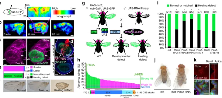
An RNAi screen identifies a role forPlexAin wing disc repair. The adult wing of Drosophila develops from a larval epithelial structure, the wing imaginal disc that can be visualizedin situby the nubbin4GFP (green fluorescent protein) reporter and bisected by applying pressure on the larval cuticle with a tungsten needle while leaving the cuticle itself intact (Fig. 1a and Supplementary Movie 1). After wounding, the two portions of the wing disc were observed to move independently but remained in proximity to each other likely because they were attached to neighbouring structures. To examine early changes following wounding, we used a GCaMP3 probe to visualize changes in intracellular calcium8. In the vicinity of the wound, we observed a rapid increase immediately after wounding, which persisted for 5–10 min (Fig. 1b and Supplementary Movie 2), which is similar to the wound-induced calcium flashes observed in the Caenorhabditis. elegans epidermis9, the zebrafish tail fin2 and theDrosophilaembryonic and pupal epithelia10,11. Fusion of the two disc fragments had occurred within 6 h of wounding. At this time, there was an accumulation of F-actin at the site of fusion (Fig. 1c) and increased activity of the Jun N-terminal kinase (JNK) as assessed by a transcriptional reporter of AP-1 activity12
(Fig. 1d). Calcium signalling, accumulation of F-actin and the increased activity of JNK are likely to be evolutionarily ancient responses to wounding since they are found in diverse organisms. Thus, screens for regulators of wound healing in imaginal discs are likely to identify mechanisms that are conserved among diverse taxa.
Approximately 95% of flies survive when one of their wing discs has been bisected (Fig. 1e,f). In these survivors, rejoining of the two fragments is highly efficient and B95% of the discs
develop into adult wings that either look completely normal or have minor notching of the wing margin. In the remaining 5% of the adult survivors, there are a variety of defects, most notably blisters, which are often seen when the two surfaces of the adult wing fail to adhere properly.
To test whether a gene has a role in wound healing, we manipulated its activity in the wing pouch and fragmented one of the two wing discs; the intact contralateral disc served as a control (Fig. 1g). A dominant-negative form of the JNK (JNKDN) elicited an increase in the frequency of wing morphology defects on the wounded side (43% versus 5% in controls; Fig. 1h) consistent with previous observations in other systems that indicated a role for JNK in wound healing4,5,13–15. We then screened 1,193 RNA interference (RNAi) lines targeting proteins that are predicted to be either on the cell surface or secreted (CSS)16because these CSS molecules are likely to play important roles in cell–cell communication and tissue remodelling during wound repair. We wounded at least 25 larvae per stock, thus screeningB30,000
flies (Fig. 1h). Of these, 34 lines (2.8%) were classified as ‘strong hits’ since they elicited wing abnormalities only on the wounded side. Plexin A(PlexA) was the top hit, withB80% of wounded
animals exhibiting wing abnormalities with only minor effects in the absence of wounding, a result confirmed using three additional RNAi lines (Fig. 1i,j and Supplementary Fig. 1a,b). In addition, to inactivatePlexAfunction in the wing disc in a way that does not depend on RNAi, we expressed the Cas9 nuclease and a guide RNA (gRNA) that targetedPlexAin cells of the wing
UAS-dcr2;
nub-Gal4, UAS-GFP UAS-RNAi library
X
Wound Wound in situin situ
Gal4 RNAi UAS
WT Developmental defect
Healing defect
F-actin
F-actin nub-GFPnub-GFP
6hpw
ctrl nub-PlexA RNAi 0%
10% 20% 30% 40% 50% 60% 70% 80% 90% 100%
ctrl PlexA RNAi 1
PlexA RNAi 4
Cas9 PlexA CRISPR PlexA
RNAi 3 PlexA RNAi 2
Normal or notched
(211) (90) (25) (20) (27) Healing defect
JNKDN
Strong hit
Weak hit
2.8 (%)
Normal Developmental defect
Lethal
8.5 60.4 23.4 4.91193 CSS stocks Normal
PlexA
Normal Notched Healing defect
nub-GFP * * * * (98) (39)*
– wound
+ wound 6hpw
nub-GFP nub-GFP
DAPI DAPI
AP1-repo AP1-repo
n= 459
Survive Lethal
n= 438
Normal/notched Healing defect Normal Notched
DAPIA BAC-plexA-myc BAC-plexA-myc
nub-GFP nub-GFP
Basal Apical
6hpw
High Low 0 Min
0 min 6 min 20 min
20 nub-gcamp3
Abnormal wings (%)
50 60 70 80
10 0 20 30 40
a
b
c
e
f
g i
h j k
d
pouch. Under these conditions, as with RNAi lines that targeted PlexA, proper repair was not observed (Fig. 1i and Supplementary Fig. 1c). PlexA is indeed expressed in wing discs, and its expression was more pronounced around wound edges (Fig. 1k and Supplementary Fig. 1d–f).
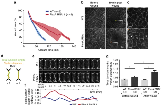
PlexAregulates wound closure in the epithelium of the notum. In order to directly visualize the effect ofPlexAinhibition on the dynamics of wound closure, we examined the closure of wounds generated by a laser in the epithelium of the pupal notum, which is amenable to live imaging. In control pupae, laser-generated wounds closed within 120 min (Fig. 2a and Supplementary Movie 3). In contrast, expression of a PlexARNAi resulted in a delay in wound closure, taking 180–240 min. In the pupal notum, it was also evident that thePlexARNAi made the cell boundaries, which were visualized using E-cadherin–GFP, more wiggly in the region around the wound (Fig. 2b,c). To better analyse this phenomenon, we measured the ratio between the total junction length and the distance between the correspondent vertexes for each condition (Fig. 2d). This ratio fluctuates along time in both control andPlexARNAi (Fig. 2e,f), but in the latter there is a peak of the ratio B5 min post wounding, when the changes in
cell outlines are more pronounced. Using this time point as a post-wounding reference we measured this ratio using cell out-lines from five independent pupae for each condition, both before and after wounding (Fig. 2g). With this analysis we observed that even before wounding,PlexAinhibition is sufficient to make the junction morphology more wiggly when compared with control,
but also that when the tissue is challenged by a laser wound this phenotype is significantly more pronounced. Wiggly cell outlines have been previously observed when cell–cell adhesion is altered17,18. Thus, in the epithelium of the notum,PlexAfunction is necessary for efficient wound closure and a reduction inPlexA function likely results in alterations in cell adhesion.
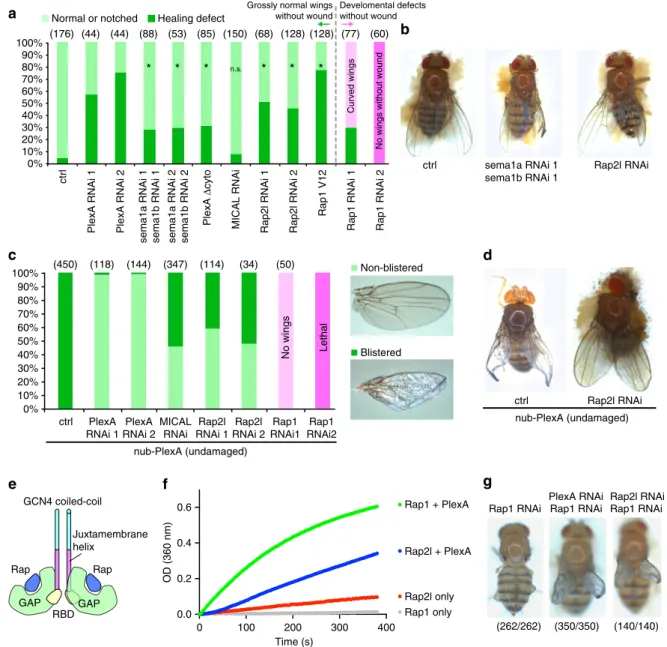
PlexA signalling regulates wound repair. The role of plexins has been best characterized in the context of axonal pathfinding19–22. Plexins are transmembrane proteins that bind to their ligands, semaphorins, and then regulate the actin cytoskeleton to mediate neurite repulsion19–21,23. The flavoprotein oxidase Mical has been shown to function downstream of plexins in this process. To determine whether these other components of the semaphorin/ plexin pathway are also involved in tissue repair in imaginal discs, we first tested whether they were expressed in imaginal discs. Expression of all the known plexins, semaphorins andMicalwas detected in discs by reverse transcriptase PCR (RT–PCR; Supplementary Fig. 2a–c). We then tested the requirement of Sema1a and Sema1b, which are ligands for PlexA19–22, in the repair of damaged discs. While knockdown of eitherSema1aor Sema1b had little effect (Supplementary Fig. 2d), simultaneous knockdown of bothSema1aandSema1bincreased the frequency of adult wings of abnormal morphology on the wounded side (Fig. 3a,b). The weaker effect of knockdown of Sema1a and Sema1b when compared withPlexA could be because of either partial knockdown or redundancy with other semaphorins. Overexpression of a truncated form of PlexA that lacks a
Wound area (%)
Closure time (min)
WT
PlexA RNAi 1
WT
PlexA RNAi 1
Total junction length / vertex distance ratio
Total junction length / vertex distance ratio Total junction length
Vertex distance
Before wound
Before
wound0 2.5 5 7.5 10 12.5 15 17.5 20 22.5 25 27.5
Time (min) Ratio
> 1 ~ 1
10 min post wound
WT (n= 6) PlexA RNAi 1 (n= 5)
*
* *
*
0 0
*
*
WT
Before wound
PlexA RNAi 1 1.00
0.95
WT (51)
WT (51) PlexA RNAi 1
(52)
PlexA RNAi 1 (51)
1.00 1.05 1.10 1.15 1.20 1.25
After wound
*
100
80
60
40
20
240
60 120 180
1.15
0.95 1.05 1.10
a b c
d e g
f
cytoplasmic domain (PlexA Dcyto)23 also resulted in wing abnormalities following wounding (Fig. 3a). This indicates that PlexADcyto likely functions in a dominant-negative capacity by competing for the ligands of endogenous PlexA and that PlexA-mediated intracellular signalling is likely important for tissue repair. Interestingly,Micalinhibition did not cause a significant disruption of adult wing morphology following wounding, even though Mical knockdown by RNAi was efficient (Fig. 3a and Supplementary Fig. 2c). Taken together, these findings suggest that PlexA functions together with Sema1a and Sema1b in tissue repair.
Vertebrate plexins function as a GTPase-activating protein (GAP) for the small GTPase Rap, thereby reducing the levels of active (GTP-bound) Rap. Semaphorin binding is predicted to
increase the GAP activity of plexins through their dimerization, thereby reducing Rap activity24,25. In Drosophila, mutations in the Rap1 gene disrupt morphogenesis in the embryo26. Rap1 regulates the stability of adherens junctions27and decreasing the levels of Rap-GTP by expression of a GAP, Rapgap1, has been shown to increase the mobility of adherens jnctions by reducing their coupling to the actin cytoskeleton28. Thus, an attractive hypothesis is that the activation of PlexA signalling in damaged tissue increases GAP activity for Rap1 and thus allows cell rearrangements and tissue remodelling.
Drosophila has two rap-family GTPases, Rap1 and Rap2l, which are orthologues of vertebrate Rap1 and Rap2, respectively. In at least some situations, such as endothelial cell adhesion in mammals, Rap1 and Rap2 function antagonistically29. Moreover,
Rap2l RNAi
ctrl sema1a RNAi 1
sema1b RNAi 1
ctrl Rap2l RNAi
0% 10% 20% 30% 40% 50% 60% 70% 80% 90% 100%
0% 10% 20% 30% 40% 50% 60% 70% 80% 90% 100%
ctrl
GCN4 coiled-coil
Rap2l RNAi 1
Rap2l RNAi 2 PlexA
RNAi 1
MICAL RNAi
nub-PlexA (undamaged) PlexA
RNAi 2
Rap1 RNAi1
Rap1 RNAi2
Blistered Non-blistered
nub-PlexA (undamaged)
* * * n.s.
(176)
(450) (118) (144) (347) (114) (34) (50)
(44) (44) (88) (53) (85) (150) (68) (128) (128) (77) (60)
* * *
Normal or notched Healing defect
No wings without wound
No wings Lethal
Curved wings
Grossly normal wings without wound
Develomental defects without wound
0 100 200 300 400
0.0 0.2 0.4 0.6
Time (s)
Rap1 RNAi
(262/262) (350/350) (140/140) Rap1 RNAi Rap2l RNAi Rap1 RNAi
PlexA RNAi
GAP GAP
RBD Rap Rap
Juxtamembrane helix
Rap1 + PlexA
Rap2l + PlexA
Rap2l only Rap1 only
OD (360 nm)
ctrl
PlexA RNAi 1 PlexA RNAi 2
sema1a RNAi 1 sema1b RNAi 1 sema1a RNAi 2 sema1b RNAi 2
Rap1 RNAi 2
Rap1 RNAi 1
Rap1 V12
Rap2l RNAi 2
Rap2l RNAi 1
MICAL RNAi
PlexA
Δ
cyto
a
b
c d
e f g
mammalian plexins have a higher GAP activity for Rap1 (ref. 24). We therefore examined the effect of reducing the levels of both Rap1 and Rap2l. Reducing the levels of Rap1 caused developmental defects even in the absence of injury (Fig. 3a), consistent with its role in embryonic morphogenesis and adherens junction formation26,27. In contrast, inhibition of Rap2l caused injury-dependent phenotypic abnormalities that were similar to those elicited by reducingPlexAlevels (Fig. 3a,b). This is consistent with the possibility that both Rap2l and PlexA antagonize Rap1 function.
To further characterize the relationship betweenPlexA and the rap genes, we manipulated activity of the rap GTPases under conditions of PlexA overexpression. When PlexA was over-expressed, the adult wings showed blistering (Fig. 3c,d), suggesting a lack of proper adhesion between the cells on the dorsal and ventral surfaces. The blistering phenotype caused by PlexA overexpression was suppressed efficiently by knockdown of eitherRap2lor Mical(Fig. 3c,d). In contrast, reducing Rap1 levels while overexpressingPlexAresulted either in a complete absence of wings or in lethality (Fig. 3c). Since the blistering phenotype elicited by overexpression of PlexA alone can be changed to phenotypes of no wings or lethality simply by increasing expression levels (Supplementary Fig. 2e), we conclude that Rap1 reduction likely enhances the effect of PlexA overexpression. If, as in mammals, PlexA reduces the activity of the rap GTPases inDrosophila, reducing Rap levels should enhance the PlexA overexpression phenotype. Indeed, we observed this kind of interaction with Rap1 but not with Rap2I. Thus, our genetic data are consistent with PlexA functioning primarily as a GAP for Rap1 in Drosophilatissues rather than for Rap2l.
To test whether Drosophila PlexA possesses bona fide GAP activity for Rap GTPases, we performed an in vitro GAP assay. We purified the cytoplasmic domain of Drosophila PlexA connected to the GCN4 coiled-coil domain to promote its dimerization and performed a photometric GAP assay using purified Drosophila Rap1 and Rap2l as substrates, as previously described24. We found that PlexA catalyses GTP hydrolysis of rap GTPases, possessing about ninefold higher activity towards Rap1 compared with Rap2l (Fig. 3e,f), which is consistent with the predictions made by the genetic interactions.
As mentioned above, mammalian Rap1 and Rap2 have antagonistic roles in the context of endothelial cell adhesion; Rap1 promotes adhesion while Rap2 reduces adhesion29. Since knockdown of either PlexA or Rap2l, each of which can antagonize Rap1, impairs wound healing, the healing defect might be caused by a failure to downregulate Rap1 activity. Consist with this possibility, expression of a constitutively active Rap1 (V12) impaired wound repair (Fig. 3a). Further, the wing-defect phenotype caused by Rap1 knockdown was similarly suppressed by inhibition of PlexA or Rap2l, which would be predicted to restore Rap1 activity to some extent (Fig. 3g). Thus, taking our genetic and biochemical data together, we propose that PlexA regulates epithelial wound repair by functioning as a GAP primarily for Rap1. Since PlexA demonstrates GAP activities towards both Rap1 and Rap2l but with a higher activity for Rap1in vitro, Rap1 could potentially suppress Rap2l regulation by PlexA in cells. Although Mical knockdown suppresses the PlexA overexpression phenotype, the absence of a healing defect with Mical knockdown might imply that Mical functions as an effector for PlexA only in a subset of functions or that the function of Mical is redundant in some contexts. Indeed,C. elegansdoes not have a recognizableMical orthologue despite a clear role for semaphorin-plexin signalling in axonal pathfinding30.
PlexA regulates delamination of epithelial cells. To examine the role of PlexA in imaginal discs, we first generated PlexARNAi -expressing clones in undamaged discs. These clones were indis-tinguishable from control clones, indicating that mutant cells proliferate normally (Supplementary Fig. 3a,b). When we compared damaged discs expressing PlexARNAi with wild-type discs, the initial fusion of the two fragments, F-actin accumula-tion at the wound edge and JNK activaaccumula-tion were indistinguishable from wounded wild-type discs (Supplementary Fig. 3c–e). In contrast, inhibition of JNK function interferes with these early events in wound healing15. However, unlike wild-type discs, healingPlexARNAidiscs had a large cleft when visualized from the side of the peripodial epithelium (Fig. 4a,b and Supplementary Movie 4). In addition, wild-type discs had many small-volume cells with intense GFP fluorescence that resembled apoptotic cells basal to the epithelial layer. In nub4PlexARNAi discs some of these cells had been basally extruded, while others were observed throughout the thickness of the epithelium (Fig. 4c–e). These cells contained activated caspase, indicating that they were undergoing apoptosis (Fig. 4f). Thus, PlexA function appears necessary for efficient extrusion of dying cells.
To test whether increasing PlexA activity is sufficient to promote cell extrusion, we overexpressed PlexA in undamaged discs and observed extensive basal extrusion of cells (Fig. 4g). Most of these cells did not express activated caspase (Supplementary Fig. 3f,g), indicating either that PlexA promotes the extrusion of live cells or that the levels of caspase activation are below the threshold of detection by this method. To test whether PlexA functions in the cells being extruded or in their neighbours, we expressed PlexA in scattered single cells. PlexA-overexpressing cells were preferentially dislocated towards the basal side of the epithelium (Fig. 4j–l), indicating a cell-autonomous role ofPlexAin this process. Dislocation of cells to the basal side by ectopic PlexA expression was enhanced by Rap1 inhibition and suppressed by Rap1 activation, consistent with the notion that PlexA promotes cell extrusion by reducing Rap1 activity (Fig. 4h,i,m). Furthermore, blocking apoptosis in damaged discs by expression of the baculoviral anti-apoptotic protein, p35, resulted in an increased number of adult wings with morphological defects upon wounding (Supplementary Fig. 3h,i). Thus, the death of damaged cells, as well as their efficient extrusion basally, might be a prerequisite for efficient repair of damaged epithelia withPlexAhaving a role in cell extrusion. This is consistent with the reported necessity for apoptotic cell death in damaged tissue before efficient regeneration in Hydra and in tadpole tails31,32.
To further explore the role ofPlexAin basal extrusion of cells, we examined its requirement in a different situation. Inhibition of Rho kinase (Rok) results in misorientation of mitotic spindles causing cells to delaminate basally from the epithelium and subsequently undergo apoptosis33. In these discs, a condensation of F-actin is seen in the basal portion of the epithelium that separates the epithelium from the basally extruded cells (Fig. 4n). Concurrent expression of PlexARNAi does not prevent either apoptosis or the basal displacement of these cells, but it results in the retention of the delaminating cells within the basal portion of the epithelium rather than in their extrusion (Fig. 4o). This indicates that PlexA is also important for the epithelial remodelling induced by Rok inhibition.
around the dying cells, which are characterized by intense GFP fluorescence as mentioned previously (Fig. 4f). At higher mag-nification, each of the F-actin foci could be seen as a ring structure surrounding the cell (Fig. 5b). Such actin rings
are typically observed around cells undergoing apoptosis and extrusion34. Upon PlexA inhibition, the F-actin accumulation associated with dying cells was significantly reduced (Fig. 5a). Conversely, ectopic expression of PlexAin the whole-wing disc A
Wound P
nub-GFP
nub-PlexA RNAi
Phalloidin
DAPI
GFP
DAPI
GFP nub-gal4
Apical
Basal
Apical
Basal Basal Apical
Apical Basal
Phalloidin
DAPI
nub-PlexA RNAi
PlexA RNAi, GFP Apical
Basal
100
80
60
40
20
0
nub-gal4 %
nub-PlexA RNAi *
Apical Apical
Basal Basal
Apical
Basal
Apical Basal Apical
Basal
500
0 1,000 1,500 2,000 2,500 (a.u.)
nub-PlexA nub-PlexA, Rap1 RNAi
nub-PlexA RNAi
Cleaved DCP1 Cleaved DCP1
Phalloidin Phalloidin
GFP GFP
GFP GFP
Rok and PlexA RNAi Rok RNAi
Rok and PlexA RNAi Rok RNAi
PlexA GFP
Apical
Apical Middle Basal
Apical Middle Basal
Apical Middle Basal
Middle Basal
GFP
100 (%) 50
0
PlexA
nub-PlexA nub-PlexA, Rap1 RNAi
Apical Apical
Basal Basal
Apical Middle Basal
Apical Middle Basal
Control
100 (%) 50
0
PlexA
Apical Middle Basal
Apical Middle Basal
Control PlexA, rap1 v12
Y x
z
z z
x
x x
Cleaved DCP1
GFP
a
b
c d
e f g h
i j
k
l
m
n o
gave rise to many extruded cells with actin rings (Fig. 5c). Further, ectopic expression of PlexA in multiple-cell or single-cell clones for a limited time also induced F-actin accumulation around cells, especially on their apical side (Fig. 5d and Supplementary Fig. 4a,b). Importantly, while F-actin accumulates in or immediately around cells that are being extruding following wounding orPlexAoverexpression, the polymerized F-actin does not induce a concomitant accumulation
of adherens junctions, which was analyzed by measuring the ratio of F-actin to b-catenin (Fig. 5b,d and Supplementary Fig. 4a). Since Rap1 strengthens the coupling of F-actin and adherens junctions28,29,35,36, the lack of coupled accumulation of these two proteins is suggestive of reduced Rap1 activity resulting from the ability of PlexA to function as a GAP for Rap1.
The finding that ectopic PlexA expression induces an uncoupling of F-actin and adherens junctions prompted us to 6hpw
6hpw Phalloidin
GFP
Control
PlexA RNAi
F-actin
β-catenin
GFP
F-actin
F-actin plexA, GFP plexA, GFP
Ratio (F-actin/β-catenin) plexA, GFP β-catenin
F-actin PlexA, GFP
Apical Basal Apical
Basal
Apical
Basal
6hpw
FRAP
0 100 200 300 400 500 600 700 800 900 (s) 0s 900s
ctrl
ctrl
Cadherin-GFP Pre-bleach
Post-bleach PlexA RNAi
PlexA RNAi
0 (%) 100
80
60
40
20
a b
c
d
e
investigate the role of endogenous PlexA in the dynamic remodelling of adherens junctions because Rap1-mediated coupling of F-actin and adherens junctions is thought to stabilize adherens junctions28,29,35,36. Since it was difficult to perform a detailed analysis of junction dynamics in the wounded wing disc, we once again used the pupal notum epithelium to visualize adherens junction dynamics. PlexA inhibition affects not only wound repair (Fig. 2a) in the notum but also its normal development. Reducing PlexA levels in the notum results in an increased width of the midline region separating the microchaetes fromB10 to 20mm (Supplementary Fig. 5a). This is the region
where the two wing discs fuse with each other during normal pupal development, a process that bears at least some superficial similarities to the rejoining of fragmented discs. The analysis of the pupal notum showed thatPlexAinhibition induces a cleft in the midline (Supplementary Fig. 5b), which is reminiscent of the healing defect of the damaged wing disc (Fig. 4b). Although cell extrusion is known to occur in the midline of the notum during development37,38, we did not see an obvious effect ofPlexARNAi in reducing cell extrusion (Supplementary Fig. 5c), suggesting that the extent to which PlexA mediates cell extrusion depends on its context. To examine the dynamics of adherens junctions, we performed a fluorescence recovery after photobleaching (FRAP) analysis of cadherin–GFP in the epithelium of the midline notum. PlexA RNAi slowed down FRAP of cadherin–GFP (Fig. 5e), indicating that cadherin is less dynamic and more stable in the PlexA RNAi epithelia. This suggests that PlexA functions to reduce the stability of adherens junctions, thereby allowing the movement of cells in the epithelium with respect to their neighbours. Such cellular movements may be necessary for the extrusion of damaged cells and the subsequent remodelling of the epithelium to enable the re-establishment of epithelial continuity following damage.
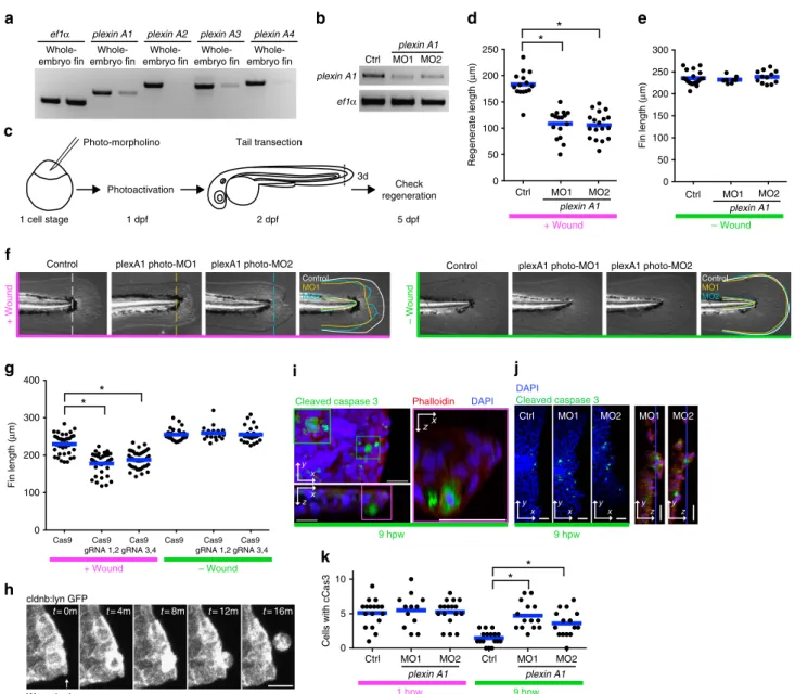
Plexin A1 regulates wound repair in zebrafish fins. Finally, to test whether plexins have a role in repairing damaged tissue in vertebrates, we utilized the well-characterized system of tail fin regeneration in zebrafish larvae, where defects in the earliest stages of wound repair impair fin regeneration at much later stages2. The zebrafish, Danio rerio, has fourPlexA orthologues, plexin A1, A2, A3andA4.plexin A1andA3are expressed highly in larval tail fins (Fig. 6a). Reducing the levels of Plexin A3 using antisense mopholino oligonucleotides (MO) did not affect regeneration (Supplementary Fig. 6a–d). Reducing levels of Plexin A1 caused defects in early embryonic development. However, by using photo-morpholinos, which can be activated by ultraviolet light, we reduced Plexin A1 function 1 day post fertilization without significantly affecting normal development (Fig. 6b,c). Under these conditions, regeneration of the amputated tail fin was impaired, thus demonstrating a role for a PlexA orthologue in tissue repair in vertebrates (Fig. 6d–f). In addition to eliciting phenotypes using MOs, we also found that Plexin A1 inhibition through injection of Cas9 mRNA and two different gRNAs forplexin A1also impaired tissue repair (Fig. 6g).
We also examined earlier stages in wound healing by live imaging. During wound repair in tail fins, we observed epithelial cell extrusion around the wound edge (Fig. 6h and Supplemen-tary Movie 5). Extruded cells had features of cells undergoing apoptosis (Fig. 6i). As wound healing proceeded, the number of apoptotic cells around wounds decreased in wild-type tail fins (Fig. 6j,k) but less so when the levels of Plexin A1 were reduced. Thus, consistent with our observations inDrosophila, reduction of Plexin A1 inhibited removal of the apoptotic cells around wounds in zebrafish. Although we have not defined the precise mechanism by which Plexin A1 regulates wound healing in
zebrafish, these observations may reflect similar functions for plexins in wound repair in Drosophilaand zebrafish.
Discussion
We have described a role for Drosophila PlexA and for its zebrafish orthologue Plexin A1 in wound repair. PlexA exerts its function as a GAP for Rap1, which promotes coupling of F-actin and adherens junctions and thereby stabilizes adherens junc-tions28,29,35,36. On the basis of our data inDrosophila, we propose the following mechanism by which PlexA regulates epithelial wound repair (Supplementary Fig. 7a,b). Injury upregulates PlexA in cells in the vicinity of the wound (Fig. 1k), which leads to inhibition of Rap1 (Fig. 3f). PlexA-mediated Rap1 inhibition uncouples F-actin and adherens junctions, thus facilitating junctional remodelling (Figs 2 and 5). This is necessary for proper cell rearrangements including cell extrusion (Fig. 4), and the subsequent epithelial remodelling that occurs during wound repair. Since an actin regulator Mical functions downstream of PlexA in a context-dependent manner23 (Fig. 3c), it is possible that PlexA could also influence wound repair and epithelial remodelling through an additional mechanism such as Mical-mediated regulation of F-actin.
A recent study reported that PlexinB and its ligands Sema4b, 4d and 4g regulate renal wound repair and mitotoic spindle orientation in mammals39. We did not observe an obvious effect of PlexA inhibition either on mitotic orientation or mitosis in Drosophila. However, given that the experimental settings and the types of plexins and semaphorins differ between the two studies, it is possible that the other plexin and semaphorins have additional functions in mediating repair ofDrosophilaepithelia.
Finally, our findings are worth examining from an evolutionary perspective. Although we do not know whether the role of Plexin A1 in wound repair in zebrafish is mechanistically the same as that inDrosophilaimaginal discs, our demonstration of a role for plexins in epithelial wound repair in metazoans that belong to two different superphyla (Ecdysozoa and Deuterosto-mia) suggests that this function could be evolutionarily ancient (Supplementary Fig. 7c). Plexins and semaphorins exist in all eumetazoa, in the placozoan Trichoplax adhaerens and in the sponge Amphimedon queenslandica. Thus, their origin predates the evolution of the nervous system. If the ancestral role for plexins is in wound repair, possibly in remodelling of adherens junctions and extruding damaged epithelial cells, it may have been subsequently modified in the nervous system to function in neurite repulsion. We therefore suggest that some of the other genes that have been studied primarily in the nervous system may also have a hitherto unsuspected role in epithelial wound repair.
Methods
Drosophilastocks.Flies were crossed and raised at 25°C unless otherwise noted. For wild-type controls, Oregon-R (Bloomington stock centre (BL) 4269) was used unless otherwise specified. For the primary screening of CSS genes, RNAi stocks were collected from the BL and the ViennaDrosophilaRNAi center (VDRC) based on a previous report16and the LOCATE database (http://locate.imb.uq.edu.au). Several RNAis were also obtained from the National Institute of Genetics (NIG).
The following fly stocks were used in this study: UAS-dcr2; nub-gal4(BL25754)
UAS-GFP
UAS-his2A::RFP(a gift from Dr Knoblich) UAS-GCaMP3(BL32116)
UAS-Rap1 RNAi2(BL35047)
UAS-Rap1 V12(Hariharan lab, unpublished) UAS-sema1a RNAi 1(BL29554)
UAS-sema1a RNAi 2(BL34320) UAS-sema1b RNAi 1(BL28588) UAS-sema1b RNAi 2(NIG6446R-3)
UAS-HA-PlexA23(a gift from Dr Terman and Dr Kolodkin) UAS-HA-PlexADcyto23(a gift from Dr Terman)
PlexA-myc BAC40(a gift from Dr Kolodkin) UAS-p35(BL5073)
UAS-Rok RNAi(VDRC104675) UAS-dcr2; pnr-Gal4/TM3(BL25758) UAS-dcr2; Act5c-Gal4/Cyo(BL25708) TRE reporter12(a gift from Dr Bohmann) Sp; Dr / SM5-TM6B
UAS-mCherry-moesin/Cyo Pnr-Gal4/TM3(BL3039)
E-Cad-GFP
For the TIE-DYE experiment, the following stocks were used as previously described41.
y, w, hsFLP; Sp; Dr / SM5-TM6b
w; Actostopolacznls, UbiostopoGFPnls; Actostopogal4, UAS-his2A::RFP/SM5-TM6b
Using above stocks, the following stable stocks were built and used for experiments.
UAS-dcr2; nub-Gal4, UAS-GFP
UAS-dcr2; nub-Gal4, UAS-GFP, UAS-HA-PlexA UAS-dcr2; nub-Gal4, UAS-GFP; UAS-PlexA RNAi 1 UAS-dcr2; nub-Gal4, UAS-GFP; UAS-PlexA RNAi 2 UAS-dcr2; nub-Gal4, UAS-GFP; UAS-sema1b RNAi 1 UAS-dcr2; nub-Gal4, UAS-GFP; UAS-sema1b RNAi 2 UAS-dcr2; nub-gal4, UAS-GFP; UAS-Rap2l RNAi 1 nub-Gal4; UAS-RFP
hsFLP;; UAS-PlexA RNAi 1
+ Wound
– Wound
0 50 100 150 200 250 300
0 50 100 150 200 250
2 dpf 5 dpf
3d
1 cell stage 1 dpf Photo-morpholino
Photoactivation
Tail transection
Check regeneration Ctrl MO1 MO2
ef1α plexin A1
plexin A1
Fin length (
μ
m)
Fin length (
μ
m)
Regenerate length (
μ
m)
* *
* *
Ctrl MO2
MO1
plexin A1
MO2 MO1
plexin A1
Ctrl
– Wound
ef1α Whole-embryo fin
plexin A1 plexin A2 plexin A3 plexin A4
Whole-embryo fin Whole-embryo fin
Whole-embryo fin
Whole-embryo fin
Control
+ Wound
plexA1 photo-MO1 plexA1 photo-MO2 plexA1 photo-MO1 plexA1 photo-MO2
Control
MO1
MO2
Control
Control
MO1
MO2
MO2
MO1 MO1 MO2
Ctrl
9 hpw 9 hpw
* *
0 5 10
MO2 MO1
Ctrl Ctrl MO1 MO2
1 hpw 9 hpw
t= 0m
Wound edge cldnb:lyn GFP
t= 4m t= 8m t= 12m t= 16m z
x
Phalloidin DAPI
DAPI
Cleaved caspase 3 Cleaved caspase 3
y x
y y y y y
x x x z z
zx
0 100 200 300 400
Cas9 Cas9 gRNA 1,2
Cas9 gRNA 3,4
Cas9 Cas9 gRNA 1,2
Cas9 gRNA 3,4
+ Wound – Wound
Cells with cCas3
plexin A1 plexin A1
a b d
c
e
f
g
h
i j
k
hsFLP; UAS-HA-PlexA
hsFLP; UAS-HA-PlexA; UAS-GFP, Actostopogal4, UAS-GFP UAS-mCherry-Moesin, E-Cadherin–GFP/Cyo; Pnr-Gal4/TM6b
In situwound of wing discs.L3 larvae of thenub4GFPstock were quickly immobilized in ice water, and the right wing pouch was bisected by applying pressure on the larval cuticle with a thin and dull-pointed tungsten needle with the help of GFP fluorescence (see Supplementary Movie 1). This procedure does not involve penetration of the larval cuticle. For each fly stock, at least 25 larvae were wounded.
Calcium imaging.L3 larvae (nub-gal4/UAS-GCaMP3; UAS-RFP/þ) were quickly immobilized in ice water and the right wing pouch was visualized using RFP and injured with a tungsten needle. Immediately after wounding, a larva was transferred on a slide glass and a coverslip was put on the larva to immobilize it completely. Live imaging of GCaMP3 fluorescence was performed with a stereomicroscope (Zeiss SteREO Lumar V12). Images were acquired every 10 s. For kymographic analysis of a calcium flash, signals in a rectangular box were summed into a 1D line and kymograph was made with ImageJ (NIH).
Immunofluorescence and confocal imaging.Wing discs in late L3 larvae were dissected in PBS, fixed with paraformaldehyde in PBS and washed in 0.1% Triton in PBS. We used the following antibodies and fluorescent dyes: mouse 9E10 myc antibody (1:100, sc-40, Santa Cruz), rabbit cleavedDrosophilaDcp-1 antibody (1:400, #9578, Cell Signaling), mouse anti-Arm (1:100, N2 7A1, DHSB), 4,6-diamidino-2-phenylindole (DAPI; 1:1,000, Molecular Probes), rhodamine-conjugated phalloidin (1:500, Sigma-Aldrich) and Alexa Fluor 633 Phalloidin (1:50, Invitrogen). Fluorescence images were acquired with a confocal microscope (Zeiss LSM700). Three-dimensional reconstruction was performed by the Zen software.zaxis analysis was performed using Volocity (PerkinElmer). For the TIE-DYE clone analysis in Supplementary Fig. 3a,b, GFP signals that overlap with RFP signals were subtracted from the GFP channel to make the analysis simple. After the subtraction, clone sizes were quantified using ImageJ. For FRAP experiments, fluorescence of cadherin–GFP in 2-mm-diameter spots were bleached in the epithelia of the midline notum at 13 after puparium formation (APF) and images were acquired every 20 s. Fluorescence recovery was quantified using ImageJ. For analysis of cell extrusion/delamination in the midline notum, time-lapse images with Ecad-GFP were acquired in the midline of the notum from 13 to 25 APF.PlexARNAi 1 was expressed in the notum usingpnr-gal4.
Laser ablation and imaging.Flies with genotype w;UAS-mCherry-Moesin, E-Cadherin–GFP; pnr-GAL4/TM6b were crossed withPlexARNAi 1 and kept at 29°C. Pupae were staged at 13 h after puparium formation, and the notum epithelium was imaged at 29°C on an Andor Revolution XD spinning disk imaging system (Nikon Eclipse Ti-E and Yokogawa CSU-x1) with a Nikon (Plan Apo VC PFS) 60 oil-immersion objective. Pupae mounting was performed as previously described11. Wounds were made using a MicroPoint (Andor Technology) ultraviolet nitrogen-pumped dye laser (435 nm)—15 pulses at maximum power. Images were acquired before and immediately after wounding every 2.5 min for 6 h.
Wound area and junction quantification inDrosophilapupae.Wound area over time was quantified using Fiji manual selection tool and area measure function. Junction ratio (total junction length/vertex distance) was obtained by dividing the total membrane length value with the distance in a straight line between each end of the corresponding membrane vertex as shown in Fig. 2d. Measurements were done using five independent pupae for both the control andPlexARNAi. At least 10 membranes from each pupa were randomly sampled and measured using the Fiji line segment tool. Data were processed in Microsoft Excel and GraphPad Prism 6.0.
RT–PCR (Drosophila).Ten wing discs in late L3 larvae were dissected in PBS and snap-frozen at80°C. RNA was prepared using the RNeasy Mini Kit (QIAGEN) and one-step RT–PCR (QIAGEN) was performed. To check efficiency of RNAi-mediated knockdown, wing discs in L3 larvae (UAS-Dcr2/þor Y; Act5c-gal4/UAS-RNAi or UAS-Dcr2/þor Y; Act5c-gal4/þ; UAS-RNAi/þ) were dissected. Oligonucleotide sequences for RT–PCR were as follows:
atub84Bforward, 50-CCTTGTCGCGTGTGAAACACTTCC-30;
atub84Breverse, 50-GTTGGCCGCATCCTCCTTACC-30; PlexAforward, 50-AAACATTCCATCAAGCCAAAGACCTCG-30; PlexAreverse, 50-TTAAGAGGACTTCCTGCGCGGC-30; PlexBforward, 50-AGACTTGGAAAAGATTCGCCGTCG-30; PlexBreverse, 50-GCAATCACCATCTAGTGTAGCAGC-30; sema1aforward, 50-ATACCAAAATGTGTCTCGTCAAGGGC-30; sema1areverse, 50-CTTAGGCTATCGTACTGCTCGGTC-30; sema1bforward, 50-CGATGAGTTTGTTCTAGTTGGTGCC-30; sema1breverse, 50-CCTCCACATCTCGGCTGACTTC-30; sema2aforward, 50-GCAGTTTCCACATTAACGAGATTCAGG-30; sema2areverse, 50-GACCCAGATTGGTGCCCACATAG-30;
sema2bforward, 50-TGCTGCCCATCATCTGCGCC-30; sema2breverse, 50-CGTTTACCCTTGGAAACACAATTCAGG-30; sema5cforward, 50-CGACAAAATCAAGTGTGGATTTGCTGG-30; sema5creverse, 50-CACGCGTACAGCTGGTTCTCAC-30; MICALforward, 50-GACGCAGTGGAGACTTGCTGCC-30; MICALreverse, 50-GCAGATCTGGTCCAAATAGTTGAGCC-30;
Clone generation with heat shocks.Clones were induced by heat shocks in a 37-°C circulating water bath. For experiments to make clones composed of mul-tiple cells (Fig. 5d and Supplementary Figs 3a,b and 4a), a heat shock of 10 min was performed at 48–72 h after egg lay and the discs were dissected from late L3 larvae. For experiments to make single-cell clones (Fig. 4j–m and Supplementary Fig. 4b), a heat shock of 14.5 min was performed at 100-h after egg lay and the discs were dissected 10–12 h after the heat shock.
In vitrorap GAP assay.The active PlexA dimer construct was generated by fusing the GCN4 coiled-coil motif to the N terminus of the cytoplasmic region (residues 1,306–1,944) ofDrosophilaPlexA. The construct corresponds to the mouse CC(d)PlexinA1cytoas described previously, which displayed the highest RapGAP activity among all the tested dimer constructs25. The fusion was subcloned into a modified pET28 vector (Novagen) containing an N-terminal tandem His6-SUMO tag42.DrosophilaRap1 and Rap2l (residues 2–166 and 2–167, respectively) were subcloned into a modified pET28 vector containing an N-terminal His6tag and a cleavage site for the C3 protease of human rhinovirus24,25. Proteins were expressed in the bacterial strain BL21 (DE3) and purified as previously described25. Removal of the SUMO and His6tags was achieved by addition of the Ulp1 and C3 protease, respectively. Rap1 and 2l were GTP-loaded before use in the activity assays. GTP loading was achieved by incubating Rap1 or 2l at a final concentration of 450mM over night at 4°C in a buffer containing 9 mM GTP, 3.4 mM EDTA, 10 mM Tris-HCl pH 8.0, 10% glycerol, 150 mM NaCl and 2 mM dithiothreitol. The exchange was quenched by the addition of MgCl2to a final concentration of 20 mM. Removal of excess GTP was achieved by passing the mixtures over a size exclusion column (Superdex 75 10/300 GL, GE Healthcare). The GAP activity of the active dimer form ofDrosophilaPlexAcytotowardsDrosophilaRap1 and 2l was measured using the photometric assay as describe by Webb and Hunter with minor modifications24,25,43. Briefly, the release of inorganic phosphate upon GTP hydrolysis is coupled to the reaction catalysed by purine nucleoside phosphorylase, which converts 2-amino-6-mercapto-7-methylpurine ribonucleoside (MESG) to its guanine base form (2-amino-6-mercapto-7-methylpurine) and ribose-1-phosphate in the presence of phosphate. MESG has almaxat 320 nm, while the guanine base product has almaxat 360 nm. The reaction was monitored spectroscopically by following the absorbance decay at 320 nm and the increase at 360 nm at 27°C. PlexinA and Rap were mixed to final concentrations of 20 and 100mM, respectively, in a reaction buffer containing 8 U ml1PNP, 300mM MESG, 20 mM Tris-HCl pH 7.6, 20 mM NaCl, 5% glycerol, 2 mM TCEP and 2 mM MgCl2. Each reaction was repeated three times. Owing to lack of the catalytic machinery, basal GTP hydrolysis of Rap1 and Rap2l in the absence of plexin is extremely slow and virtually undetectable under these conditions24,25. Slow increases of the absorbance at 360 nm in reactions of either Rap1 or Rap2l without plexin were produced partially by spontaneous conversion of MESG. The absorbance increase was faster in reactions with Rap2l, which was because of precipitation of the protein, as indicated by the simultaneous increase at 320 nm. To eliminate these artefacts, the plexin-catalysed rates were calculated by subtracting the slopes of the Rap-alone reactions from the slopes of the linear portions of the corresponding reactions in the presence of plexin.
Conditional gene inactivation by tissue-specific Cas9 expression.To induce wing-specific gene knockout, we generated flies that simultaneously carried the 11F02-GAL4 driver transgene44, a UAS-Cas9 transgene and a double-gRNA transgene. The UAS-Cas9 transgene was generated in a custom UAS vector that has a 10 UAS sequence, gypsy insulators, a phiC31 attB sequence and a vermiliontransgene. It was integrated into the attP40-landing site by phiC31-mediated recombination45. Double-gRNA vectors were generated as described46. The gene-specific 20-bp sequences of gRNAs used in the present study are shown below for each target gene.
plexA: 50-GAGCTGTAGTACGTTGTAGC-30, 50-AATGCGAGAACGAGT
TAATG-30
e(ebony): 50-GCCATCGTGCTCTACACCTC-30, 50-GACTCTCAGGTGA
AGATCCG-30
wg(wingless): 50-GAGAAGGAAACAGCGACGCC-30, 50-GGTGACCCA
CTCGATTGCCA-30
ct(cut): 50-GGACGAACTGCAGCGCTTGA-30, 50-GAGCAGTGTGGCC
CAGCATC-30
cv(crossveinless): 50-GAAGAACTGCTCCTGCTGCA-30, 50-GCTGTCCAA
GAAGTCCCACG-30.
0.2 mg ml1Tricaine (ethyl 3-aminobenzoate; Sigma-Aldrich) and the tail fin was resected with a razor blade2. Regenerate length was quantified by measuring the distance between the caudal tip of the notochord and the caudal tip of the tail fin at 3 days after wounding. For time-lapse imaging, tail fins ofTg(cldnb:lynGFP)48were transected at 3 d.p.f. and time-lapse imaging was started immediately. Zebrafish immunostaining was performed as previously described2. A rabbit cleaved caspase-3 antibody (1:caspase-300, #9661, Cell Signaling) was used with DAPI (1:1,000, Molecular Probes) and rhodamine-conjugated phalloidin (1:500, Sigma-Aldrich).
MO injection and RT–PCR.MO oligonucleotides (Gene Tools) in Danieau buffer (58 mM NaCl, 0.7 mM KCl, 0.4 mM MgSO4, 0.6 mM Ca(NO3)2and 5.0 mM HEPES, pH 7.1–7.3) were injected (3 nl) into one-cell stage embryos. Danieau buffer was used as a control. For Plexin A3 knockdown, 500mMplexin A3splice MO1 (50-AGGCATGACATTGACTCACTTGTTA-30) or 500mMplexin A3splice
MO2 (50-ATCTGAATGGTGTCTTACCATATCC-30) was used. For Plexin A1
knockdown, 150mMplexin A1splice MO1 (50-AGCAGATAATTCTCTTACCGA
GATC-30) or 150mMplexin A1splice MO2 (50-ACCAGTCGTTTGCTACCGTA
CCTCC-30) was used. For ultraviolet-mediated Plexin A1 knockdown,
combina-tion of 471-mMplexin A1Photo-MO1 (50
-ATCTCGGTAAGPGAATTATCTGC-30) and 429-mMplexin A1splice MO1 or combination of 471mMplexin A1
Photo-MO2 (50-GGTACGGTAPCAAACGACTGG-30) and 429-mMplexin A1splice
MO2 was used. Photoactivation of Photo-MOs was performed with the Gene Tools UV light box (Gene Tools) in 1 d.p.f. embryos as described previously49. For morphotyping of the splicing MOs, RNA was prepared from 2.5 d.p.f. larvae using TRIZOL (Invitrogen), and one-step RT–PCR (QIAGEN) was performed.
Oligonucleotide sequences used for morphotyping RT–PCR were as follows: plexin A1forward, 50-GCTGCAGACTGATATCCATGAGC-30;
plexin A1reverse, 50-GTGACATCCTCGTCCTGGAG-30; plexin A3forward, 50-TGAATGGTGTTCCTCCGCCG-30; plexin A3reverse, 50-TTGGACTTCAGAGAAAGCTTCACG-30.
For expression analysis of plexins in larval tail fins, tail fins of 50 larvae at 3 d.p.f. were resected, RNA was prepared using TRIZOL and one-step RT–PCR was performed. Oligonucleotide sequences for RT–PCR were as follows: ef1aforward, 50-TACGCCTGGGTGTTGGACAAA-30;
ef1areverse, 50-TCTTCTTGATGTATCCGCTGA-30; plexin A1forward, 50-GGAACGTGGCATCCCTCATC-30; plexin A1reverse, 50-GTGACATCCTCGTCCTGGAG-30; plexin A2forward, 50-GACTACTTTCCAACAATCTCCAGCCG-30; plexin A2reverse, 50-CGCAGAAGCCATTATAGACATAGGAGG-30; plexin A3forward, 50-GTAAACTGGTGGCGGATGAGG-30; plexin A3reverse, 50-AACACCACCGAATGGTCTCCG-30; plexin A4forward, 50-TTGTTCGTGGCCACAGCTGTTG-30; plexin A4reverse, 50-GAGTGGTTCTTGTACACGTAGGC-30.
CRISPR-Cas9 in zebrafish.We designed single guide RNAs (sgRNAs) by seeking sequences corresponding to GGN18NGG on the sense or antisense strand of
plexin A1DNA. To generate sgRNAs, we performed PCR with Phusion polymerase without a template50; a unique oligonucleotide encoding the T7 polymerase-binding site and the sgRNA target sequence GGN18N (CRISPRF¼GAAATTAAT
ACGACTCACTATA GGN18GTTTTAGAGCTAGAAATAGC); and a common
oligonucleotide encoding the remainder of the sgRNA sequence (sgRNAR¼AAA AGCACCGACTCGGTGCCACTTTTTCAAGTTGATAACGGACTAGCCTTATT TTAACTTGCTATTTCTAGCTCTAAAAC). The DNA templates were used for in vitrotranscription by T7 RNA polymerase (Ambion). Four unique sgRNAs were generated using the following CRISPRF oligonucleotides:
sgRNA 1, GAAATTAATACGACTCACTATAGG GAGATGGAGTACGCCA CTGTTTTAGAGCTAGAAATAGC;
sgRNA 2, GAAATTAATACGA CTCACTATAGGTAGCAAACGACTGGTT TAGTTTTAGAG CTAGAAATAGC;
sgRNA 3, GAAATTAATACGACTCACTAT AGGACAGAACCAAAGCGA GAGGTTTTA GAGCTAGAAATAGC
sgRNA 4,
GAAATTAATACGACTCACTATAGGACCGATCGACTCCATCACGTTTT AGAGCTAGAAATAGC.
For making Cas9 RNA, pCS2-nCas9n (Addgene 47929)51was linearized by Not1 andin vitrotranscribed by SP6 RNA polymerase (Ambion). RNA of Cas9 (250 ngml1) and two sgRNAs (15 ngml1each) in Danieau buffer were injected (3 nl) into one-cell stage embryos. Cas9 only was used as a control.
Statistics.Figure 1i: *Po0.05, two-tailedw2-test with the Bonferroni correction
(RNAi), two-tailedw2-test (CRISPR), ctrl:n¼211, PlexA RNAi 1:n¼90, PlexA RNAi 2:n¼25, PlexA RNAi 3:n¼20, PlexA RNAi 4:n¼27, Cas9:n¼98, PlexA CRISPR:n¼39.
Figure 2a: shadowed areas represent s.d. WT:n¼6, PlexA RNAi:n¼3. Figure 2g: error bars represent s.e.m. *Po0.05; Kruskal–Wallis test with Dunn’s
post-test, WT before wound:n¼51, PlexA RNAi before wound:n¼52, WT after wound:n¼51, PlexA RNAi after wound:n¼51.
Figure 3a: *Po0.05, two-tailedw2-test with the Bonferroni correction, ctrl: n¼176, PlexA RNAi 1:n¼44, PlexA RNAi 2:n¼44, sema1a RNAi 1, sema1b
RNAi 1:n¼88, sema1a RNAi 2, sema1b RNAi 2:n¼53, PlexADcyto:n¼85, MICAL RNAi:n¼150, Rap2l RNAi 1:n¼68, Rap2l RNAi 2:n¼128, Rap1 V12: n¼128, Rap1 RNAi 1:n¼77, Rap1 RNAi 2:n¼60.
Figure 4e: *Po0.05; two-tailed Mann–WhitneyU-test, nub-gal4: 10 discs, 150
cells, nub-plexA RNAi: 10 discs, 166 cells.
Figure 4i: error bars represent s.e.m. (nub-PlexA:n¼6, nub-PlexA, Rap1 RNAi:n¼6).
Figure 4l: error bars represent s.e.m. (n¼17 discs).
Figure 4m: error bars represent s.e.m. (top:n¼5, bottom:n¼6). Figure 5e: error bars represent s.e.m. (ctrl:n¼20, PlexA RNAi:n¼18). Figure 6d: *Po0.05; one-way analysis of variance (ANOVA) with Dunnett’s
post-test, Ctrl: 15 larvae, MO1: 16 larvae, MO2: 18 larvae.
Figure 6g: *Po0.05; one-way ANOVA with Dunnett’s post-test, Cas9, þwound: 38 larvae, Cas9, gRNA 1,2,þwound: 38 larvae, Cas9, gRNA 3,4, þwound: 42 larvae, Cas9, wound: 25 larvae, Cas9, gRNA 1,2, wound: 17 larvae, Cas9, gRNA 3,4, wound: 17 larvae.
Figure 6k: *Po0.05; one-way ANOVA with Dunnett’s post-test, Ctrl 1 h.p.w.:
17 larvae, MO1: 1 h.p.w.: 12 larvae, MO2: 1 h.p.w.: 17 larvae, Ctrl 9 h.p.w.: 17 larvae, MO1: 9 h.p.w.: 14 larvae, MO2: 9 h.p.w.: 15 larvae.
Supplementary Fig. 3h: *Po0.05, two-tailedw2-test, þwound/ctrl:n¼99, þwound/p35:n¼91, wound/ctrl:n¼573,wound/p35:n¼310.
Supplementary Fig. 5a: *Po0.05, two-tailed unpairedt-test, pnr-gal4:n¼7,
pnr-PlexA RNAi:n¼6.
Data availability.The data that support the findings and reagents used in this study are available from one of the corresponding authors (S.K.Y.) upon request.
References
1. Niethammer, P., Grabher, C., Look, A. T. & Mitchison, T. J. A tissue-scale gradient of hydrogen peroxide mediates rapid wound detection in zebrafish. Nature459,996–999 (2009).
2. Yoo, S. K., Freisinger, C. M., LeBert, D. C. & Huttenlocher, A. Early redox, Src family kinase, and calcium signaling integrate wound responses and tissue regeneration in zebrafish.J. Cell Biol.199,225–234 (2012).
3. Poss, K. D. Advances in understanding tissue regenerative capacity and mechanisms in animals.Nat. Rev. Genet.11,710–722 (2010).
4. Campos, I., Geiger, J. A., Santos, A. C., Carlos, V. & Jacinto, A. Genetic screen inDrosophila melanogasteruncovers a novel set of genes required for embryonic epithelial repair.Genetics184,129–140 (2010).
5. Lesch, C., Jo, J., Wu, Y., Fish, G. S. & Galko, M. J. A targeted UAS-RNAi screen inDrosophilalarvae identifies wound closure genes regulating distinct cellular processes.Genetics186,943–957 (2010).
6. Losick, V. P., Fox, D. T. & Spradling, A. C. Polyploidization and cell fusion contribute to wound healing in the adultDrosophilaepithelium.Curr. Biol.23,
2224–2232 (2013).
7. Tamori, Y. & Deng, W. M. Tissue repair through cell competition and compensatory cellular hypertrophy in postmitotic epithelia.Dev. Cell25,
350–363 (2013).
8. Tian, L.et al.Imaging neural activity in worms, flies and mice with improved GCaMP calcium indicators.Nat. Methods6,875–881 (2009).
9. Xu, S. & Chisholm, A. D. A Galphaq-Ca(2)(þ) signaling pathway promotes actin-mediated epidermal wound closure inC. elegans.Curr. Biol.21,
1960–1967 (2011).
10. Razzell, W., Evans, I. R., Martin, P. & Wood, W. Calcium flashes orchestrate the wound inflammatory response through DUOX activation and hydrogen peroxide release.Curr. Biol.23,424–429 (2013).
11. Antunes, M., Pereira, T., Cordeiro, J. V., Almeida, L. & Jacinto, A. Coordinated waves of actomyosin flow and apical cell constriction immediately after wounding.J. Cell Biol.202,365–379 (2013).
12. Chatterjee, N. & Bohmann, D. A versatile PhiC31 based reporter system for measuring AP-1 and Nrf2 signaling inDrosophilaand in tissue culture.PLoS ONE7,e34063 (2012).
13. Galko, M. J. & Krasnow, M. A. Cellular and genetic analysis of wound healing inDrosophilalarvae.PLoS Biol.2,E239 (2004).
14. Bosch, M., Serras, F., Martin-Blanco, E. & Baguna, J. JNK signaling pathway required for wound healing in regeneratingDrosophilawing imaginal discs. Dev. Biol.280,73–86 (2005).
15. Ramet, M., Lanot, R., Zachary, D. & Manfruelli, P. JNK signaling pathway is required for efficient wound healing in Drosophila.Dev. Biol.241,145–156 (2002).
16. Kurusu, M.et al.A screen of cell-surface molecules identifies leucine-rich repeat proteins as key mediators of synaptic target selection.Neuron59,
972–985 (2008).
17. Desai, R.et al.Monomeric alpha-catenin links cadherin to the actin cytoskeleton.Nat. Cell Biol.15,261–273 (2013).
19. Tran, T. S., Kolodkin, A. L. & Bharadwaj, R. Semaphorin regulation of cellular morphology.Annu. Rev. Cell Dev. Biol.23,263–292 (2007).
20. Winberg, M. L.et al.Plexin A is a neuronal semaphorin receptor that controls axon guidance.Cell95,903–916 (1998).
21. Yazdani, U. & Terman, J. R. The semaphorins.Genome Biol.7,211 (2006). 22. Sweeney, L. B.et al.Temporal target restriction of olfactory receptor neurons
by Semaphorin-1a/PlexinA-mediated axon-axon interactions.Neuron53,
185–200 (2007).
23. Hung, R. J.et al.Mical links semaphorins to F-actin disassembly.Nature463,
823–827 (2010).
24. Wang, Y.et al.Plexins are GTPase-activating proteins for Rap and are activated by induced dimerization.Sci. Signal.5,ra6 (2012).
25. Wang, Y., Pascoe, H. G., Brautigam, C. A., He, H. & Zhang, X. Structural basis for activation and non-canonical catalysis of the Rap GTPase activating protein domain of plexin.eLife2,e01279 (2013).
26. Asha, H., de Ruiter, N. D., Wang, M. G. & Hariharan, I. K. The Rap1 GTPase functions as a regulator of morphogenesisin vivo.EMBO J.18,605–615 (1999). 27. Knox, A. L. & Brown, N. H. Rap1 GTPase regulation of adherens junction
positioning and cell adhesion.Science295,1285–1288 (2002).
28. Wang, Y. C., Khan, Z. & Wieschaus, E. F. Distinct Rap1 activity states control the extent of epithelial invagination via alpha-catenin.Dev. Cell25,299–309 (2013).
29. Pannekoek, W. J., Linnemann, J. R., Brouwer, P. M., Bos, J. L. & Rehmann, H. Rap1 and Rap2 antagonistically control endothelial barrier resistance.PLoS ONE8,e57903 (2013).
30. Mizumoto, K. & Shen, K. Interaxonal interaction defines tiled presynaptic innervation inC. elegans.Neuron77,655–666 (2013).
31. Chera, S.et al.Apoptotic cells provide an unexpected source of Wnt3 signaling to drive hydra head regeneration.Dev. Cell17,279–289 (2009).
32. Tseng, A. S. & Levin, M. Tail regeneration in Xenopus laevis as a model for understanding tissue repair.J. Dental Res.87,806–816 (2008).
33. Nakajima, Y., Meyer, E. J., Kroesen, A., McKinney, S. A. & Gibson, M. C. Epithelial junctions maintain tissue architecture by directing planar spindle orientation.Nature500,359–362 (2013).
34. Eisenhoffer, G. T. & Rosenblatt, J. Bringing balance by force: live cell extrusion controls epithelial cell numbers.Trends Cell Biol.23,185–192 (2013). 35. Sawyer, J. K., Harris, N. J., Slep, K. C., Gaul, U. & Peifer, M. TheDrosophila
afadin homologue Canoe regulates linkage of the actin cytoskeleton to adherens junctions during apical constriction.J. Cell Biol.186,57–73 (2009). 36. Ando, K.et al.Rap1 potentiates endothelial cell junctions by spatially
controlling myosin II activity and actin organization.J. Cell Biol.202,901–916 (2013).
37. Koto, A., Kuranaga, E. & Miura, M. Apoptosis ensures spacing pattern formation ofDrosophilasensory organs.Curr. Biol.21,278–287 (2011). 38. Marinari, E.et al.Live-cell delamination counterbalances epithelial growth to
limit tissue overcrowding.Nature484,542–545 (2012).
39. Xia, J.et al.Semaphorin-Plexin signaling controls mitotic spindle orientation during epithelial morphogenesis and repair.Dev. Cell33,299–313 (2015). 40. Pecot, M. Y.et al.Multiple interactions control synaptic layer specificity in the
Drosophilavisual system.Neuron77,299–310 (2013).
41. Worley, M. I., Setiawan, L. & Hariharan, I. K. TIE-DYE: a combinatorial marking system to visualize and genetically manipulate clones during development inDrosophila melanogaster.Development140,3275–3284 (2013). 42. He, H., Yang, T., Terman, J. R. & Zhang, X. Crystal structure of the plexin A3 intracellular region reveals an autoinhibited conformation through active site sequestration.Proc. Natl Acad. Sci. USA106,15610–15615 (2009). 43. Webb, M. R. & Hunter, J. L. Interaction of GTPase-activating protein with
p21ras, measured using a continuous assay for inorganic phosphate release. Biochem. J.287(Pt 2): 555–559 (1992).
44. Jory, A.et al.A survey of 6,300 genomic fragments for cis-regulatory activity in the imaginal discs ofDrosophila melanogaster.Cell Rep.2,1014–1024 ð2012Þ:
45. Markstein, M., Pitsouli, C., Villalta, C., Celniker, S. E. & Perrimon, N. Exploiting position effects and the gypsy retrovirus insulator to engineer precisely expressed transgenes.Nat. Genet.40,476–483 (2008).
46. Kondo, S. & Ueda, R. Highly improved gene targeting by germline-specific Cas9 expression in Drosophila.Genetics195,715–721 (2013).
47. Nusslein-Volhard, C. & Dahm, R.Zebrafish: a Practical Approach1st edn (Oxford University Press, 2002).
48. Haas, P. & Gilmour, D. Chemokine signaling mediates self-organizing tissue migration in the zebrafish lateral line.Dev. Cell10,673–680 (2006). 49. Tallafuss, A.et al.Turning gene function ON and OFF using sense and
antisense photo-morpholinos in zebrafish.Development139,1691–1699 (2012).
50. Bassett, A. R., Tibbit, C., Ponting, C. P. & Liu, J. L. Highly efficient targeted mutagenesis ofDrosophilawith the CRISPR/Cas9 system.Cell Rep.4,220–228 (2013).
51. Jao, L. E., Wente, S. R. & Chen, W. Efficient multiplex biallelic zebrafish genome editing using a CRISPR nuclease system.Proc. Natl Acad. Sci. USA110,
13904–13909 (2013).
Acknowledgements
We thank D. Bohmann, J. Knoblich, A. Kolodkin, J. Terman and TRiP at Harvard Medical School, the Bloomington Stock Center, the ViennaDrosophilaRNAi center and NIG for transgenic fly stocks. We thank J. Bosch for sharing RNAi stocks, other Har-iharan laboratory members for help with fly work, S. Amacher and N. Shih for help with zebrafish work and D. Bilder, R. Harris, N. Patel and M. Worley for comments on the manuscript. S.K.Y. was supported by the Miller Institute; I.K.H. by R01GM085576 from NIGMS and an American Cancer Society Research Professor Award 120366-RP-11-078-01-DDC; X.Z. and H.G.P. by GM088197 from NIGMS and I-1702 from the Welch Foundation; A.J. and T.P. by European Research Council (2007-StG-208631); and S.K. by Mochida Memorial Foundation for Medical and Pharmaceutical Research.
Author contributions
S.K.Y. and I.K.H. conceived and oversaw the project. S.K.Y., H.G.P. and T.P. executed the experiments: S.K.Y. executed most experiments except laser wounding inDrosophila
pupae (T.P.) and GAP assays (H.G.P.). H.G.P. and X.Z. contributed expertise in protein purification andin vitroGAP assays. T.P. and A.J. contributed expertise in laser wounding inDrosophilapupae. S.K. contributed expertise and reagents inDrosophila
CRISPR. S.K.Y., T.P., S.K., X.Z. and I.K.H. wrote the paper.
Additional information
Supplementary Informationaccompanies this paper at http://www.nature.com/ naturecommunications
Competing financial interests:The authors declare no competing financial interests.
Reprints and permissioninformation is available online at http://npg.nature.com/ reprintsandpermissions/
How to cite this article:Yoo, S. K.et al.Plexins function in epithelial repair in both
Drosophilaand zebrafish.Nat. Commun.7:12282 doi: 10.1038/ncomms12282 (2016). This work is licensed under a Creative Commons Attribution 4.0 International License. The images or other third party material in this article are included in the article’s Creative Commons license, unless indicated otherwise in the credit line; if the material is not included under the Creative Commons license, users will need to obtain permission from the license holder to reproduce the material. To view a copy of this license, visit http://creativecommons.org/licenses/by/4.0/