Detection of
Talaromyces marneffei
from
Fresh Tissue of an Inhalational Murine
Pulmonary Model Using Nested PCR
Yinghui Liu1, Xiaowen Huang2, Xiuwen Yi1, Ya He1, Eleftherios Mylonakis3, Liyan Xi1*
1Department of Dermatology, Sun Yat-Sen Memorial Hospital, Sun Yat-Sen University, Guangzhou, China,
2Department of Dermatology, General Hospital of Guangzhou Military Command of PLA, Guangzhou, China,3Division of Infectious Disease, Rhode Island Hospital, Waren Alpert Medical School of Brown University, Providence, Rhode Island, United States of America
Abstract
Penicilliosis marneffei, often consecutive to the aspiration ofTalaromyces marneffei( Peni-cillium marneffei), continues to be one of the significant causes of morbidity and mortality in immunocompromised patients in endemic regions such as Southeast Asia. Improving the accuracy of diagnosing this disease would aid in reducing the mortality of associated infec-tions. In this study, we developed a stable and reproducible murine pulmonary model that mimics human penicilliosis marneffei using a nebulizer to deliverTalaromyces marneffei
(SUMS0152) conidia to the lungs of BALB/c nude mice housed in exposure chamber. Using this model, we further revealed that nested PCR was sensitive and specific for detect-ingTalaromyces marneffeiin bronchoalveolar lavage fluid and fresh tissues. This inhalation model may provide a more representative analysis tool for studying the development of penicilliosis marneffei, in addition to revealing that nested PCR has a predictive value in reflecting pulmonary infection.
Introduction
Talaromyces marneffei(T.marneffei) which former name wasPenicillium marneffei[1], is a thermally dimorphic fungus that causes lethal penicilliosis marneffei [1,2]. The last four decades have witnessed an increasing incidence of infection since the first case was reported in 1984 [3,4]. Despite advancements in medical mycology, mortality of penicilliosis marneffei remains high, about 51% in untreated patients and 24.3% in treated patients [4]. Hence, further study focused on penicilliosis marneffei is urgently needed to reduce mortality associated with this disease.
To date, several murine models have been developed to study this disease. N. Kudeken et al. infected mice by intratracheal instillation ofT.marneffeito define the host immune response against this pathogen [5,6]. Sun et al. used a systemic murine model which relies on the injection of a suspension ofT.marneffeiyeast cells into the lateral tail vein of mice to assess the virulence of different strains [7]. Though these studies provided some new insights, the methodologies are time consuming or don’t represent typical human exposures routes. A OPEN ACCESS
Citation:Liu Y, Huang X, Yi X, He Y, Mylonakis E, Xi L (2016) Detection ofTalaromyces marneffeifrom Fresh Tissue of an Inhalational Murine Pulmonary Model Using Nested PCR. PLoS ONE 11(2): e0149634. doi:10.1371/journal.pone.0149634
Editor:Eliseo A Eugenin, Rutgers University, UNITED STATES
Received:October 5, 2015
Accepted:February 3, 2016
Published:February 17, 2016
Copyright:This is an open access article, free of all copyright, and may be freely reproduced, distributed, transmitted, modified, built upon, or otherwise used by anyone for any lawful purpose. The work is made available under theCreative Commons CC0public domain dedication.
Data Availability Statement:All relevant data are within the paper.
Funding:The study was supported by the funding of the establishment of early diagnosis of modern technology platform of AIDS, tuberculosis and other chronic diseases with invasive fungal infections—the
stablishment of early diagnosis of technology platform of Penicilliosis marneffei (sub-programed)
sensitive and rapid identification, nested PCR was employed. However, the resource of sam-pling DNA is always limited to lab cultures, tissue embedded samples [10] or whole blood sam-ples [12]. Up to now, nested PCR has not been evaluated in fresh tissues. Hoping to spread its application in a clinical setting, we constructed a murine infection model in this study by utiliz-ing an inhalation chamber. We then evaluated the performance of a nested PCR assay in identi-fyingT.marneffeiin fresh tissue samples and bronchoalveolar lavage fluid (BALF).
Materials and Methods
Strains and Mice
T.marneffeistrain (SUMS0152) was used for all experiments [13,14]. It was grown on potato dextrose agar (PDA) (Becton, Dickinson and Company, USA) at 25°C for 2 weeks, conidia were then collected by flooding the culture surface with phosphate buffer solution (PBS). The resulting fungal suspensions were adjusted to the required concentrations using a hemocytom-eter [12].
Specific pathogen-free female BALB/c nude mice weighting 20 to 22 g, (the experiment ani-mal center of Sun Yat-sen University, Guangzhou), were acclimated for one week prior to exposures. Mice were housed in individual ventilated cage with irradiated food and sterile water available and their body weight were monitored everyday. Mice were euthanized by inhaling gradually increasing concentrations carbon dioxide for 5 min when one of the follow-ing symptoms showed: inability to ambulate, inability to access food or water, emaciation (the main sign of emaciation is more than 20% weight loss from start along with hunched posture). All procedures involving mice were supervised and approved by the Institutional Animal Care and Use Committee (IACUC) and Ethics Committee of Sun Yat-Sen University. The permit number for the animal ethics is 2013–1101.
Aerosol exposure system
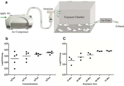
An aerosol exposure apparatus were assembled according to the description of Donald C. Sheppard (Fig 1A) [15]. Mice were introduced to the exposure chamber (Yuyan instruments company, Shanghai, China) via a hinged door on the top of chamber. Conidia suspensions were aerosolized using a Nebulizer (DeVilbiss, Shanghai, China) driven by compressed air. The generated aerosol was connected to the exposure chamber, which was connected to an air filter (Yuyan instruments company, Shanghai, China) though another tube to prevent spores from contaminating the environment. The entire apparatus can be completely sealed.
Determination of exposure time and inoculum concentration
The infection process
Forty-four mice received aerosols generated from 8 mL of a suspension containing 108conidia/ ml for a fixed exposure time of 60 min for complete and uniform exposure of all the mice. Twenty mice received aerosols generated from PBS as control. Twenty mice were used for sur-vival assay. The rest were randomly selected at select time points for further study.
Collection of BALF
BALF was collected as described previously [16]. Briefly, mice were sacrificed and a small inci-sion was made at the neck to expose trachea. A SURFLO catheter was cannulated into the tra-chea under the larynx. Sterile PBS were flushed through the animal lung, and then transferred to a 15 mL centrifuge tube (Biofil Guangzhou China). This process was repeated until 4 mL of BALF was collected. BALF was centrifuged at 12,000 rpm for 10 min. Remaining sediments were resuspended with PBS with the final volume of 0.5 mL.
Histology
On days 0, 3, 7, 15, 21, and 30, four mice were randomly selected. Organ samples were removed from sacrificed animals. For histopathological study, half of each organ was fixed in 4% neutral buffered formalin, processed, and embedded in paraffin. Tissue sections were stained with hematoxylin and eosin (H&E) or schiff periodic acid shiff (PAS) stain. The presence or absence of yeast cells, their morphology and location (intracellular or extracellular), and the host inflammatory response were recorded.
Colony forming units (CFU) determination
For determination of CFU, primary homogenates of each half of organ were prepared by add-ing 1 ml sterile saline per gram of tissue. Serial 10-fold dilutions of primary homogenates were plated in triplicate on PDA at 25°C for 3 days before colonies were counted.
Fig 1. Preliminary experiments of aerosol exposure system.Illustration shows the inhalation exposure system. Air is directed into the compressed air-driven nebulizer, and then directed into a multi-animal exposure chamber. After passing through the exposure chamber the air is filtered before being sent into the exhaust system (a). Fungal burdens in lung of each mouse after exposure to conidia suspensions of various concentrations for 60 min (b) and a suspension containing 108/mL for different times (c).
Nested PCR
Nested PCR assay were performed as described previously [8,13]. Briefly, the out primers were RRF1: 5’ATCTAAATCCCTTAACGAGGAACA3’and RRH1: 5’CCGTCAATTTCTTTAA GTTTCAGCCTT3’. The inner primers were Pm1: 5’ATGGGCCTTTCTTTCTGGG3’and Pm2: 5’GCGGGTCATCATAGAAACC3’(synthesized by Shenggong company, Shanghai, China). In the primary PCR, 2.5μL template DNA were added to a 25μL reaction system
con-taining 12.5μL Premix Taq DNA polymerase (Takara, Dalian, China), and 1μM of out primers
and 8μL of RNase-free water. The parameter for PCR reaction was: 95°C for 5 min; 35 cycles of
95°C for 30 s, 55°C for 30 s, 72°C for 2 min and final extension at 72°C for 10 min. One micro liter of the primary PCR product was subjected to second PCR amplification. The parameters of amplification in nested PCR were the same as described in the primary PCR except that the annealing temperature of 68°C and specific inner primer. Five microliters of nested PCR ampli-fication products were analyzed by electrophoresis (Bio-Rad, California, USA) and 1% agarose gels (Invitrogen, USA). Expected sizes of the nested PCR products were about 400-bp.
DNA from selected fungal species and mice were used to test specificity of the nested PCR. The sensitivity of nested PCR was determined by comparing it with single PCR assay carried out under the same condition described above with primer pair Pm1 and Pm2. Briefly, 1μl of
the DNA solution from BALF sample (8.4×10⁹fg/μl) was used as a template for PCR and
seri-ally diluted 10-fold in RNase-free water. The amplification products were analyzed by electrophoresis.
Statistical analysis
Graphs were plotted by using GraphPad Prism 5 software (GraphPad Software, La Jolla, CA). Survival data were analyzed by means of log-rank comparisons of Kaplan–Meier survival curves. Coefficient of variation was used to access the variation of CFU of lung within each group. Fungal burden were statistically processed by t-test. Statistical analyses were considered significant whenPvalue of less than 0.05 was produced.
Results
Characterization of the fungal aerosol exposure system and titration of
infectious inoculum
Survival assay and analysis of fungal burden within infected mice
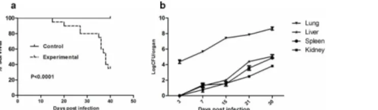
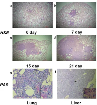
Aerosolizing 8 mL of the suspension containing 108/mL conidia for 60 min resulted in lethal infection with mortality of 65% in 40 days (Fig 2A). Death most often occurred during late infec-tion, from the fifteenth day after inoculation. We counted the colonies ofT.marneffeithat were present in the lung, liver, spleen and kidney tissues at 3 days, 7 days, 15 days, 21 days and 30 days after inhalation of conidia. Differences between the numbers of CFU in lungs versus livers, spleens or kidneys were significant (P= 0.0087, 0.0045, 0.0011 respectively) (Fig 2B). We also found thatT.marneffeiwas confined to lung tissues until the seventh day after infection, and then diffused to remote organs. We speculated that the liver might be the first infected remote organ followed by spleen and kidney. This speculation is based on the observation that we can’t find fungal infection evidence in other organs except for the lung and the liver in the mice. Histo-pathological examination by H&E and PAS staining showed inflammatory cells infiltration and fungal burden in these tissues. At day 7, large numbers of macrophages loaded with yeast cells and multinucleated giant cells were identified in lung tissues (Fig 3E). By day 11, similar patho-logical changes were observed in liver tissues (Fig 3F). HE staining revealed the complete process of lung inflammation, primarily characterized by leukocyte infiltration and granuloma formation with tissue destruction caused by robust fungal invasion (Fig 3A–3D).
Specificity and Sensitivity of nested PCR
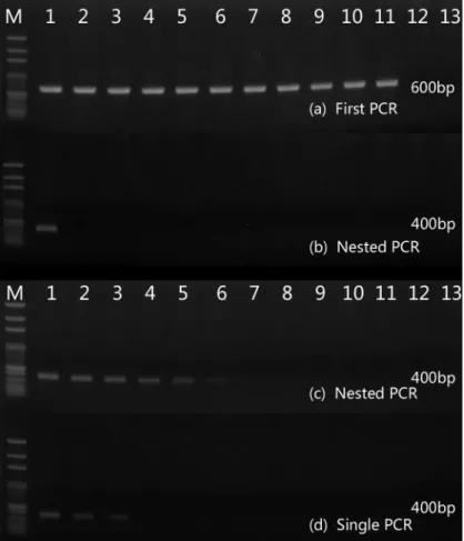
To determine the specificity of nested PCR, DNA from selected fungal species and mice were analyzed. Primers RRF1 and RRH1 specific to fungi result in approximately 600-bp PCR prod-ucts in all fungal samples (Fig 4A). Primers pm1 and pm2 specific toT.marneffeiproduce approximately 400-bp PCR products inT.marneffeisamples. While mice and other fungal control samples gave negative PCR results (Fig 4B). Sensitivity of the nested PCR with primer pairs RRF1 and RRH1 and Pm1 and Pm2 and single PCR with specific primers Pm1 and Pm2 was 8.4×10⁴and 8.4×10⁷fg/μl, respectively (Fig 4C and 4D).
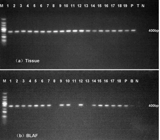
Detection of T. marneffei in fresh tissue and BALF by Nested PCR
A total of 19 samples of lung tissues and 18 samples of BALF were obtained from 19 infected mice confirmed by CFU assay. Sterile water, BALF and fresh tissues from healthy mice were used as the negative control. Purified genomic DNA fromT.marneffeiwas used as the positive control. We performed nested PCR in all these samples and detected a 400-bp product by aga-rose gel electrophoresis (Fig 5).
Discussion
Murine models are critical for studying the pathogenesis and diagnosis of clinically important fungi. In this study, we first constructed a murine pulmonary model that would mimic human Fig 2. Survival and fungal burdens after inhalational infection withT.marneffei.Survival curves (a) and fungal loads in lung, spleen, liver and kidney at selected time post infection (b) of BALB/c nude mice received 5×10⁸conidia ofT.marneffeistrain 152.
penicilliosis marneffei utilizing inhalation exposure. We found that a nested PCR assay employed in this model might contribute to the development of new diagnostic methods in detectingT.marneffeiin fresh tissues.
Infection with penicilliosis marneffei begins by inhalation of the infectious conidia ofT. marneffei. Thus, the pulmonary route of infection would be optimal to initiate the model, for it can closely mimic the natural route of infection into human. Recently, Buskirk et al. developed a nose-only, acoustical generator exposure system to aerosolize fungal conidia to recapitulate human exposures, but this system was unavailable for most of laboratory in developing country due to the high cost [17]. In this study, we adapted a new inhalation apparatus, which is rela-tively simple, low cost and timesaving. It could deliver fungal conidia directly to the alveoli of mice, and allow for infection of up to 30 mice simultaneously. The relatively small infectious inoculum (5×10³) could result in infection with long duration of 40 days, which provided a rea-sonable window for further study of the disease. Survival and CFU assay revealed that invasive pulmonary infection occurred in about 65% of infected mice. CFU assay also showed that the lung appears to be the predominant organ during infection and the liver might be the first infected remote organ, consistent with the findings in our previous study [7]. Furthermore, we observed similar histopathological changes of infected mice with human penicilliosis marneffei by using this model. The progressive lung inflammation displayed in this model was consistent with the histopathological findings of reported cases of penicilliosis marneffei [18]. Our results indicated that the model could faithfully simulate human infection of penicilliosis marneffei.
In addition, our study revealed that nested PCR had high specificity and sensitivity in detectingT.marneffeiin fresh tissues and BALF. Till now, diagnosis of fungal infections has been a significant challenge, although nested PCR assays have been reported as powerful tools for detecting pathogenic fungi [9,12]. To our knowledge, it was previously used to identifyT. Fig 3. Lung sections of mice infected withT.marneffei.Progressive inflammatory reaction presented with leukocyte infiltration and granuloma formation with tissue destruction, HE-stains 100X (a-d).
Macrophages loaded with yeast cells and formation of multinucleated giant cells in lung 7 days post infection, PAS stains 400X, partial enlarged panel (1000X) depicts fission yeast (e). Macrophages loaded with yeast cells in liver 11 days post infection (f).
marneffeiin paraffin embedded tissue and whole blood samples [9,12]. This is the first time that nested PCR has been used for the detection ofT.marneffeifrom fresh tissues and BALF and proved to be suitable for detectingT.marneffei. Compared with CFU results, we found equivalence between nested PCR and CFU in fresh tissues, but two samples revealed negative results in BALF. The discrepancies might be explained by the aforementioned, unsuccessful DNA extraction. These results prompted us to hypothesize that BALF can’t entirely replace lung tissues to reflect pulmonary infection. While in the clinic, lung biopsies are not routinely performed because this invasive examination may make patient’s respiratory function worse. BALF could be obtained by non-invasive procedure, most likely proving favorable for patients. According to the results, we could conclude that nested PCR assay on BALF has high predictive value in reflecting pulmonary infection with a positive rate of 83.3%.
In summary, we successfully developed a novel inhalation exposure system to initiate the model by directly delivering conidia to alveoli, and verified that nested PCR on BALF may serve as a good alternative tool to conventional diagnosis of penicilliosis marneffei. This simple Fig 4. Specificity and sensibility of nested PCR.A 600-bp PCR product were amplified from all fungal samples (a) and a 400-bp PCR product was amplified fromT.marneffeisamples (b). M, 100-bp-ladder DNA; Lane1 to 13,T.marneffei,Aspergillus flavus,Aspergillus fumigatus,Aspergillus niger,Cryptococcus neoformans,Candida albicans,Candida krusei,Fonsecaea pedrosoi,Fonsecaea monophora,Histoplasma capsulatum,Paracoccidioides brasiliensis, Negative control (mice DNA), Negative control (water) respectively. Sensitivity of nested PCR (c) and single PCR (d) was 8.4×10⁴and 8.4×10⁷fg/μl, respectively.
M, 100-bp-ladder DNA; lanes 1 to 13, 8.4×10⁹, 8.4×10⁸, 8.4×10⁷, 8.4×10⁶, 8.4×10⁵, 8.4×10⁴, 8.4×10³,
8.4×10², 8.4×101, 8.4×100, 8.4×10⁻¹, 8.4×10⁻², 8.4×10⁻³f g /μl, respectively.
model provides an alternative to existing intranasal models and may contribute to deeper insight into penicilliosis marneffei.
Acknowledgments
We are grateful to Boyou Li for assistance with histopathological studies and Joshua Engle, Dedong Li for English revision and helpful comments.
Author Contributions
Conceived and designed the experiments: LX EM. Performed the experiments: YL XH XY. Analyzed the data: YL XH YH. Contributed reagents/materials/analysis tools: YL XH YH. Wrote the paper: YL XH.
References
1. Houbraken J, de Vries RP, Samson RA (2014) Modern taxonomy of biotechnologically important Aspergillus and Penicillium species. Adv Appl Microbiol 86:199–249. doi: 10.1016/B978-0-12-800262-9.00004-4PMID:24377856
2. Huang X, Li D, Xi L, Mylonakis E (2014) Caenorhabditis elegans: a simple nematode infection model for Penicillium marneffei. PLoS One 9: e108764. doi:10.1371/journal.pone.0108764PMID:25268236
3. DiSalvo AF, Fickling AM, Ajello L (1973) Infection caused by Penicillium marneffei: description of first natural infection in man. Am J Clin Pathol 60: 259–263. PMID:4720403
Fig 5. Nested PCR assays in fresh lung tissues (a) and BALF (b).A 400-bp specific product was amplified in most of samples. M: 100-bp-ladder DNA, P, purified genomic DNA fromT.marneffei, T, lung tissue from healthy mice, B, BALF from healthy mice, N, water.
4. Hu Y, Zhang J, Li X, Yang Y, Zhang Y, Ma J, et al. (2013) Penicillium marneffei infection: an emerging disease in mainland China. Mycopathologia 175: 57–67. doi:10.1007/s11046-012-9577-0PMID: 22983901
5. Kudeken N, Kawakami K, Kusano N, Saito A (1996) Cell-mediated immunity in host resistance against infection caused by Penicillium marneffei. J Med Vet Mycol 34: 371–378. PMID:8971625
6. Kudeken N, Kawakami K, Saito A (1997) CD4+ T cell-mediated fatal hyperinflammatory reactions in mice infected with Penicillium marneffei. Clin Exp Immunol 107: 468–473. PMID:9067519
7. Sun J, Li X, Feng P, Zhang J, Xie Z, Song E, et al. (2014) RNAi-mediated silencing of fungal acuD gene attenuates the virulence of Penicillium marneffei. Med Mycol 52: 167–178. doi:10.1093/mmy/myt006 PMID:24577002
8. Zeng H, Li X, Chen X, Zhang J, Sun J, Xie Z, et al. (2009) Identification of Penicillium marneffei in Paraf-fin-Embedded Tissue Using Nested PCR. Mycopathologia 168: 31–35. doi: 10.1007/s11046-009-9195-7PMID:19308671
9. Sun J, Li X, Zeng H, Xie Z, Lu C, Xi L, et al. (2010) Development and evaluation of loop-mediated iso-thermal amplification (LAMP) for the rapid diagnosis of Penicillium marneffei in archived tissue sam-ples. FEMS Immunol Med Microbiol 58: 381–388. doi:10.1111/j.1574-695X.2010.00647.xPMID: 20113352
10. Sun J, Najafzadeh MJ, Zhang J, Vicente VA, Xi L, de Hong GS (2011) Molecular identification of Peni-cillium marneffei using rolling circle amplification. Mycoses 54: e751–759. doi:10.1111/j.1439-0507. 2011.02017.xPMID:21929692
11. Zhang JM, Sun JF, Feng PY, Li XQ, Lu CM, Lu S, et al. (2011) Rapid identification and characterization of Penicillium marneffei using multiplex ligation-dependent probe amplification (MLPA) in paraffin-embedded tissue samples. J Microbiol Methods 85: 33–39. doi:10.1016/j.mimet.2011.01.022PMID: 21277339
12. Pongpom M, Sirisanthana T, Vanittanakom N (2009) Application of nested PCR to detect Penicillium marneffei in serum samples. Med Mycol 47: 549–553. doi:10.1080/13693780802484875PMID: 19353372
13. Chen R, Li X, Lu S, Ma T, Huang X, Mylonakis E, et al. (2014) Role of extracellular signal-regulated kinases 1 and 2 and p38 mitogen-activated protein kinase pathways in regulating replication of Penicil-lium marneffei in human macrophages. Microbes Infect 16: 401–408. doi:10.1016/j.micinf.2014.02. 005PMID:24583279
14. Lu S, Hu Y, Lu C, Zhang J, Li X, Xi L (2013) Development of in vitro macrophage system to evaluate phagocytosis and intracellular fate of Penicillium marneffei conidia. Mycopathologia 176: 11–22. doi: 10.1007/s11046-013-9650-3PMID:23645133
15. Sheppard DC, Rieg G, Chiang LY, Filler SG, Edwards JE Jr., Ibrahim AS (2004) Novel inhalational murine model of invasive pulmonary aspergillosis. Antimicrob Agents Chemother 48: 1908–1911. PMID:15105158
16. Stevens WW, Kim TS, Pujanauski LM, Hao X, Braciale TJ (2007) Detection and quantitation of eosino-phils in the murine respiratory tract by flow cytometry. J Immunol Methods 327: 63–74. PMID: 17716680
17. Buskirk AD, Templeton SP, Nayak AP, Hettick JM, Law BF, Green BJ, et al. (2014) Pulmonary immune responses to Aspergillus fumigatus in an immunocompetent mouse model of repeated exposures. J Immunotoxicol 11: 180–189. doi:10.3109/1547691X.2013.819054PMID:23919459