Prognostic Value of Tissue Inhibitor of
Metalloproteinase-2 Expression in Patients
with Non
–
Small Cell Lung Cancer: A
Systematic Review and Meta-Analysis
Lin Zhu1, Hong Yu1*, Shi-Yuan Liu1, Xiang-Sheng Xiao1, Wei-Hua Dong1, Yi-Nan Chen1, Wei Xu2, Tong Zhu3
1Department of Medical imaging, Shanghai Changzheng Hospital affiliated to The Second Military Medical University, Shanghai, China,2Department of Periodicals, The Second Military Medical University Library, Shanghai, China,3Department of Business Marketing, Shanghai United Imaging Healthcare Co., Ltd, Shanghai, China
Abstract
Background and Objectives
Tissue inhibitor of metalloproteinase-2 (TIMP-2) is a small secretory glycoprotein with anti– matrix metalloproteinase activity. Data on the value of TIMP-2 as a prognostic factor in non– small cell lung cancer (NSCLC) are discordant and remain controversial. A systematic re-view and meta-analysis was performed to explore this issue.
Methods
We identified the relevant literature by searching the PubMed, EMBASE, Web of Science, China National Knowledge Infrastructure, SinoMed, and Wanfang Data databases (search terms:“non-small cell lung cancer”or“NSCLC”or“Lung Carcinoma, Non-Small-Cell”, “Tis-sue Inhibitor of Metalloproteinase-2”or“TIMP-2”, and“prognosis”or“prognostic”or “sur-vive”) for updates prior to March 1, 2014. The pooled hazard ratio (HR) of overall survival with a 95% confidence interval (95% CI) was used to evaluate the strength of the associa-tion between positive TIMP-2 expression and survival in patients with NSCLC.
Results
We included 12 studies in our systematic review; five studies involving 399 patients with NSCLC were meta-analyzed. The pooled HR of all included patients was 0.57 (95% CI: 0.43–0.77), and the HRs of subgroup analysis according to stage (I–IV), testing method (immunohistochemistry) and high TIMP-2 expression percentage (<50%) were 0.63 (95% CI: 0.43–0.92), 0.55 (95% CI: 0.41–0.74), and 0.50 (95% CI: 0.28–0.88), respectively. These data suggested that high TIMP-2 expression is associated with favorable prognosis in NSCLC. The meta-analysis did not reveal heterogeneity or publication bias.
OPEN ACCESS
Citation:Zhu L, Yu H, Liu S-Y, Xiao X-S, Dong W-H, Chen Y-N, et al. (2015) Prognostic Value of Tissue Inhibitor of Metalloproteinase-2 Expression in Patients with Non–Small Cell Lung Cancer: A Systematic Review and Meta-Analysis. PLoS ONE 10(4): e0124230. doi:10.1371/journal.pone.0124230
Academic Editor:Rafael Rosell, Catalan Institute of Oncology, SPAIN
Received:September 15, 2014
Accepted:February 27, 2015
Published:April 23, 2015
Copyright:© 2015 Zhu et al. This is an open access article distributed under the terms of theCreative Commons Attribution License, which permits unrestricted use, distribution, and reproduction in any medium, provided the original author and source are credited.
Data Availability Statement:All relevant data are within the paper and its Supporting Information files.
Conclusions
TIMP-2 expression indicates favorable prognosis in patients with NSCLC; as a protective factor, it could help predict outcome and may guide clinical therapy in the future.
Introduction
The mortality rate of lung cancer (LC) is one of the highest worldwide. Based on GLOBOCAN 2008 estimates, there were an estimated 12.7 million cancer cases and 7.6 million cancer deaths in 2008. Of these, LC was the leading cancer in males, comprising 17% of total new cancer cases and 23% of total cancer deaths [1]. Lozano et al. [2] also reported that 8 million people died from cancer in 2010, the cause of death of 1.5 million of which was tracheal, bronchial, and lung cancer. For treatment purposes, LC is always divided into two major histological sub-types: small cell lung cancer (SCLC) and non–small cell lung cancer (NSCLC) [3]. NSCLC ac-counts for approximately 85% of LCs, but the 5-year survival rate is only 15%, even though treatments such as surgical management have developed rapidly in recent years [4]. Therefore, diagnosis and treatment at the early stage is important for patients with NSCLC. Understand-ing the mechanism of invasion and metastasis and explorUnderstand-ing means of preventUnderstand-ing NSCLC in-vasion and metastasis at the molecular level is the key to future therapy.
Early studies of NSCLC focused on identifying somatic mutations in genes involved in LC development and led to the discovery of crucial activated oncogenes and abnormal signaling pathways [5]. The development of NSCLC is also considered a multi-step process [6]. In recent years, researchers have realized that NSCLC is not merely caused by gene mutations, but also biological factors in the microenvironment. Currently, much research is focused on this hot-spot, and it has been confirmed that many biological factors are associated with NSCLC devel-opment and prognosis, such as matrix metalloproteinase (MMP)-2[7], MMP-9 [8],
transforming growth factor (TGF) [9], and cyclophilin A (CypA) [10].
MMPs are enzymes that degrade almost all of the protein and collagen in the extracellular ma-trix (ECM), destroying the histological barrier against tumors and playing a key role in tumor in-vasion and metastasis [11]. Tissue inhibitor of metalloproteinase (TIMP), an MMP inhibitor, is a promising biomarker for reducing cancer occurrence and improving prognosis. It is a glycopro-tein with an apparent molecular size of 28.5 kDa that forms a complex of 1:1 stoichiometry with activated interstitial collagenase [12]. TIMP-2 is a TIMP family member, and is a multifunctional protein that is secreted into the ECM. Increased TIMP-2 levels are considered a favorable prog-nostic indicator in NSCLC, because they are correlated with the inhibition of endothelial cell pro-liferation and lung cancer cell angiogenesis in vivo [13]. Therefore, TIMP-2 could have significant prognostic value for patients with NSCLC. At the same time, Zhu et al [14]. and Michael et al [15]. reported that it holds no value as a marker, and results have been contradictory. The differ-ence in findings may be due to individual study limitations such as small sample size and low sta-tistical power. Therefore, we aimed to perform a more precise evaluation of the relationship between TIMP-2 expression and survival in patients with NSCLC through meta-analysis.
Materials and Methods
Search strategy and study selection
We searched for relevant studies on TIMP-2 expression and survival in NSCLC patients in the PubMed, EMBASE, Web of Science, China National Knowledge Infrastructure (CNKI),
Prognostic Value of TIMP-2 in NSCLC
PLOS ONE | DOI:10.1371/journal.pone.0124230 April 23, 2015 2 / 13
TZ, but did not have any additional role in the study design, data collection and analysis, decision to publish, or preparation of the manuscript. The specific role of this author is articulated in the‘author contributions’section.
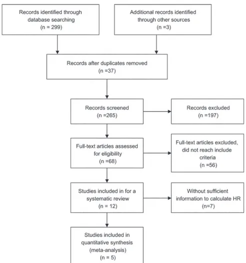
SinoMed, and Wanfang Data databases up until they were updated on March 1, 2014. We also reviewed the reference lists of relevant articles. The search terms were (“non-small cell lung cancer”or“NSCLC”or“Lung Carcinoma, Non-Small-Cell”), (“Tissue Inhibitor of Metallopro-teinase-2”or“TIMP-2”), and (“prognosis”or“prognostic”or“survive”). We did not impose language limits on the literature search. To ensure a high-quality systematic review and meta-analysis, we used the following criteria: (1) patients with cytologically or histologically con-firmed NSCLC, (2) measured TIMP-2 protein expression, (3) assessed the association between TIMP-2 and survival in NSCLC, (4) a control group to compare survival times between high and low TIMP-2 expression, (5) follow-up was>3 years, (6) for full papers, the hazard ratio (HR) and 95% confidence interval (95% CI) could be obtained from the article or calculated from information therein, (7) in duplicate articles published by the same author, we selected the most complete and newest article.Fig 1depicts the paper selection process.
Data extraction
Two authors assessed and selected potential articles independently; disagreements were re-solved by debate and discussions with another expert. The data extracted included name of the first author, publication year, number of patients, median or mean patient age, disease stage, Fig 1. Flowchart of article selection.Flowchart of article selection for systematic review and meta-analysis. HR: hazard ratio.
TIMP-2 test method, percentage of positive TIMP-2 expression, and estimated HR. Items not included directly in an article were calculated using relevant information provided therein. If the article contained insufficient data, we contacted the authors by email to obtain as much useful information as possible. Items that still could not be obtained were described as“not re-ported (NR)”.
Statistical analysis
We used the HR of overall survival and 95% CI to combine the data; if the original article con-tained the HR and 95% CI, then we used them directly. If not, we calculated the HR and 95% CI using available information in the papers or obtained the information by contacting the au-thors; Kaplan–Meier survival curves could also be used to estimate a relatively accurate HR [16,17]. If an article described both univariate and multivariate analysis, such as the report of Lv et al. [18], we chose the latter because survival in NSCLC is affected by a combination of fac-tors. Then, we calculated the logHR and standard error of logHR (SElogHR) of every article and combined them to obtain the HR and 95% CI of all included papers. Heterogeneity was as-sessed using the I² statistic and chi-square test. If study heterogeneity was considered absent, we used a fixed effects model. By convention, if I²50%, heterogeneity was considered present and we used the random effects model instead [19]. Publication bias was assessed using funnel plots; evidence of reliability was derived using sensitivity analysis.
All p-values were 2-sided; p<0.05 was considered statistically significant. Statistical calcu-lations were performed using Review Manager Version 5.2 provided by the Cochrane
Collaboration.
Results
Study characteristics
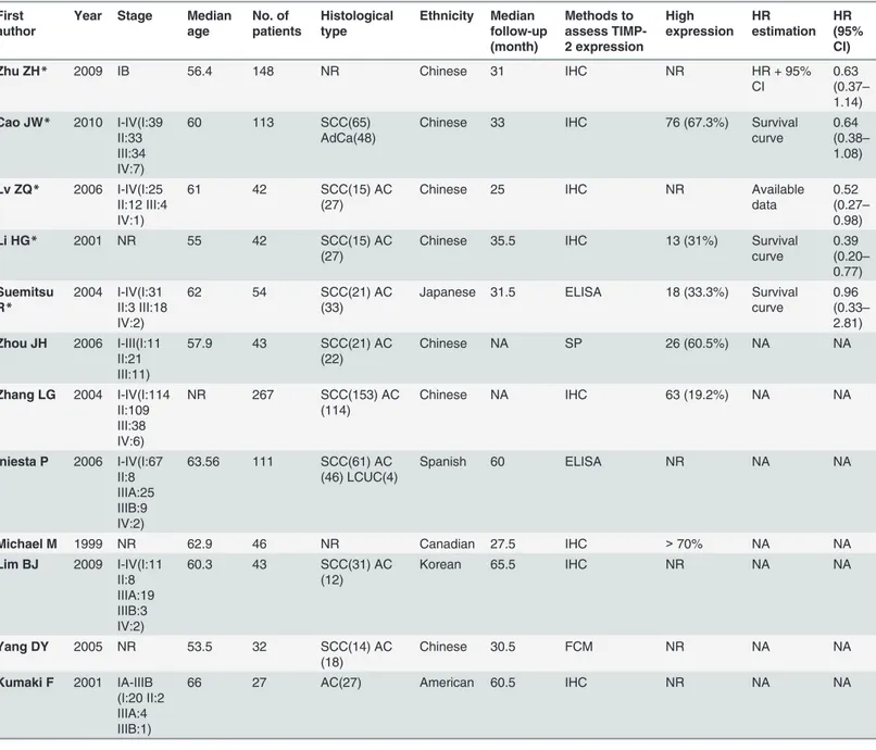
We identified 302 potential articles from the initial search. We excluded 290 articles that did not meet our inclusion criteria. The abstract of one paper, in which the authors stated that they had used tissue microarray and immunohistochemistry (IHC) to detect the expression of 31 molecular markers in patients with NSCLC and had used the Kaplan–Meier method to esti-mate survival curves, rendered it eligible for inclusion. However, it was a dissertation and we could not find the full text in all of the searched databases for further confirmation, therefore we did not know whether the full details in the article met the inclusion criteria; therefore, we excluded it [20]. We selected 12 articles for the systematic review, as they were full articles and assessed the association between TIMP-2 and survival both in the patients with NSCLC and in the control groups using different methods. At the same time, they followed all included sub-jects for at least three years. Then, we excluded seven of the 12 eligible studies from the meta-analysis due to insufficient information for calculating the HR. The information provided by two of the excluded articles could only be used to calculate the relative risk (RR) [21,22]: one found no association between TIMP-2 expression and outcome [21]; the other concluded that TIMP-2 was a harmful factor in NSCLC patients [22]. Four papers reported no significant as-sociation between prognosis of NSCLC and TIMP-2 expression [15,23–25]. By contrast, Yang et al. [26] performed a flow cytometry study and concluded that high TIMP-2 expression was a significant indicator of long survival in patients with NSCLC. Therefore, the remaining five studies with sufficient information for obtaining HR and that involved 399 patients with NSCLC [14,18,27–29] were meta-analyzed. They were all published full papers, the sample size of these studies ranged 42–148 patients, and the reported median age was 55–62 years. The percentage of positive TIMP-2 expression ranged from 31% to 67.3%. However, the five includ-ed articles all happeninclud-ed to be from Asian countries.Table 1lists the major characteristics of the Prognostic Value of TIMP-2 in NSCLC
eligible studies. We also used the GRADE profiler to evaluate the quality of the included stud-ies, which were all assessed as moderate;Table 2presents the findings in detail.
Main
meta-analysis results
Following screening, five studies were included in the final meta-analysis. They had been pub-lished between 2001 and 2010. The minimum sample size was 42 and the maximum was 148. Table 1. Main characteristics of the eligible studies.
First author
Year Stage Median age No. of patients Histological type Ethnicity Median follow-up (month) Methods to assess TIMP-2 expression High expression HR estimation HR (95% CI)
Zhu ZH* 2009 IB 56.4 148 NR Chinese 31 IHC NR HR + 95%
CI
0.63 (0.37–
1.14) Cao JW* 2010 I-IV(Ⅰ:39
Ⅱ:33
Ⅲ:34 IV:7)
60 113 SCC(65)
AdCa(48)
Chinese 33 IHC 76 (67.3%) Survival
curve
0.64 (0.38–
1.08)
Lv ZQ* 2006 I-IV(Ⅰ:25
Ⅱ:12Ⅲ:4 IV:1)
61 42 SCC(15) AC
(27)
Chinese 25 IHC NR Available
data
0.52 (0.27–
0.98)
Li HG* 2001 NR 55 42 SCC(15) AC
(27)
Chinese 35.5 IHC 13 (31%) Survival
curve 0.39 (0.20– 0.77) Suemitsu R*
2004 I-IV(Ⅰ:31
Ⅱ:3Ⅲ:18 IV:2)
62 54 SCC(21) AC
(33)
Japanese 31.5 ELISA 18 (33.3%) Survival curve
0.96 (0.33–
2.81) Zhou JH 2006 I-III(Ⅰ:11
Ⅱ:21
Ⅲ:11)
57.9 43 SCC(21) AC
(22)
Chinese NA SP 26 (60.5%) NA NA
Zhang LG 2004 I-IV(Ⅰ:114
Ⅱ:109
Ⅲ:38 IV:6)
NR 267 SCC(153) AC
(114)
Chinese NA IHC 63 (19.2%) NA NA
Iniesta P 2006 I-IV(Ⅰ:67
Ⅱ:8
ⅢA:25
ⅢB:9 IV:2)
63.56 111 SCC(61) AC
(46) LCUC(4)
Spanish 60 ELISA NR NA NA
Michael M 1999 NR 62.9 46 NR Canadian 27.5 IHC >70% NA NA
Lim BJ 2009 I-IV(Ⅰ:11
Ⅱ:8
ⅢA:19
ⅢB:3 IV:2)
60.3 43 SCC(31) AC
(12)
Korean 65.5 IHC NR NA NA
Yang DY 2005 NR 53.5 32 SCC(14) AC
(18)
Chinese 30.5 FCM NR NA NA
Kumaki F 2001 IA-IIIB (Ⅰ:20Ⅱ:2
ⅢA:4
ⅢB:1)
66 27 AC(27) American 60.5 IHC NR NA NA
IHC: Immunohistochemistry; SP: streptavidin–peroxidase; ELISA: enzyme-linked immunosorbent assay; FCM:flow cytometry; TIMP-2: tissue inhibitor of metalloproteinase-2; HR: hazard ratio; CI: confidence interval; NR: not reported; NA: not available; SCC: squamous cell carcinoma; AdCa:
adenosquamous carcinoma; AC: Adenocarcinoma; LCUC: Large Cell Carcinoma;
*studies included in the meta-analysis
Table 2. GRADE profiler evaluation of the quality of included studies.
Prognostic value of high TIMP-2 expression in patients with NSCLC Patient or population: Patients with NSCLC
Intervention group: High TIMP-2 expression Comparison: Low TIMP-2 expression
Outcomes Illustrative comparative risks* (95% CI) Relative effect(95% CI) No of Participants(studies) Quality of the evidence (GRADE) Comments Assumed risk Corresponding risk
Hazard ratio (HR) Study population HR 0.57 386 ⊕⊕⊕⊝
Dichotomous 777 per 1000 575 per 1000(476 to 685) (0.43 to 0.77) (5 studies) Moderate1,2,3,4
Follow-up: 3 years Moderate
*The basis for theassumed risk(i.e., median control group risk across studies) is described in the footnotes. Thecorresponding risk(and its 95% CI) is based on the assumed
risk in the comparison group and therelative effectof the intervention (and its 95% CI).
CI:Confidence interval;HR:Hazard ratio.
GRADE Working Group grades of evidence:
High quality:Further research is very unlikely to change our confidence in the estimate of effect.
Moderate quality:Further research is likely to have an important impact on our confidence in the estimate of effect and may change the estimate.
Low quality:Further research is very likely to have an important impact on our confidence in the estimate of effect and is likely to change the estimate.
Very low quality:We are very uncertain about the estimate. Footnotes:
1Negative results without suf
ficient information. 2
No unified standard. 3Large sample.
4Methods used to assess TIMP-2 expression, etc., and multivariate analysis.
doi:10.1371/journal.pone.0124230.t002
Prognostic
Value
of
TIMP-2
in
NSCLC
PLOS
ONE
|DOI:10.137
1/journal.p
one.0124230
April
23,
2015
6/1
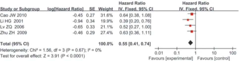
The median age of the included patients ranged 55–62 years, and all included patients under-went surgical resection. Most of the included studies reported on stage I–IV disease, but Zhu et al. [14] only included patients with stage IB disease, and Li et al. [28] did not report this in-formation. Specific numbers of included patients with different tumor stages in each article were summarized and listed inTable 1. Four of the five studies were from China, and the re-maining study was from Japan. Histological types of most included patients consist of squa-mous cell carcinoma and adenocarcinoma and histology of patients included in the article from Zhu et al. were not reported. The detail information were summarized in theTable 1. The median follow-up of these studies ranged 25–35.5 years, and the reported percentage of posi-tive TIMP-2 expression was 31–67.3%. Suemitsu et al. [29] sampled blood to perform enzyme-linked immunosorbent assay (ELISA) analysis, and the remaining four studies analyzed tumor and normal tissues using IHC. The HR reported by Lv et al. [18] referred to multivariate analy-sis, and that of the other four studies referred to univariate analysis.Table 3summarizes the main meta-analysis results; Figs2–5depict the forest plots. The overall combined HR of the in-cluded studies was 0.57 (95% CI: 0.43–0.77); we used the fixed effect model because heteroge-neity was considered absent (p = 0.64, I² = 0%). This suggests that positive TIMP-2 expression may be a significant predictor of good prognosis in NSCLC.
In subgroup analysis, we analyzed the studies according to disease stage (I–IV), testing method for TIMP-2 expression (IHC), and percentage of high TIMP-2 expression (<50%). When we limited the first two subgroup analysis factors, both meta-analyses showed that high TIMP-2 expression improved survival in NSCLC. The combined HRs were 0.63 (95% CI: 0.43–0.92) and 0.55 (95% CI: 0.41–0.74), respectively. When we analyzed the two studies in which the percentage of positive TIMP-2 expression was<50% separately, we found that TIMP-2 still had positive prognostic value in NSCLC; the HR was 0.50 (95% CI: 0.28–0.88). Table 3. Main meta-analysis results.
No. of studies
No. of included patients
Combined HR (95% CI)
Heterogeneity test (x2,p,I2)
Overall 5 399 0.57 (0.43, 0.77) 2.50, 0.64, 0%
TIMP-2 in NSCLC tested by IHC
4 345 0.55 (0.41, 0.74) 1.56, 0.67, 0%
TIMP-2 in stages I-IV 3 209 0.63 (0.43, 0.92) 0.91, 0.63, 0% TIMP-2 in high
expression:<50%
2 96 0.50 (0.28, 0.88) 1.94, 0.16, 48%
TIMP-2: tissue inhibitor of metalloproteinase-2; HR: hazard ratio; CI: confidence interval; IHC: Immunohistochemistry;
doi:10.1371/journal.pone.0124230.t003
Fig 2. Forest plot of the association between positive TIMP-2 expression and survival in patients with NSCLC.Positive TIMP-2 expression significantly predicted good prognosis in NSCLC (HR = 0.57,
P = 0.0002).
The heterogeneity of every subgroup meta-analysis was non-significant, and we used the fixed effect model for all.
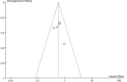
Publication bias
Funnel plots were constructed to examine publication bias. The obtained funnel plot presented no proof of obvious publication bias for the included studies (Fig 6).
Sensitivity analysis
Sensitivity analysis was performed to examine the reliability of our conclusion. We removed one study at a time and the results were not significantly changed (Table 4). This suggests that the conclusion of our meta-analysis is reliable.
Discussion
LC, especially NSCLC, is the most common cancer; mortality is high. In recent years, the inci-dence of LC has increased sharply in both men and women [30]. Therefore, identifying an ef-fective early diagnostic biological marker is vital for patients with NSCLC. At present, the reported biological markers for NSCLC include carcinoembryonic antigen (CEA) [31] and CD133 [32]. A new research hotspot, MMPs destroy the ECM barrier to promote LC angiogen-esis and metastasis, and has an unfavorable effect on prognosis [33]. TIMPs are MMP inhibi-tors that can reduce MMP destruction of the cell matrix and then maintain a complete
connection between cells to decrease cancer metastasis and improve prognosis. Some research-ers reported that TIMPs induce apoptosis and inhibit invasion by inhibiting signal transducer and activator of transcription-3 (STAT3) by activating the mitogen-activated protein (MAP) kinases such as c-Jun N-terminal kinase (JNK), p38, and extracellular signal–regulated kinase (ERK) in human lung cancer A549 cells [34]. At the same time, Wu et al. [35] reported that when an anti-tumor drug was used on A549 cells, MMP expression was reduced while TIMP-1 and TIMP-2 expression was increased, and the drug suppressed cell invasion and migration by Fig 3. Forest plot of subgroup analysis by disease stage (I–IV).TIMP-2 expression was a statistically
significant favorable factor in patients with stage I-IV NSCLC (HR = 0.63, P = 0.02).
doi:10.1371/journal.pone.0124230.g003
Fig 4. Forest plot of subgroup analysis by test method for TIMP-2 expression (IHC).High TIMP-2 expression remain predicted good prognosis in patients with NSCLC when only IHC was used to assess TIMP-2 expression levels (HR = 0.55, P<0.0001).
doi:10.1371/journal.pone.0124230.g004
Prognostic Value of TIMP-2 in NSCLC
down-regulating MMP-2 and p38 MAPK signaling. Other studies have confirmed that other TIMP family members are associated with cancer prognosis, e.g., TIMP-1 [36,37] and TIMP-3 [38]. TIMP-2 has garnered much attention in recent years; many researchers have reported its prognostic value in NSCLC. However, the conclusions from individual studies on the relation-ship between TIMP-2 and NSCLC are inconsistent. Some researchers suggest that it is a protec-tive factor expressed at lower levels in patients with NSCLC, while others have found that expression remains the same or is increased in NSCLC. Therefore, we conducted this meta-analysis to clarify the exact relationship between TIMP-2 and prognosis in NSCLC.
We obtained articles by a comprehensive search strategy and also defined strict inclusion criteria. HR was used as the indicator of time-to-event outcomes for combining individual studies. Compared to the RR or odds ratio, HR measures not only the number of events but also takes into account when they occurred, and is more appropriate for analyzing time-to-event outcomes [16]. The methods for extrapolating the HR are as follows: When the original article did not contain the HR and 95% CI, we calculated them from available data therein or contacted the authors to obtain them. If the data were still insufficient but the original article provided survival curves, we were able to extrapolate the HR using the methods reported by Tierney et al. and Williamson et al. [16,17]. Although this approach may be less accurate than the methods described earlier, we had two authors read the curve independently to minimize
Fig 6. Funnel plot of meta-analysis assessing TIMP-2 expression in patients with NSCLC.Funnel plots were constructed to examine publication bias of all included researches.
doi:10.1371/journal.pone.0124230.g006
Fig 5. Forest plot of subgroup analysis by percentage of high TIMP-2 expression (<50%).TIMP-2 expression in NSCLC patients was remain a significant predictor of good survival (HR = 0.50, P = 0.02).
inaccuracy; we found no apparent contradiction when we compared the estimated HRs with the published results in the original articles.
Subgroup analysis is a method for exploring the sources of heterogeneity and for increasing the reliability of an article. Tumor stage is an important factor that affects the prognosis of NSCLC [39]. While the patients in the included studies were of different disease stages, Zhu et al. studied only stage IB NSCLC patients [14], and Li et al. did not report the disease stage of the pa-tients they enrolled [28]. Therefore, we performed subgroup analysis according to stage, and found that TIMP-2 expression was a statistically significant favorable factor in patients with stage I-IV NSCLC. From this, we may assume that high TIMP-2 expression is not only related to out-come in early-stage NSCLC, but also the late stages. The methods used to measure TIMP-2 ex-pression varied between the studies, which may have reduced the reliability of our result. We selected the most commonly applied method, i.e., IHC, for the subgroup analysis and found that the conclusion that high TIMP-2 expression is associated with good prognosis in NSCLC re-mained the same if only IHC was used to assess TIMP-2 expression levels. The percentage of high TIMP-2 expression of patients in the included studies ranged 31–67.3%. The reasons for this may be the differing states of illness, definitions of positive expression, and race or sex com-position of the sampled populations in the included studies. Therefore, we selected studies where the percentage of high TIMP-2 expression was<50% for subgroup analysis to determine the strength of the previous conclusion. This subgroup analysis also suggested that high TIMP-2 ex-pression in NSCLC patients was a significant predictor of good survival. Thus, we believe that TIMP-2 may be a relatively stable indicator of favorable prognosis in NSCLC.
Although we discovered no significant heterogeneity in the included studies, potential het-erogeneity we did not discover might still affect the quality of this meta-analysis, and should be taken into account. The possible contributors to heterogeneity are as follows: Although one in-clusion criterion was that the time of follow-up must be>3 years, the details of follow-up times in the studies differed; the methods used to assess TIMP-2 expression varied, and even if different studies used the same method, the antibody type and manufacturer would differ; pa-tients selected for each study differed in terms of illness, weight, sex ratio, racial/ethnic compo-sition; each study had its own definition of positive TIMP-2 expression, and there were no unified standards for defining the boundaries of positive expression. When we combined the studies according to the percentage of high TIMP-2 expression being<50%, there was compar-atively large heterogeneity (I² = 48%). This may have resulted from the different methods used to assess TIMP-2 expression and the differing patient characteristics.
As an important influencing factor, there has always been publication bias in meta-analysis. This meta-analysis did not detect publication bias, but we realize that we cannot avoid it completely. For example, the included studies were published in different languages, and posi-tive results are likely to be accepted by journals easily; on the contrary, negaposi-tive results tend to be rejected or are not even submitted. At the same time, a published paper with negative results Table 4. Sensitivity analysis results.
Excluded studies HR (95% CI)
Zhu ZH 0.56 (0.40, 0.78)
Cao JW 0.55 (0.39, 0.77)
Lv ZQ 0.59 (0.43, 0.81)
Li HG 0.63 (0.46, 0.86)
Suemitsu R 0.55 (0.41, 0.74)
HR: hazard ratio; CI: confidence interval;
doi:10.1371/journal.pone.0124230.t004
Prognostic Value of TIMP-2 in NSCLC
does not contain detailed data of the negative aspects. We could only include papers that met all of the inclusion criteria but had insufficient data for the systematic review. All this may have contributed to publication bias in our meta-analysis.
To understand our findings better, some limitations should be considered. The disease stage of the included patients was not homogeneous: Zhu et al. [14] studied patients with early-stage disease, and Li et al. [28] did not report this information; Suemitsu et al. [29] performed ELISA analysis on blood samples; another four studies performed IHC analysis of tumor and normal tissues. IHC results are very difficult to reproduce due to several factors, which include the lack of standardized tissue fixation and staining protocols, difficulty in obtaining a consensus stan-dard for microscopic evaluation, and cutoff determination among different laboratories. These factors all could lead to different experimental outcomes. Moreover, four of the included stud-ies used univariate analysis to explore the relationship between TIMP-2 expression and NSCLC outcome [14,27–29], which is less credible than multivariate analysis such as that by Lv et al. [18]. Multivariate analysis was more important when defining the independent prog-nostic role of TIMP-2 when assessed together with other potential progprog-nostic factors such as MMP-2 and TIMP-1, as there was likely a certain relationship between the expression of these molecules, and univariate analysis always ignores this. In addition, all patients in the included studies were Asian; the median follow-up duration for each included study differed; the pa-tients included in the five studies all underwent surgical resection and one study [18] reported that the included patients underwent surgical resection but without chemotherapy or radio-therapy; the remaining four studies did not include this information, all of which could have af-fected survival. Despite our utmost efforts to contact the authors, we were unable to obtain some data on negative results, greatly reducing the number of articles that could have been in-cluded in the meta-analysis and details such as disease stage and percentage of high TIMP-2 ex-pression in the included studies were also unknown; moreover, the quality of some of the included studies was not wholly satisfactory. These factors could also have affected the out-come of our evaluation of the prognostic value of TIMP-2, and should not be ignored.
In conclusion, despite the limitations of the present study and heterogeneity across the in-cluded studies, our systematic review and meta-analysis suggest that high TIMP-2 expression is a protective factor against the development of NSCLC, and is associated with favorable prog-nosis in NSCLC. It is a potential new biological marker for the early detection and diagprog-nosis of patients with NSCLC, and can guide clinical therapy in the future or act as a predictor of che-motherapy [40] or a target of inhibitory antibodies [41]. In addition, further investigation in-volving more high-quality studies and adopting the appropriate multivariate analysis are required to verify and expand on our conclusion.
Supporting Information
S1 PRISMA Checklist.(DOC)
Author Contributions
Conceived and designed the experiments: HY LZ SYL XSX. Performed the experiments: HY LZ TZ YNC. Analyzed the data: LZ SYL WHD WX. Contributed reagents/materials/analysis tools: TZ YNC WHD WX XSX. Wrote the paper: LZ HY YNC TZ.
References
2. Lozano R, Naghavi M, Foreman K, Lim S, Shibuya K, Aboyans V, et al. Global and regional mortality from 235 causes of death for 20 age groups in 1990 and 2010: a systematic analysis for the Global Bur-den of Disease Study 2010. Lancet. 2012; 380(9859):2095–128. doi: 10.1016/S0140-6736(12)61728-0PMID:23245604
3. Sakashita S, Sakashita M, Sound Tsao M. Genes and pathology of non-small cell lung carcinoma. Semin Oncol. 2014; 41(1):28–39. doi:10.1053/j.seminoncol.2013.12.008PMID:24565579
4. Buyukcelik A, Yalcin B, Utkan G. Multidisciplinary management of lung cancer. N Engl J Med. 2004; 350(19):2008–10; author reply -10. PMID:15131843
5. Johnson BE, Kelley MJ. Overview of genetic and molecular events in the pathogenesis of lung cancer. Chest. 1993; 103(1 Suppl):1s–3s. PMID:8380130
6. Hanahan D, Weinberg RA. Hallmarks of cancer: the next generation. Cell. 2011; 144(5):646–74. doi:
10.1016/j.cell.2011.02.013PMID:21376230
7. Qian Q, Wang Q, Zhan P, Peng L, Wei SZ, Shi Y, et al. The role of matrix metalloproteinase 2 on the survival of patients with non-small cell lung cancer: a systematic review with meta-analysis. Cancer In-vest. 2010; 28(6):661–9. doi:10.3109/07357901003735634PMID:20394501
8. Peng WJ, Zhang JQ, Wang BX, Pan HF, Lu MM, Wang J. Prognostic value of matrix metalloproteinase 9 expression in patients with non-small cell lung cancer. Clin Chim Acta. 2012; 413(13–14):1121–6. doi:10.1016/j.cca.2012.06.008PMID:22732508
9. Huang AL, Liu SG, Qi WJ, Zhao YF, Li YM, Lei B, et al. TGF-beta1 protein expression in non-small cell lung cancers is correlated with prognosis. Asian Pac J Cancer Prev. 2014; 15(19):8143–7. PMID:
25338997
10. Qian Z, Zhao X, Jiang M, Jia W, Zhang C, Wang Y, et al. Downregulation of cyclophilin A by siRNA di-minishes non-small cell lung cancer cell growth and metastasis via the regulation of matrix metallopepti-dase 9. BMC Cancer. 2012; 12:442. doi:10.1186/1471-2407-12-442PMID:23031673
11. Stetler-Stevenson WG, Krutzsch HC, Liotta LA. Tissue inhibitor of metalloproteinase (TIMP-2). A new member of the metalloproteinase inhibitor family. J Biol Chem. 1989; 264(29):17374–8. PMID:
2793861
12. Kessenbrock K, Plaks V, Werb Z. Matrix metalloproteinases: regulators of the tumor microenvironment. Cell. 2010; 141(1):52–67. doi:10.1016/j.cell.2010.03.015PMID:20371345
13. Guedez L, Jensen-Taubman S, Bourboulia D, Kwityn CJ, Wei B, Caterina J, et al. TIMP-2 targets tumor-associated myeloid suppressor cells with effects in cancer immune dysfunction and angiogene-sis. J Immunother. 2012; 35(6):502–12. doi:10.1097/CJI.0b013e3182619c8ePMID:22735808
14. Zhu ZH, Sun BY, Ma Y, Shao JY, Long H, Zhang X, et al. Three immunomarker support vector ma-chines-based prognostic classifiers for stage IB non-small-cell lung cancer. J Clin Oncol. 2009; 27 (7):1091–9. doi:10.1200/JCO.2008.16.6991PMID:19188679
15. Michael M, Babic B, Khokha R, Tsao M, Ho J, Pintilie M, et al. Expression and prognostic significance of metalloproteinases and their tissue inhibitors in patients with small-cell lung cancer. J Clin Oncol. 1999; 17(6):1802–8. PMID:10561218
16. Tierney JF, Stewart LA, Ghersi D, Burdett S, Sydes MR. Practical methods for incorporating summary time-to-event data into meta-analysis. Trials. 2007; 8:16. PMID:17555582
17. Williamson PR, Smith CT, Hutton JL, Marson AG. Aggregate data meta-analysis with time-to-event out-comes. Stat Med. 2002; 21(22):3337–51. PMID:12407676
18. Lv ZQ, Li HG, Zhang W, Jiang SP, Zeng H, Zeng YJ, et al. Prognosis of non-small cell lung cancer and molecular biological factors. J Sun Yat-Sen Univ (Med Sci). 2006(S2: ): 200–202.
19. Higgins JP, Thompson SG. Quantifying heterogeneity in a meta-analysis. Stat Med. 2002; 21 (11):1539–58. PMID:12111919
20. Xie ZM. Prediction prognosis ofⅠstage non-small cell lung cancer Based on the theory of molecular in-formation and chaos. J Sun Yat-Sen Univ; 2007.
21. Zhang LG, Ma Y, Liang YF, Zhang QG. Relaxion between expression of matrix metalloproteinases, tis-sue inhibitors of metalloproteinases and cell apoptosis, suvival in hunam lung caicinoma. Chin J Anat. 2004(01: ):31–5.
22. Zhou JH, Wang WX, Wen JF, Cao HQ, Shen M. Expression of MMP-2、MMP-9、TIMP-1、TIMP-2 and clinical pathological significance in non-small cell lung cancer. Pract Prevent Med. 2006(01: ):23–
6.
23. Iniesta P, Moran A, De Juan C, Gomez A, Hernando F, Garcia-Aranda C, et al. Biological and clinical significance of MMP-2, MMP-9, TIMP-1 and TIMP-2 in non-small cell lung cancer. Oncol Rep. 2007; 17(1):217–23. PMID:17143501
Prognostic Value of TIMP-2 in NSCLC
24. Lim BJ, Jung SS, Choi SY, Lee CS. Expression of metastasis-associated molecules in non-small cell lung cancer and their prognostic significance. Mol Med Rep. 2010; 3(1):43–9. doi:10.3892/mmr_ 00000216PMID:21472198
25. Kumaki F, Matsui K, Kawai T, Ozeki Y, Yu ZX, Ferrans VJ, et al. Expression of matrix metalloprotei-nases in invasive pulmonary adenocarcinoma with bronchioloalveolar component and atypical adeno-matous hyperplasia. Am J Pathol. 2001; 159(6):2125–35. PMID:11733363
26. Yang DY, Ding CM, Sun ZX, Jing PY, Wang P. Relationship between MMP-2 as well as TIMP-2 and the invation metastasis of non-small cell lung carcinoma. Foreign Med Sci: Respir System. 2005(06: ):407–9.
27. Cao JW. Expression and clinical significance of MMP-9 and its inhibitors TIMP-1, TIMP-2 in non-small cell lung cancer. J Guangxi Med Univ; 2010.
28. Li HG, Dong SK, Sheng XM, Zeng yj. Expression of MMP-2 and TIMP-2 in lung carcinoma and its me-tastasis and prognosis. Chin J Clin Oncol. 2001; 28(1):31–3.
29. Suemitsu R, Yoshino I, Tomiyasu M, Fukuyama S, Okamoto T, Maehara Y. Serum tissue inhibitors of metalloproteinase-1 and -2 in patients with non-small cell lung cancer. Surg Today. 2004; 34(11):896–
901. PMID:15526122
30. Le Chevalier T. Adjuvant chemotherapy for resectable non-small-cell lung cancer: where is it going? Ann Oncol. 2010; 21 Suppl 7:vii196–8. doi:10.1093/annonc/mdq376PMID:20943614
31. Grunnet M, Sorensen JB. Carcinoembryonic antigen (CEA) as tumor marker in lung cancer. Lung Can-cer. 2012; 76(2):138–43. doi:10.1016/j.lungcan.2011.11.012PMID:22153832
32. Tirino V, Camerlingo R, Franco R, Malanga D, La Rocca A, Viglietto G, et al. The role of CD133 in the identification and characterisation of tumour-initiating cells in non-small-cell lung cancer. Eur J Cardi-othorac Surg. 2009; 36(3):446–53. doi:10.1016/j.ejcts.2009.03.063PMID:19464919
33. Davies KJ. The Complex Interaction of Matrix Metalloproteinases in the Migration of Cancer Cells through Breast Tissue Stroma. Int J Breast Cancer. 2014; 2014:839094. doi:10.1155/2014/839094
PMID:24800085
34. Xue P, Zhao Y, Liu Y, Yuan Q, Xiong C, Ruan J. A novel compound RY10-4 induces apoptosis and in-hibits invasion via inhibiting STAT3 through ERK-, p38-dependent pathways in human lung adenocarci-noma A549 cells. Chem Biol Interact. 2014; 209:25–34. doi:10.1016/j.cbi.2013.11.014PMID:
24300195
35. Wu KC, Yang ST, Hsia TC, Yang JS, Chiou SM, Lu CC, et al. Suppression of cell invasion and migra-tion by propofol are involved in down-regulating matrix metalloproteinase-2 and p38 MAPK signaling in A549 human lung adenocarcinoma epithelial cells. Anticancer Res. 2012; 32(11):4833–42. PMID:
23155249
36. Ylisirnio S, Hoyhtya M, Turpeenniemi-Hujanen T. Serum matrix metalloproteinases -2, -9 and tissue in-hibitors of metalloproteinases -1, -2 in lung cancer—TIMP-1 as a prognostic marker. Anticancer Res. 2000; 20(2b):1311–6. PMID:10810441
37. Ma J, Wang J, Fan W, Pu X, Zhang D, Fan C, et al. Upregulated TIMP-1 correlates with poor prognosis of laryngeal squamous cell carcinoma. Int J Clin Exp Pathol. 2014; 7(1):246–54. PMID:24427345
38. Wu DW, Tsai LH, Chen PM, Lee MC, Wang L, Chen CY, et al. Loss of TIMP-3 promotes tumor invasion via elevated IL-6 production and predicts poor survival and relapse in HPV-infected non-small cell lung cancer. Am J Pathol. 2012; 181(5):1796–806. doi:10.1016/j.ajpath.2012.07.032PMID:22982189
39. Giedl J, Hohenberger W, Meister R. The pTNM classification of carcinomas of the lung, and its prognos-tic significance. Thorac Cardiovasc Surg. 1983; 31(2):71–5. PMID:6190254
40. Miyake H, Nishikawa M, Tei H, Furukawa J, Harada K, Fujisawa M. Significance of circulating matrix metalloproteinase-9 to tissue inhibitor of metalloproteinases-2 ratio as a predictor of disease progres-sion in patients with metastatic renal cell carcinoma receiving sunitinib. Urol Oncol. 2014; 32(5):584–8. doi:10.1016/j.urolonc.2014.01.016PMID:24680659