Universidade Federal de Minas Gerais Instituto de Ciências Exatas
Departamento de Química
Juliana Cecília de Mendonça Silva
PROPRIEDADES QUÍMICAS DA ARSENOPIRITA, ADSORÇÃO DE AGENTES LIXIVIANTES E SEU MECANISMO DE OXIDAÇÃO A PARTIR DE CÁLCULOS DFT
UFMG/ICEx/DQ. 1.161ª T. 522ª
Juliana Cecília de Mendonça Silva
PROPRIEDADES QUÍMICAS DA ARSENOPIRITA, ADSORÇÃO DE AGENTES LIXIVIANTES E SEU MECANISMO DE OXIDAÇÃO A PARTIR DE CÁLCULOS DFT
Tese apresentada ao Departamento de Química do Instituto de Ciências Exatas da Universidade Federal de Minas Gerais como requisito parcial para obtenção do grau de Doutor em Ciências – Química
iii “A bit of science distances one from God, but much science nears one to Him. The more I study nature, the more I stand amazed at the work of the Creator.”
iv
Agradecimentos
A Deus, por ter me criado, sempre me amado e sustentado. A Nossa Senhora e meu anjo da guarda por sempre me protegerem e guiarem.
Aos meus pais, Júlio e Luciene, pelo amor, carinho e suporte em todos os momentos.
Ao meu namorado, Daniel, que foi o melhor presente do meu doutorado e me ajudou tanto no final da tese.
Aos meus orientadores, professor Hélio e Heitor, pela oportunidade de fazer o doutorado e sua orientação.
Aos amigos do grupo GPQIT, Antônio, Aline, Mirele, Egon, Maicon, Fernando, Paulo, Guilherme, Mateus e Jessyca e àqueles que fizeram parte do grupo, Maurício, Cláudio e Angel, pela amizade, auxílio e boas discussões.
Ao professor Thomas Heine e seu grupo pela oportunidade de estudar por um período de um ano na Alemanha. E a todos os amigos que fiz nesse país, especialmente Vladimir, Darka, Martin, Augusto, Aga, Rossalba, Wenqing, Bärbel, Heike, Christine, Clarisse e Bernd.
Às Gen, Sâmara, Cynthia, Cássia, Rebeca, Suzana e Sara, às focolarinas, Walnete, Nives, Eli, Rosa, Lílian, Fabiana, Cíntia, Tes e Vanessa, e aos membros do Movimento dos Focolares de Belo Horizonte e da Alemanha, além dos amigos June e Jim Gladden, que também contribuíram para essa tese com suas orações.
A todos os meus amigos e familiares. Aos meus avós in memoriam.
Aos professores, colegas e funcionários do Departamento de Química da UFMG. Em especial às secretárias de pós-graduação Paulete e Fernanda, ao bibliotecário Sérgio e ao porteiro Luís pela gentileza e solicitude.
v
Contents
Abbreviation list ... vii
List of Figures ... viii
List of Tables ... xiii
Resumo ... xiv
Abstract ... xv
1 Introduction ... 1
1.1 Sulfide Minerals ... 1
1.2 Crystal Structure of Sulfides ... 5
1.3 Arsenopyrite and Acid Rock Drainage ... 8
1.4 Arsenopyrite and the Chemical Reactivity of Sulfides ... 10
1.5 References ... 15
2 Methodology ... 20
2.1 Density Functional Theory (DFT) ... 20
2.2 Plane Waves (PW) ... 25
2.3 Pseudopotentials ... 27
2.4 Quantum Theory of Atoms in Molecules (QTAIM) ... 29
2.5 Electron Localization Function (ELF) ... 32
2.6 Nudged Elastic Band (NEB) ... 33
2.7 Computational Aspects of the Arsenopyrite Calculations ... 36
2.8 Final Considerations ... 40
2.9 References ... 41
3 Structural, Mechanical and Electronical Properties of Arsenopyrite and its Surfaces. ... 44
3.1 Arsenopyrite Structure ... 44
3.2 Electronic Structure of Arsenopyrite ... 50
3.3 Topological Analysis of the Electron Density. ... 54
3.4 Arsenopyrite Surfaces ... 60
3.5 Final Considerations ... 70
3.6 References ... 71
vi
4.1 Water Adsorption on Arsenopyrite Surface ... 76
4.1.1 Water adsorption on (101) Surface ... 76
4.1.2 Water adsorption on (001) Surface ... 82
4.2 Adsorption of HCl ... 95
1.6 Adsorption of H2SO4 ... 97
4.3 Final Considerations ... 108
4.4 References: ... 109
5 Oxidation of Arsenopyrite ... 111
5.1 Adsorption of Oxygen ... 112
5.2 Adsorption of Oxygen and Water ... 126
5.3 Oxidation Reaction ... 130
5.4 Final Considerations ... 137
5.5 References ... 137
6 Concluding Remarks and Perspectives ... 139
6.1 References ... 143
vii
Abbreviation list
AFM (Atomic Force Microscopy) AMD (Acid Mine Drainage) ARD (Acid Rock Drainage) BB (bidentate binuclear) BCP (Bond Critical Point) BM (bidentate mononuclear)
BSSE (Basis Set Superposition Error) CCP (Cage Critical Point)
CI (Climbing Image)
CPMD (Car-Parrinello Molecular Dynamics) DFT (Density Functional Theory)
DOS (Density of States)
ELF (Electron Localization Function)
EXAFS (Extended X-ray Absorption Fine Structure) GGA (Generalized Gradient Approximation)
HF (Hartree-Fock)
LDA (Local Density Approximation) LEED (Low Energy Electron Diffraction) MEP (Minimum Energy Path)
MB (Monodentate binuclear) MM (monodentate mononuclear) NCP (Nuclear Critical Point) NEB (Nudged Elastic Band) PES (Potential Energy Surface) PW (Plane Waves)
QTAIM (Quantum Theory of Atoms in Molecules) RCP (ring critical point)
STM (Scanning Tunneling Microscopy) TS (Transition State)
hTST (harmonic Transition State Theory) WHO (World Health Organization)
XANES (X-ray Absorption Near-Edge Structure) XC (Exchange and Correlation)
viii
List of Figures
Figure 1.1: Rio Tinto in Spain ... 2
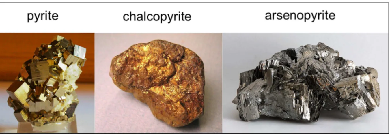
Figure 1.2: Sulfide mineral samples. ... 4
Figure 1.3: Unit cell of pyrite. ... 6
Figure 1.4: Unit cell of chalcopyrite. ... 6
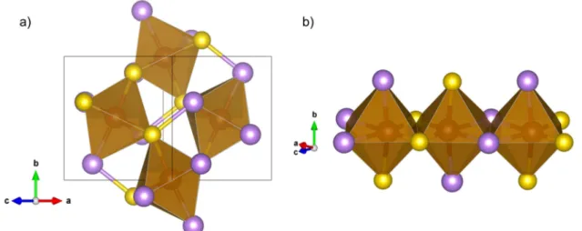
Figure 1.5: Unit cell of arsenopyrite ... 7
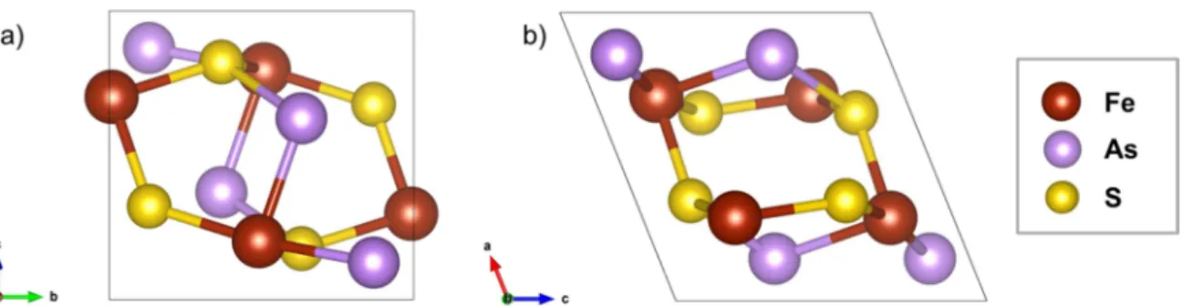
Figure 1.6: a) The As–S dianions coordinated octahedrally to Fe; b) neighboring octahedra share an edge. ... 8
Figure 1.7: Eh-pH diagram for Arsenopyrite. ... 11
Figure 1.8: Pyrite oxidation mechanism. ... 14
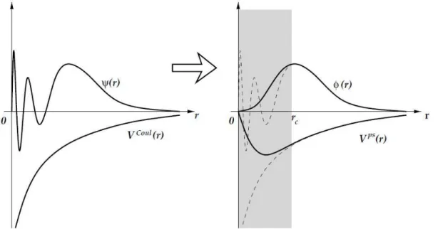
Figure 2.1: Representation of the construction of a pseudo wave function ϕ(r) and its respective pseudopotential Vps(r) from the wave function ψ(r) and the Coulomb potential VCoul(r). ... 28
Figure 2.2: a) Map of the trajectory vector
r for ethylene; b) Map of the trajectory vector
r showing the atomic interaction line. ... 31Figure 2.3: Scheme of a minimum energy path found by the NEB method. ... 35
Figure 2.4: a) Convergence test for the cutoff energy Ecut. b) Calculation time as a function of the cutoff energy. ... 37
Figure 2.5: a) K-points mesh convergence test.. ... 38
Figure 2.6: Magnetization test of bulk arsenopyrite. ... 40
Figure 3.1: Arsenopyrite monoclinic unit cell used in bulk calculations. a) view along a axis; b) view along b axis. ... 44
Figure 3.2: Structure of sulfides: a) pyrite; b) marcasite; c) loellingite; d) arsenopyrite. The filled circles represent the cations and the open ones the anions. From reference [9]. ... 47
Figure 3.3: Molecular orbital diagram of the Fe–Fe bond [9]. ... 47
Figure 3.4: Path through K-space for band structure calculation for a monoclinic cell suggested in reference [18]. ... 50
Figure 3.5: Band structure calculated for arsenopyrite. ... 51
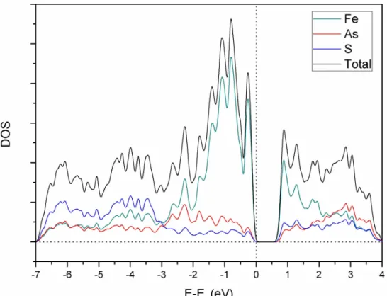
Figure 3.6: Total and projected DOS of the atoms of arsenopyrite plotted using a Gaussian width of 0.005 Ry. ... 52
Figure 3.7: Total and projected DOS over the atomic orbitals of arsenopyrite plotted using a Gaussian width of 0.005 Ry. ... 52
ix Figure 3.9: a) As–S dianions octahedrally coordinated to Fe. b) Neighbor octahedrons sharing one edge. Yellow atoms are sulfur, purple are arsenic and brown are iron. ... 53 Figure 3.10: Electron density map of the arsenopyrite (010) plane. Brown balls are iron atoms. 54 Figure 3.11: Bond critical points (BCP) in green, Ring critical points (RCP) in blue, and Cage critical points (CCP) in pink for arsenopyrite QTAIM analysis. Atoms in brown are iron, in yellow are sulfur and in purple are arsenic. ... 56 Figure 3.12: a) Bond critical points and b) Ring critical points in detail. Yellow atoms are sulfur, purple are arsenic and brown are iron. ... 57 Figure 3.13: ELF of arsenopyrite As–S bond. brown is iron, purple is arsenic and yellow is sulfur. ... 58 Figure 3.14: ELF of the arsenopyrite (010) plane. Iron atoms are represented by brown spheres. ... 58 Figure 3.15: Surface Types according to Tasker. Adapted from [39]. ... 61 Figure 3.16: Arsenopyrite cleavage planes. Iron is in brown, sulfur in yellow and arsenic in
x Figure 4.6: Adsorption of a water molecule to the sulfur site on arsenopyrite (101) surface. a) Side view and b) Top view. Brown is iron, purple is arsenic, yellow is sulfur, red is oxygen and white hydrogen. ... 80 Figure 4.7: Adsorption of a water molecule to two neighbor arsenic atoms on arsenopyrite (101) surface. a) Side view and b) Top view. Brown is iron, purple is arsenic, yellow is sulfur, red is oxygen and white hydrogen. ... 81 Figure 4.8: Adsorption of a water molecule to two neighbor sulfur atoms on arsenopyrite (101) surface. a) Side view and b) Top view. Brown is iron, purple is arsenic, yellow is sulfur, red is oxygen and white hydrogen. ... 81 Figure 4.9: Adsorption of a water molecule to two neighbor arsenic and sulfur atoms on
arsenopyrite (101) surface. a) Side view and b) Top view. Brown is iron, purple is arsenic, yellow is sulfur, red is oxygen and white hydrogen. ... 82 Figure 4.10: Arsenopyrite (001) surface: a) top view; b) side view. ... 83 Figure 4.11: First layer of atoms in arsenopyrite (001) surface showing the two different Fe sites: Fe1, more exposed, and Fe2, less exposed. a) top view; b) side view. ... 83 Figure 4.12: Adsorption of one water molecule on two different Fe sites on the (001) arsenopyrite surface: a) Fe1 top view; b) Fe1 side view; c) Fe2 top view; Fe2 side view. Yellow atoms are sulfur, purple are arsenic, brown are iron, red are oxygen and white are hydrogen. ... 85 Figure 4.13: Adsorption of one water molecule on Fe sites on (100), (110) and (101) arsenopyrite surfaces: a) (100) top view; b) (100) side view; c) (110) top view; d) (110) side view; e) (101) top view; (101) side view. Yellow atoms are sulfur, purple are arsenic, brown are iron, red are oxygen and white are hydrogen. ... 87 Figure 4.14: Adsorption of one water molecule on the As and S sites of (001) arsenopyrite
xi Figure 4.20: Optimized structure of 4 water molecules adsorbed on arsenopyrite (001) surface. Yellow atoms are sulfur, purple are arsenic, brown are iron, red are oxygen and white are
hydrogen. ... 94 Figure 4.21: Dissociative adsorption of HCl on arsenopyrite (001) surface: a), b) on Fe1 and S sites c), d) on Fe1 and As sites. Yellow atoms are sulfur, purple are arsenic, brown are iron, green are chlorine and white are hydrogen. ... 96 Figure 4.22: Figure 9: Dissociative adsorption of HCl on Fe2 site of arsenopyrite (001) surface. Yellow atoms are sulfur, purple are arsenic, brown are iron, green are chlorine and white are hydrogen. ... 97 Figure 4.23: Most stable structures for adsorption of H2SO4 on arsenopyrite (001) surface: a),b) H2SO4 MM; c),d) H+, HSO4- BB, e), f) 2H+, SO42- BB on Fe sites; g), h) 2H+, SO42- BB on Fe and As sites. Yellow atoms are sulfur, purple are arsenic, brown are iron, red are oxygen and white are hydrogen. ... 100 Figure 4.24: Adsorption of H2SO4 on arsenopyrite (001) surface: a), b) 2H+, SO42- BM; c), d) H+, HSO4- MM. Yellow atoms are sulfur, purple are arsenic, brown are iron, red are oxygen and white are hydrogen. ... 101 Figure 4.25: Adsorption of H2SO4 on Fe and As atom of arsenopyrite (001) surface: a), b) 2H+, SO42-; c), d) H+, HSO4-. Yellow atoms are sulfur, purple are arsenic, brown are iron, red are oxygen and white are hydrogen. ... 102 Figure 4.26: Figure S7: Adsorption of HSO4- on Fe and S atom of arsenopyrite (001) surface: a), b) S1; c), d) S2. Yellow atoms are sulfur, purple are arsenic, brown are iron, red are oxygen and white are hydrogen. ... 103 Figure 4.27: Adsorption of H2SO4 on arsenopyrite (001) surface: a), b) SO42- BB; c),d) HSO4 -MM. Yellow atoms are sulfur, purple are arsenic, brown are iron, red are oxygen and white are hydrogen. ... 104 Figure 4.28: Adsorption of H2O on pyrite (001) surface. a),b) molecular adsorption; c),d)
xii Figure 5.5: Dissociative adsorption of O2 on the arsenopyrite (001) surface: a) Fe1 and Fe2 sites top view, b) Fe1 and Fe2 sites side view; c) Fe2 and Fe3 sites top view, d) Fe2 and Fe3 sites side view ; e) Fe3 and Fe4 sites top view, f) Fe3 and Fe4 sites side view; g) Fe4 and Fe1 sites
top view, h) Fe4 and Fe1 sites side view ... 119
Figure 5.6: Dissociative adsorption of O2 on the arsenopyrite (001) surface: a) Fe1 and Fe2 sites top view, b) Fe1 and Fe2 sites side view; c) Fe2 and Fe3 sites top view, d) Fe2 and Fe3 sites side view... 120
Figure 5.7: Dissociative adsorption of O2 on the arsenopyrite (001) surface: a) Fe1 and Fe4 sites top view, b) Fe1 and Fe4 sites side view; c) equivalent Fe3 sites top view, d) equivalent Fe3 sites side view; e) Fe3 and Fe4 sites top view, f) Fe3 and Fe4 sites side view ... 121
Figure 5.8: Dissociative adsorption of O2 on the arsenopyrite (001) surface on As atoms. ... 122
Figure 5.9: Dissociative adsorption of O2 on the arsenopyrite (001) surface on S atoms ... 123
Figure 5.10: Dissociation of the O2 molecule on the arsenopyrite (001) surface.. ... 125
Figure 5.11: Energy barrier graph for the O2 dissociation on the arsenopyrite (001) surface. ... 125
Figure 5.12: Co-adsorption of water and dissociated oxygen to the arsenopyrite (001) surface. ... 127
Figure 5.13: Co-adsorption of water and oxygen end-on to the arsenopyrite (001) surface. ... 128
Figure 5.14: Substitution of one water molecule for an O2 molecule on the arsenopyrite (001) surface... 129
Figure 5.15: Substitution of two water molecules for an O2 molecule on the arsenopyrite (001) surface ... 129
Figure 5.16: Proposed oxidation reaction for the arsenopyrite (001) surface. ... 131
Figure 5.17: Minimum Energy Paths for the H donation reactions in the oxidation of arsenopyrite.. ... 132
Figure 5.18: OH groups bound to S atoms in the oxidation reaction of the arsenopyrite (001) surface... 133
Figure 5.19: Migration of the OH group from an Fe atom to an As atom on the arsenopyrite surface... 134
Figure 5.20: Reaction steps for the formation of AsO43- on the FeAsS (001) surface. ... 136
xiii
List of Tables
Table 2.1:Total energy variation with respect to the cutoff energy for the XC functionals PBE e
PW91. ... 37
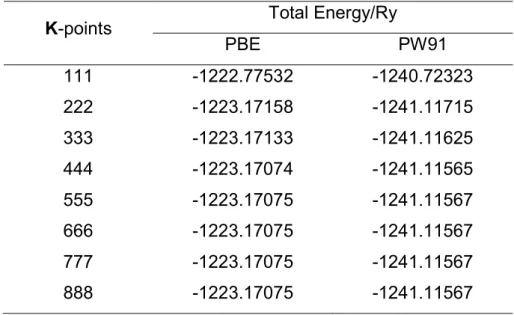
Table 2.2: K-points mesh test for two XC functionals: PBE and PW91. ... 39
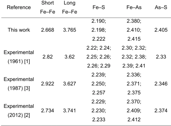
Table 3.1: Interatomic distances for arsenopyrite bulk. ... 45
Table 3.2: Crystallographic data of bulk arsenopyrite. ... 46
Table 3.3: Critical points in QTAIM analysis. ... 55
Table 3.4: Atomic charges in AIM analysis ... 59
Table 3.5: Calculated and experimental bulk moduli for arsenopyrite, marcasite and pyrite. ... 60
Table 3.6: Surfaces correspondence in C21/d and P21/c unit cells. ... 61
Table 3.7: Surface energies and coordination numbers of surface atoms of different arsenopyrite surfaces. ... 63
Table 3.8: Comparison of Arsenopyrite and Pyrite’s surface energies. ... 67
Table 4.1: Atomic distances on arsenopyrite (001) surface as effect of water adsorption. ... 85
Table 4.2: Dissociative adsorption of water on arsenopyrite surface. ... 90
Table 4.3: Adsorption Energy of water calculated for different sulfide surfaces. ... 95
Table 4.4: Atomic distances on arsenopyrite (001) surface as effect of HCl adsorption. ... 97
Table 4.5: Adsorption energies of aqueous H2SO4 species in different positions on arsenopyrite (001) surface in kcal mol-1. ... 99
Table 4.6: Atomic distances on arsenopyrite (001) surface as effect of H2SO4 adsorption ... 99
Table 4.7: Dissociative adsorption of SO42- and HSO4- on arsenopyrite surface. ... 99
Table 4.8: Adsorption Energy of leaching agents calculated for different sulfide surfaces. ... 105
Table 5.1: O–O distances calculated for different O2 bond types. ... 114
Table 5.2: Adsorption energies and M–O distances for the O2 adsorption on the arsenopyrite (001) surface. ... 123
Table 5.3: Atomic distances on the arsenopyrite (001) surface as effect of oxygen adsorption. 126 Table 5.4:Adsorption energies and Fe–O distances in O2 and H2O adsorption on the arsenopyrite (001) surface. ... 128
xiv
Resumo
Os sulfetos minerais, dentre os quais a arsenopirita, estão comumente presentes em rejeitos de mineração e são responsáveis pela drenagem ácida de rocha (DAR). Nesse fenômeno, o mineral sofre oxidação em contato com oxigênio e água e produz um líquido contendo ácido e metais pesados capaz de contaminar o ambiente. A mitigação do problema da DAR e o desenvolvimento de contramedidas são facilitados pela compreensão de seu mecanismo. Por isso o conhecimento do processo de oxidação de sulfetos é muito importante, especialmente da arsenopirita, que, além da DAR, também libera o elemento tóxico arsênio em sua oxidação. Esse processo ainda não é completamente compreendido, pois as reações envolvidas são muito complexas, o que leva a uma falta de consenso na literatura. Nesse contexto, cálculos de primeiros princípios podem contribuir para o entendimento e interpretação dos dados experimentais e na elucidação do mecanismo de reação ao nível molecular.
Nesta tese foram investigadas por cálculos DFT as propriedades estruturais, eletrônicas e mecânicas da arsenopirita, suas ligações químicas e suas superfícies de clivagem, além da adsorção de agentes lixiviantes e seu mecanismo de oxidação. Evidências mostrando que não há ligação Fe–Fe na arsenopirita e que a ligação As–S possui forte caráter covalente foram fornecidas. Também foi mostrado que essa forte ligação As–S é improvável de ser quebrada durante a formação da superfície. As clivagens preferenciais são previstas de ocorrer nos planos (001), (010) e (100). A superfície (001) é a que apresenta menor energia de formação e possui propriedades magnéticas. A adsorção de água sobre essa superfície é mais favorável na forma molecular e sobre os sítios de Fe mais expostos. O ácido clorídrico favorece a adsorção dissociativa e o ácido sulfúrico tem preferência pela forma molecular monodentada, enquanto o bissulfato se adsorve em coordenação bidentada binuclear. A adsorção de oxigênio sobre o sítio de Fe é bastante favorável, principalmente de forma dissociativa, quando é formada uma ponte Fe–O–As. Nesse processo tanto os átomos de Fe quanto de As são oxidados. A co-adsorção de oxigênio e água sobre a superfície também é estável. Um mecanismo de oxidação da arsenopirita foi proposto em que o oxigênio se adsorve sobre a superfície e recebe átomos de H de moléculas de água, que reduzem novamente o Fe. Nesse mecanismo, o arsênio é o elemento que se oxida preferencialmente, o que explica os dados experimentais disponíveis. Cálculos para a pirita também foram realizados para comparar com a arsenopirita, com o objetivo de prover uma compreensão significativa sobre ambos os sistemas.
xv
Abstract – Chemical Properties of Arsenopyrite, Adsorption of
Leaching Agents and its Oxidation Mechanism from DFT
Calculations
Sulfide minerals, among them arsenopyrite, are commonly present in mining tailings and are responsible for acid rock drainage (ARD). In this phenomenon, the mineral undergoes oxidation in contact with oxygen and water and produces a liquid containing acid and heavy metals capable of contaminating the environment. Mitigation of the ARD problem and the development of countermeasures are facilitated by understanding its mechanism. Therefore the knowledge of the sulfides oxidation process is very important, especially of arsenopyrite, which, in addition to ARD, also releases the toxic element arsenic by its oxidation. This process is not yet completely understood, since the reactions involved are very complex, which leads to a lack of consensus in the literature. In this context, first principle calculations can contribute to the understanding and interpretation of the experimental data in the elucidation of the reaction mechanism at a molecular level.
In this thesis, the structural, electronic and mechanical properties of arsenopyrite, its chemical bonds and its surfaces cleavage, as well as the adsorption of leaching agents and its oxidation mechanism have been investigated by DFT calculations. Evidences showing that there is no Fe–Fe bond in arsenopyrite and that the As–S bond has strong covalent character have been provided. It has been also shown that this strong As–S bond is unlikely to be broken during the surface formation. The preferential cleavages are predicted to occur in the planes (001), (010) and (100). The (001) surface is the one that presents the lowest surface energy, and has magnetic properties. The adsorption of water on this surface is most favorable in molecular form on the most exposed Fe sites. Hydrochloric acid favors dissociative adsorption and sulfuric acid prefers the molecular monodentate adsorption form, while bisulfate adsorbs in bidentate binuclear coordination. The adsorption of sulfate is predicted to be not favored. The adsorption of oxygen on the Fe site is very favorable, especially dissociatively, when a Fe–O–As bridge is formed. In this process, both Fe and As atoms are oxidized. The co-adsorption of oxygen and water on the surface is also stable. An oxidation mechanism for arsenopyrite has been proposed, in which the oxygen is adsorbed to the surface and receives H atoms from water molecules, which again reduces the Fe. In the proposed mechanism, arsenic is the element that preferentially oxidizes, which explains the available experimental data. Calculations for pyrite have also been performed and compared with arsenopyrite, aiming to provide significant insights about both systems.
1
1
Introduction
1.1
Sulfide Minerals
Sulfide minerals are considered the most important group of minerals of the earth's crust [1] for being the main natural sources of non-ferrous metals, such as copper, zinc and lead. These metals are softer acids that easily bind to the soft base S2-, while harder acids, such as iron, aluminum and chromium, prefer to form oxides.
The definition of sulfide mineral is very wide due to the great number of existing compounds and their different properties. However, it can be said that they are natural binary or ternary crystalline sulfur compounds or their synthetic analogues [2]. Although there are hundreds of known sulfide minerals, most of them are secondary minerals and only six exist in abundance to form rocks: pyrite (FeS2),
pyrrhotite (Fe1-xS2), galena (PbS), sphalerite (ZnS), chalcopyrite (CuFeS2) and
chalcocite (Cu2S) [1].
The metal sulfides – sulfides that have metals in their composition – have aroused the interest of the scientific community due to their potential applications as semiconductor materials. The use of these minerals in solar cells [3-5], solid batteries [6] and catalysis [7] has also been investigated.
Another interest in the study of sulfides came from the discovery of living organisms in hydrothermal systems on the ocean floor, whose metabolic pathway is chemosynthetic instead of photosynthetic [1]. These organisms use the surface of sulfide minerals as a catalyst for the synthesis of complex molecules necessary for life, which raised a theory of the origin of life from sulfide minerals [8, 9]. In fact, acetic acid has been synthesized from carbon monoxide (CO) and methanethiol (CH3SH) on sulfide surfaces [10] in conditions similar to the primitive terrestrial
atmosphere, which is thought to have existed prior to the origin of life.
2 these ores. In this process, the sulfides present in mining tailings, in contact with oxygen and environmental water, oxidize producing sulfuric acid. The generated solution acts as a leaching agent, in other words, as a mixture that solubilizes the solid constituent mineral, producing a liquid containing dissolved metals and sulfuric acid, which can contaminate soil and aquifers [11].
ARD is a spontaneous process that arises whenever a sulfide rock is exposed to the atmosphere and moisture, without special conditions. A classic example is the ARD that occurs in Rio Tinto, a river in Spain (Figure 1.1) which received this name because of its red color. Along the river there is a large deposit of pyrite, with a pH of the order of 2 and high concentrations of heavy metals. The ARD process in this region is lasting for about 50 centuries, since mining activities started there. The extreme local conditions pointed to environmental studies that simulate what might be found on planet Mars [12]. But the crucial cause of ARD is anthropogenic, mainly due to mining activity, which aggravates the problem because of the amount of sulfides exposed to the atmosphere [11]. For this reason, it is a concern for the state of Minas Gerais, Brazil, due to the intense mining activity in its territory, where a serious mining accident happened in the town of Mariana in 2015 with huge social, economic and environmental impacts.
3 Acid rock drainage can be accelerated in the presence of microorganisms of the species Acidithiobacillus ferrooxidans, Acidithiobacillus thiooxidans and Leptospirillum ferrooxidans [14], and this is difficult to control because each mine has different characteristics. There is no standard treatment and costs can be enormous [11]. In the United States, the costs for the mining industry to treat ARD are more than a million dollars a day [15] and around US$100 billion has been or will be spent in ARD remediation in the whole world [14]. The separation of pyrite from other economically important minerals by flotation and leaching methods also generates costs for the mining industry [15], which explains the interest in its study.
Pyrite (FeS2) is the most geologically important, widespread and abundant
sulfide on Earth. For this reason, it is the one that contributes the most to ARD. Other sulfides reported to also cause ARD are pyrrhotite, bornite, arsenopyrite, enargite, tetrahedrite, realgar, orpiment, stibnite, sphalerite, galena, chalcopyrite and covellite [16]. The name pyrite has Greek origin and means fire, probably due to the sparks it produces when struck against steel. It is popularly known as "fool's gold" because of its typical golden glow. This mineral has little economic value, but is often associated with more valuable minerals, as sphalerite, chalcopyrite, galena and gold [15]. It is used as a raw material in industrial production of sulfuric acid and as iron ore only in places where iron oxides are not available [17]. When exposed to oxygen and water, pyrite is oxidized to form sulfuric acid:
2 2
2 2 2 4
2FeS (s)7O (g)2H O(l)2Fe (aq)4SO (aq)4H(aq). (1.1)
If the environmental conditions are sufficiently oxidizing, the ferrous ion will oxidize to a ferric ion:
2 3
2 2
4Fe (aq)O (g)4H(aq)4Fe (aq)2H O(l). (1.2)
In pH between 2.3 and 3.5, the ferric ion will precipitate as Fe(OH)3, leaving
the medium even more acidic:
3
2 3
4 The Fe3+ ions that do not precipitate in the reaction of equation 1.3 can
oxidize pyrite again:
3 2 2
2 2 4
FeS (s)14Fe (aq)8H O(l)15Fe (aq)2SO (aq)16H(aq). (1.4)
Chalcopyrite (CuFeS2) is another sulfide that also contributes to ARD, though
economically important, because it is the main source of copper in the world. Copper is a metal widely used in the electric industry, electronics and construction [18]. Obtaining metallic copper from the ore is traditionally made by a pyrometallurgical route, wherein the mineral is concentrated, then smelted in an oxidizing atmosphere and, at last, copper is reduced by electrolysis [19]. However, when the concentration of copper in the ore is low, the hydrometallurgical route is applied. In this process, a low-cost chemical agent is used as a leaching agent to extract the Cu2+ ion in ore
piles, followed by electrochemical reduction thereof to metallic copper [19]. The problem of the hydrometallurgical route is the low leaching kinetics. The reaction stops at a certain point, which complicates its industrial use, and several attempts were made to explain this phenomenon [20].
Arsenopyrite (FeAsS) is the most common arsenic mineral in nature, and it can be found in many ore deposits. Because of its association with rocks containing gold, silver, copper and lead [17] and its little economic importance [21], arsenopyrite is a mineral commonly present in mining waste of these metals [22]. Although stable under reducing conditions, this mineral also generates acid rock drainage. An aggravating aspect is the release of arsenic – a toxic element – to the environment, whose mechanism is still not completely understood.
Figure 1.2 shows a picture of each of the sulfides described above.
5
1.2
Crystal Structure of Sulfides
The crystal structures of all common and many rare sulfide minerals are well known. They range from the simple crystal structure of salt, such as PbS, or structures involving anion-anion bonds, as CuS, to complex structures where there is uncertainty about the occupation sites and oxidation states of the atoms, as in tetrahedrites, economically important minerals [26] for being a source of copper and silver [27]. The electric and magnetic properties of these minerals are also more diverse than in any other group, ranging from diamagnetic insulators (ZnS), diamagnetic semiconductors (PbS), antiferromagnetic semiconductors (CuFeS2), to
antiferromagnetic and ferromagnetic conductors (Fe7S8 and Fe9S10) [26]. Therefore
these materials can have a nonzero electron density located between the cations and anions, forming chemical bonds, as well as delocalized electronic states, which makes describing the chemical bonds a challenging task [26]. Pauling [28] has observed that many sulfides have mostly covalent bonds, with a small ionic character. Moreover, it is common to observe deviations in the stoichiometric composition of sulfides, defects, impurities and association with other minerals.
Pyrite was one of the first solids analyzed by X-ray diffraction. It crystallizes in a cubic face-centered structure with space group Pa3, in a structure similar to NaCl, as shown in Figure 1.3, exchanging Na+ with Fe2+ and Cl- for the center of the S–S
bond in 2-2
S . The orientation of the S–S dimers in dumbbell shape is respectively parallel to the four diagonals of the cell body [29]. The sulfur atoms are coordinated to a S atom and three Fe atoms in a tetrahedral way, while the iron atoms are octahedrally coordinated to six S atoms. The unit cell of pyrite contains 4 FeS2
6 Figure 1.3: Unit cell of pyrite. The iron atoms are in brown color and the sulfur atoms in yellow.
Chalcopyrite crystallizes in a tetragonal system, with space group I d42 and 4 CuFeS2 formulas per unit cell, as shown in Figure 1.4. Each metal atom is
coordinated to four sulfur atoms forming a tetrahedron, whereas the sulfur atoms are bonded to two iron and two copper atoms also in tetrahedral shape. In chalcopyrite the oxidation states of the atoms are (Cu1+)(Fe3+)(S
22-) [33]. The solid is an
antiferromagnetic semiconductor with alternating atomic layers of iron atoms with spin density up and down along the c direction [29]. Chalcopyrite has no preferred cleavage plane [31, 34].
7 Arsenopyrite has a monoclinic unit cell belonging to the space group P21/c
derived from marcasite (orthorhombic FeS2) [17] with 4 FeAsS formulas per unit cell,
as shown in Figure 1.5a-b. However, a refinement of the structure of arsenopyrite in the space group C21/d has also been performed [35], in a pseudo-orthorhombic unit
cell as shown in Figure 1.5c. Nevertheless the first refinement, in the P21/c group,
seems to be preferred by most authors [17, 36, 37]. Its structure contains arsenic and sulfur dianions (As–S) in the shape of dumbbells coordinated to iron in an octahedral way (Figure 1.6a). The adjacent octahedra in a row share one edge and their coordinations are related to each other by an inversion operation. This results in alternating short and long distances between cations (Figure 1.6b).
8 Figure 1.6: a) The As–S dianions coordinated octahedrally to Fe; b) neighboring octahedra share an edge. The iron atoms are in brown color, the arsenic atoms in purple, and the sulfur atoms in yellow.
Natural arsenopyrite has a composition ranging from FeAs0.9S1.1 to FeAs1.1S0.9
[36]. It is a diamagnetic semiconductor mineral [30]. About the cleavage plane, the literature informations do not agree with each other and vary among the (100) [38], (001) [39], (101) [17, 40] and (110) [27, 41] planes. Many of the published studies do not clearly define which unit cell was used as a reference, creating ambiguities in the definition of a preferential cleavage plane. Therefore, there may be discrepancies due to two different unit cells, since the cleavage plane changes accordingly.
1.3
Arsenopyrite and Acid Rock Drainage
In atmospheric conditions, arsenopyrite is a stable phase in reducing environments, but it is oxidized in the presence of common oxidants, such as ferric ion (Fe3+) and dissolved oxygen in aqueous media [22]. Under these conditions, the
oxidation of arsenopyrite has slow kinetics, which can be catalyzed by microorganisms present in the environment [22]. Its oxidation by weathering effects releases the chemical species H2SO4, H3AsO3 and H3AsO4 [21] to the environment.
9 release, as a result of arsenopyrite oxidation, increases environmental risk and can become a public health problem [42]. For this reason, understanding the kinetics and dissolution mechanisms of this mineral in different conditions is extremely important in environmental, social and economic ways [42].
Arsenic is a ubiquitous element found naturally in the atmosphere, soil, rocks, natural waters and organisms. However anthropogenic activity such as mining, fossil fuel burning, and the use of arsenic as a pesticide and herbicide in the past, besides its current use in wood preservation, can increase its availability in the environment [43]. Arsenic is often found in penta- and trivalent states, in which arsenite, As(III), is more toxic, more soluble and more mobile than arsenate, As(V) [44]. To humans, the toxicity of As(III) is related to its irreversible complexation with sulfhydryl (R–SH) groups present in enzymes and aminoacids, while As(V) competes with phosphate ions, interrupting the phosphorylation process [45]. Arsenate is more abundant under oxidizing conditions, while arsenite is prevalent in a reducing environment [42].
A study in the scope of INCT-Acqua investigated the possibility of contamination by arsenic in a community close to the mining area in the Brazilian town of Paracatu/MG [46]. The amount of As present in air, water, soil/dust and food was measured and, considering all sources, the calculated risk associated with arsenic exposure to the population was 0.44 μg/kg/day for adults. The maximum dose established by the WHO (BMDL0.5) is 3 μg/kg/day, higher than the dose found
in Paracatu. Still the environmental risks of mining waste cannot be neglected, requiring chemical procedures to immobilize arsenic and mitigate environmental problems.
In the gold mine Cuiabá, located in the town of Sabará/MG, arsenopyrite present in the sulfide concentrate is volatilized in the form of arsenic trioxide (As2O3)
in the calcination process, then As is adsorbed to water in the gas scrubbers and finally removed by coprecipitation/adsorption with the addition of oxide or iron salts and lime [47]. The waste of this process is arranged in tailings dams located near the metallurgical plant.
10 degraded. Subsequently, anaerobic and aerobic processes naturally neutralize the water. Limestone drains are a series of tubes containing lime that neutralizes the acid drainage while the liquid flows within them. Water covers serve as a barrier to atmospheric oxygen, slowing down the oxidation process. In natural processes, the waste is dumped into water bodies containing limestone and a high buffering capacity that is able to neutralize it. Oxide minerals present in the system can avoid ARD because they are able to receive H+, buffering the medium and slowing down
the oxidation reaction [49]. In this case, tests are necessary to measure the ecological sensitivity of the environment. Active methods involve investments into technology and require the addition of bases, followed by a separation of solids and liquids to remove metal hydroxides and finally the disposal of the sludge. These processes are expensive and involve products that may require additional cleaning. Lastly, the use of bacteria and algae capable of decomposing sulfides has been the best solution from an economic and environmental point of view, but it is not always technically applicable [48].
Although there is no general pattern to predict ARD, the procedures commonly used in engineering do not deviate from Morin and Hutt’s wheel [14]. They are procedures done in practice in the field and laboratory, but the knowledge about sulfides oxidation mechanism is still scarce. This comprehension allows the definition of more efficient and effective processes to reduce the environmental problem of ARD, as well as the costs of waste treatment. This understanding is highly enriched from the study of the chemical reactivity of the sulfide surfaces, a subject which has been given attention to in the literature in recent years [50-52].
1.4
Arsenopyrite and the Chemical Reactivity of Sulfides
11 presents near-surface conditions. It indicates that arsenopyrite can be oxidized when exposed to the atmosphere.
Figure 1.7: Eh-pH diagram for Arsenopyrite. Reference [22] adapted from [53].
Experimental researches use techniques such as LEED (Low Energy Electron Diffraction), Infra-Red Spectroscopy, XPS (X-ray Photoelectron Spectroscopy) and Auger Spectroscopy, besides Cyclic Voltammetry and other methods, to study the chemical composition and reactivity of surfaces [1, 52]. In particular the use of high-intensity photon sources, together with synchrotron radiation, has increased the sensitivity of analysis regarding surfaces and the spatial resolution of existing techniques. The use of synchrotron radiation allowed for extending the application of X-ray Absorption Spectroscopy techniques such as EXAFS (Extended X-ray Absorption Fine Structure) and XANES (X-ray Absorption Near-Edge Structure), which can be used to investigate the environment of surface atoms of a particulate sample in contact with a fluid at a molecular scale [31].
12 reactions on a surface in real time [31]. However, these techniques cannot always reach only the surface atoms. In the LEED technique, for example, electrons can penetrate between 5 and 20 Å into the surface [31], in XPS, about six atomic monolayers for Fe analysis and 15 layers for As analysis [54], and in Auger Spectroscopy about 10 Å [55]. To get a molecular point of view of the reaction, precisely on the surface atoms, it is possible to combine experimental results with results obtained by computer simulation [56-58], in particular Density Functional Theory (DFT) methods. The increased computing capacity and the improvement of numerical methods and computer programs made systems such as sulfide minerals accessible for numerical calculations. Theoretical methods help in understanding the geometry of both solid and surface, and also its electronic, electrical and mechanical properties.
In order to understand the mechanism of chalcopyrite leaching, many experimental [59-62] and theoretical [34, 63-66] studies have been published. Also pyrite has been widely investigated by both experimental [15, 50, 67, 68] and theoretical [50, 69-74] techniques, besides two theoretical works about arsenic incorporation into pyrite [75, 76]. About arsenopyrite, there are several studies in the literature concerning the products formed in its oxidation in different media [21, 22, 38, 77-81] and one theoretical study about the electronic characteristic of arsenopyrite (110) surface [41]. But since the natural samples used in these studies may vary in composition and the solutions formed in the process are too complex, as well as the reactions involved, the results found for products, kinetics and mechanism of arsenopyrite oxidation are not always in agreement, and a lack of consensus is observed [22].
Buckley and Walker [77], as well as Mikhlin et al. [81], suggested a depletion in Fe and As on arsenopyrite surface after acid treatment. Costa et al. [82] observed elemental S when arsenopyrite was reacted in acid. However Richardson and Vaughan [55] found the opposite: a surface enriched in Fe and As after reaction with H2SO4. Nesbitt and Muir [79] reported the absence of S on arsenopyrite surface
13 observed that As is the most rapidly oxidized element under these conditions. Nesbitt et al. [21] and Schaufuss et al. [83] agreed that As is more readily oxidized than Fe, and S, while McKibben et al.’s [84] results show that Fe is dissolved quicker than As. Walker et al. [85] proposed that the rate-determining step of the oxidation reaction in FeAsS is the attachment of oxygen from water to As and S species, while Corkhill et al. [22] suggested that it could be the transference of electrons to the oxidant agent. Nesbitt et al. [21] also observed the same type of chemical species in air-oxidation and water-oxidation of arsenopyrite, however greater proportions of oxide species were found by water-oxidation, especially S-oxides, which indicates that the oxidation process is more intense in this medium. McKibben et al. [84] found that arsenopyrite dissolution is three to four times faster than pyrite and Blanchard et al. [76] noticed that the presence of arsenic in pyrite could accelerate its dissolution, and consequently the ARD.
The observed differences might originate from the variety of media the studies have been performed in. Therefore, more information about arsenopyrite and its chemical reactivity is necessary. In this context, first-principles calculations emerge as a tool providing information about the surface reactivity of arsenopyrite and insights about its oxidation mechanism. One advantage of theoretical studies is that the results can be carried out in more controlled situations than what can be done experimentally. The understanding of the kinetics and the mechanism of dissolution of this material in different conditions is essential for assessing the stability of the arsenic-containing tailings and the development of more efficient processes to control its remobilization with great environmental, social and economic consequences.
A proposed mechanism for the oxidation reaction of pyrite was studied by Sit and coworkers using the DFT method [73]. In this study, the proposed mechanism starts at the dissociative adsorption of O2 on two adjacent Fe atoms on the (100)
surface of pyrite, forming the species Fe4+=O2-. Simultaneously, an incoming water
14 and the reaction repeats until the surface S is completely oxidized to SO42- and all
proposed steps are energetically favorable. Yet another proposed mechanism, possibly more favorable, was made by Duarte et al. [52], which takes into account the formation of more common chemical species and is diagrammed in Figure 1.8. For the other sulfides, there is no theoretical study in the literature considering the oxidation mechanism.
Figure 1.8: Pyrite oxidation mechanism. From reference [52].
15 breakthrough in this field and to envisage more efficient strategies to mitigate the ARD in the mining regions rich in arsenopyrite.
In the next chapter, the fundamentals of the methodology used in this work are reviewed. The following chapters will be concerned with the structure and electronic properties of arsenopyrite and its cleavage surfaces and with the adsorption of water and other leaching agents. A proposal for the oxidation mechanism in the presence of oxygen and water is presented. In the final considerations, we highlight the main achievements and the perspectives of the field. We advance that the oxidation mechanism is closely related to the pyrite oxidation mechanism which we have also investigated [74].
1.5
References
1. Vaughan, D.J., Sulfide Mineralogy and Geochemistry: Introduction and Overview.
Reviews in Mineralogy and Geochemistry, 2006. 61(1): p. 147.
2. Vaughan, D.J. and J.R. Craig, Mineral chemistry of metal sulfides. Cambridge earth science series. 1978, Cambridge Eng. ; New York: Cambridge University Press.
3. Ennaoui, A., et al., Photoactive Synthetic Polycrystalline Pyrite ( FeS2 ) Journal of the
Electrochemical Society, 1985. 132(7): p. 1579-1582.
4. Ennaoui, A., et al., Photoelectrochemistry of Highly Quantum Efficient Single-Crystalline n-FeS2 (Pyrite). Journal of the Electrochemical Society, 1986. 133(1): p.
97-106.
5. Ennaoui, A. and H. Tributsch, Iron sulphide solar cells. Solar Cells, 1984. 13(2): p. 197-200.
6. Wang, S.S. and R.N. Seefurth, Electrochemical Studies of FeS2 Electrodes in Various
Sulfide-Containing Molten Salts. Journal of The Electrochemical Society, 1987.
134(3): p. 530-535.
7. Cody, G.D., et al., Assaying the catalytic potential of transition metal sulfides for abiotic carbon fixation. Geochimica et Cosmochimica Acta, 2004. 68(10): p. 2185-2196.
8. Wächtershäuser, G., Before enzymes and templates: theory of surface metabolism.
Microbiological Reviews, 1988. 52(4): p. 452-484.
9. Russell, M.J., A.J. Hall, and A.P. Gize, Pyrite and the origin of life. Nature, 1990.
344(6265): p. 387-387.
10. Huber, C. and G. Wächtershäuser, Activated Acetic Acid by Carbon Fixation on (Fe,Ni)S Under Primordial Conditions. Science, 1997. 276(5310): p. 245-247.
16
12. Fernández-Remolar, D.C., et al., The Río Tinto Basin, Spain: Mineralogy, sedimentary geobiology, and implications for interpretation of outcrop rocks at Meridiani Planum, Mars. Earth and Planetary Science Letters, 2005. 240(1): p. 149-167.
13. 02/14/2014]; Available from:
http://upload.wikimedia.org/wikipedia/commons/thumb/9/91/Riotintoagua.jpg/20 0px-Riotintoagua.jpg.
14. Parbhakar-Fox, A. and B.G. Lottermoser, A critical review of acid rock drainage prediction methods and practices. Minerals Engineering, 2015. 82: p. 107-124.
15. Chandra, A.P. and A.R. Gerson, The mechanisms of pyrite oxidation and leaching: A fundamental perspective. Surface Science Reports, 2010. 65(9): p. 293-315.
16. The Environmental Geochemistry of Mineral Deposits. Part A: Processes, Techniques, and Health Issues Part B: Case Studies and Research Topics, ed. G.S. Plumlee, M.J. Logsdon, and L.F. Filipek. 1996: Society of Economic Geologists. 588.
17. Klein, C., C.S. Hurlbut, and J.D. Dana, Manual of mineralogy : (after James D. Dana). 21st ed. 1999, New York: J. Wiley.
18. Informações e Análises da Economia Mineral Brasileira Available from: http://www.ibram.org.br/sites/1300/1382/00001145.pdf.
19. Davenport, W.G., et al., Extractive Metallurgy of Copper. 2002: Elsevier Science. 20. Córdoba, E.M., et al., Leaching of chalcopyrite with ferric ion. Part I: General aspects.
Hydrometallurgy, 2008. 93(3–4): p. 81-87.
21. Nesbitt, H.W., I.J. Muir, and A.R. Prarr, Oxidation of arsenopyrite by air and air-saturated, distilled water, and implications for mechanism of oxidation. Geochimica et Cosmochimica Acta, 1995. 59(9): p. 1773-1786.
22. Corkhill, C.L. and D.J. Vaughan, Arsenopyrite oxidation – A review. Applied Geochemistry, 2009. 24(12): p. 2342-2361.
23. SHB. 01/03/2014]; Available from: http://www.mineral-s.com/imagenes/pirita5-8599.gif.
24. Weller, R. 2009 01/03/2014]; Available from:
http://skywalker.cochise.edu/wellerr/mineral/chalcopyrite/6chalcopyrite98.jpg.
25. Harrison, J. 2009 01/03/2014]; Available from:
http://upload.wikimedia.org/wikipedia/commons/thumb/c/c4/Arsenopyrite%2C_Pa
nasqueira_Mine%2C_Portugal.jpg/800px-Arsenopyrite%2C_Panasqueira_Mine%2C_Portugal.jpg.
26. Vaughan, D.J., U. Becker, and K. Wright, Sulphide mineral surfaces: theory and experiment. International Journal of Mineral Processing, 1997. 51(1-4): p. 1-14. 27. Dana, E.S. and W.E. Ford, A Text-book of Mineralogy: With an Extended Treatise on
Crystallography and Physical Mineralogy. 4 ed. 1966, New York: Wiley.
28. Pauling, L., Crystallography and Chemical Bonding in Sulfide Minerals. Mineralogical Society of America Special Paper, 1970. 3: p. 125-131.
29. Von Oertzen, G.U., S.L. Harmer, and W.M. Skinner, XPS and ab initio calculation of surface states of sulfide minerals: pyrite, chalcopyrite and molybdenite. Molecular Simulation, 2006. 32(15): p. 1207-1212.
30. Pearce, C.I., R.A.D. Pattrick, and D.J. Vaughan, Electrical and Magnetic Properties of Sulfides. Reviews in Mineralogy and Geochemistry, 2006. 61(1): p. 127-180.
17
32. Chase, M.W., et al., Janaf Thermochemical Tables - 3rd Edition .2. Journal of Physical and Chemical Reference Data, 1985. 14: p. 927-1856.
33. Mikhlin, Y.L., et al., Spectroscopic and electrochemical characterization of the surface layers of chalcopyrite (CuFeS2) reacted in acidic solutions. Applied Surface Science,
2004. 225(1–4): p. 395-409.
34. de Oliveira, C., et al., Reconstruction of the Chalcopyrite Surfaces—A DFT Study. The Journal of Physical Chemistry C, 2012. 116(10): p. 6357-6366.
35. Fuess, H., et al., Crystal-Structure Refinement and Electron-Microscopy of Arsenopyrite. Zeitschrift Fur Kristallographie, 1987. 179(1-4): p. 335-346.
36. Morimoto, N. and L.A. Clark, Arsenopyrite Crystal-Chemical Relations. American Mineralogist, 1961. 46(11-2): p. 1448-1469.
37. Bindi, L., et al., Stoichiometric Arsenopyrite, FeAsS, from La Roche-Balue Quarry, Loire-Atlantique, France: Crystal Structure and Mossbauer Study. The Canadian Mineralogist, 2012. 50(2): p. 471-479.
38. Corkhill, C.L., et al., The oxidative dissolution of arsenopyrite (FeAsS) and enargite (Cu3AsS4) by Leptospirillum ferrooxidans. Geochimica et Cosmochimica Acta, 2008.
72(23): p. 5616-5633.
39. Ford, M. and C.C. Ferguson, Cleavage Strain in the Variscan Fold Belt, County Cork, Ireland, Estimated from Stretched Arsenopyrite Rosettes. Journal of Structural Geology, 1985. 7(2): p. 217-223.
40. Wolff, G.A. and J.D. Broder, Cleavage and the Identification of Minerals. American Mineralogist, 1960. 45(11-2): p. 1230-1242.
41. Corkhill, C.L., M.C. Warren, and D.J. Vaughan, Investigation of the electronic and geometric structures of the (110) surfaces of arsenopyrite (FeAsS) and enargite (Cu3AsS4). Mineralogical Magazine, 2011. 75(1): p. 45-63.
42. Cheng, H.F., et al., Geochemical processes controlling fate and transport of arsenic in acid mine drainage (AMD) and natural systems. Journal of Hazardous Materials, 2009. 165(1-3): p. 13-26.
43. Smedley, P.L. and D.G. Kinniburgh, A review of the source, behaviour and distribution of arsenic in natural waters. Applied Geochemistry, 2002. 17(5): p. 517-568.
44. Mandal, B.K. and K.T. Suzuki, Arsenic round the world: a review. Talanta, 2002. 58(1): p. 201-235.
45. Ladeira, A.C.Q., et al., Chemical speciation and its importance for the mineral extraction processes and environmental remediation. Cadernos Temáticos de Química Nova na Escola, 2014. 8: p. 18-23.
46. Ciminelli, V.S.T., et al., Annual Activity Report. 2012, INCT - Acqua: Belo Horizonte. p. 30-34.
47. Pantuzzo, F.L. and V.S.T. Ciminelli, Arsenic association and stability in long-term disposed arsenic residues. Water Research, 2010. 44(19): p. 5631-5640.
48. Hilson, G. and B. Murck, Progress toward pollution prevention and waste minimization in the North American gold mining industry. Journal of Cleaner Production, 2001. 9(5): p. 405-415.
49. Brough, C.P., et al., The process mineralogy of mine wastes. Minerals Engineering, 2013. 52: p. 125-135.
18
51. Chen, J.-h., et al., DFT calculation on relaxation and electronic structure of sulfide minerals surfaces in presence of H2O molecule. Journal of Central South University,
2014. 21(10): p. 3945-3954.
52. de Lima, G.F., H. Avelino de Abreu, and H. Anderson Duarte, Chapter 6 Surface reactivity of the sulfide minerals, in Chemical Modelling: Volume 10. 2014, The Royal Society of Chemistry. p. 153-182.
53. Craw, D., D. Falconer, and J.H. Youngson, Environmental arsenopyrite stability and dissolution: theory, experiment, and field observations. Chemical Geology, 2003.
199(1–2): p. 71-82.
54. Jones, R.A. and H.W. Nesbitt, XPS evidence for Fe and As oxidation states and electronic states in loellingite (FeAs2). American Mineralogist, 2002. 87(11-12): p.
1692-1698.
55. Richardson, S. and D.J. Vaughan, Arsenopyrite - a Spectroscopic Investigation of Altered Surfaces. Mineralogical Magazine, 1989. 53(370): p. 223-229.
56. Ladeira, A.C.Q., et al., Mechanism of anion retention from EXAFS and density functional calculations: arsenic (V) adsorbed on gibbsite. Geochimica et Cosmochimica Acta, 2001. 65(8): p. 1211-1217.
57. Teixeira, M.C., et al., Raman spectroscopy and DFT calculations of As(III) complexation with a cysteine-rich biomaterial. Journal of Colloid and Interface Science, 2007. 315(1): p. 128-134.
58. Duarte, G., et al., As(III) immobilization on gibbsite: Investigation of the complexation mechanism by combining EXAFS analyses and DFT calculations. Geochimica et Cosmochimica Acta, 2012. 83: p. 205-216.
59. Munoz, P.B., J.D. Miller, and M.E. Wadsworth, Reaction mechanism for the acid ferric sulfate leaching of chalcopyrite. Metallurgical Transactions B, 1979. 10(2): p. 149-158.
60. Parker, A., et al., An X-ray photoelectron spectroscopy study of the mechanism of oxidative dissolution of chalcopyrite. Hydrometallurgy, 2003. 71(1–2): p. 265-276. 61. Hackl, R.P., et al., Passivation of chalcopyrite during oxidative leaching in sulfate
media. Hydrometallurgy, 1995. 39(1–3): p. 25-48.
62. Baláž, P., et al., Combined chemical and bacterial leaching of ultrafine ground chalcopyrite. Hydrometallurgy, 1996. 42(2): p. 237-244.
63. Edelbro, R., Å. Sandström, and J. Paul, Full potential calculations on the electron bandstructures of Sphalerite, Pyrite and Chalcopyrite. Applied Surface Science, 2003.
206(1–4): p. 300-313.
64. de Oliveira, C. and H.A. Duarte, Disulphide and metal sulphide formation on the reconstructed (001) surface of chalcopyrite: A DFT study. Applied Surface Science, 2010. 257(4): p. 1319-1324.
65. de Lima, G.F., et al., Water Adsorption on the Reconstructed (001) Chalcopyrite Surfaces. The Journal of Physical Chemistry C, 2011. 115(21): p. 10709-10717.
66. de Lima, G.F., et al., Sulfuric and hydrochloric acid adsorption on the reconstructed sulfur terminated (001) chalcopyrite surface. International Journal of Quantum Chemistry, 2012. 112(19): p. 3216-3222.
19
68. Rimstidt, J.D. and D.J. Vaughan, Pyrite oxidation: A state-of-the-art assessment of the reaction mechanism. Geochimica et Cosmochimica Acta, 2003. 67(5): p. 873-880. 69. Hung, A., et al., Density-functional theory studies of pyrite FeS2(100) and (110)
surfaces. Surface Science, 2002. 513(3): p. 511-524.
70. Hung, A., et al., Density-functional theory studies of pyrite FeS2 (111) and (210)
surfaces. Surface Science, 2002. 520(1–2): p. 111-119.
71. Stirling, A., M. Bernasconi, and M. Parrinello, Ab initio simulation of water interaction with the (100) surface of pyrite. Journal of Chemical Physics, 2003. 118(19): p. 8917-8926.
72. Stirling, A., M. Bernasconi, and M. Parrinello, Defective pyrite (100) surface: An ab initio study. Physical Review B, 2007. 75(16): p. 165406.
73. Sit, P.H.L., M.H. Cohen, and A. Selloni, Interaction of Oxygen and Water with the (100) Surface of Pyrite: Mechanism of Sulfur Oxidation. Journal of Physical Chemistry Letters, 2012. 3(17): p. 2409-2414.
74. Dos Santos, E.C., J.C. de Mendonça Silva, and H.A. Duarte, Pyrite Oxidation Mechanism by Oxygen in Aqueous Medium. The Journal of Physical Chemistry C, 2016. 120(5): p. 2760-2768.
75. Reich, M. and U. Becker, First-principles calculations of the thermodynamic mixing properties of arsenic incorporation into pyrite and marcasite. Chemical Geology, 2006. 225(3-4): p. 278-290.
76. Blanchard, M., et al., Arsenic incorporation into FeS2 pyrite and its influence on
dissolution: A DFT study. Geochimica et Cosmochimica Acta, 2007. 71(3): p. 624-630. 77. Buckley, A.N. and G.W. Walker, The Surface-Composition of Arsenopyrite Exposed to
Oxidizing Environments. Applied Surface Science, 1988. 35(2): p. 227-240.
78. Fernandez, P.G., H.G. Linge, and M.W. Wadsley, Oxidation of arsenopyrite (FeAsS) in acid .1. Reactivity of arsenopyrite. Journal of Applied Electrochemistry, 1996. 26(6): p. 575-583.
79. Nesbitt, H.W. and I.J. Muir, Oxidation states and speciation of secondary products on pyrite and arsenopyrite reacted with mine waste waters and air. Mineralogy and Petrology, 1998. 62(1-2): p. 123-144.
80. Almeida, C.M.V.B. and B.F. Giannetti, Electrochemical study of arsenopyrite weathering. Physical Chemistry Chemical Physics, 2003. 5(3): p. 604-610.
81. Mikhlin, Y.L., A.S. Romanchenko, and I.P. Asanov, Oxidation of arsenopyrite and deposition of gold on the oxidized surfaces: A scanning probe microscopy, tunneling spectroscopy and XPS study. Geochimica et Cosmochimica Acta, 2006. 70(19): p. 4874-4888.
82. Costa, M.C., A.M. Botelho do Rego, and L.M. Abrantes, Characterization of a natural and an electro-oxidized arsenopyrite: a study on electrochemical and X-ray photoelectron spectroscopy. International Journal of Mineral Processing, 2002. 65(2): p. 83-108.
83. Schaufuss, A.G., et al., Reactivity of surface sites on fractured arsenopyrite (FeAsS) toward oxygen. American Mineralogist, 2000. 85(11-12): p. 1754-1766.
20
2
Methodology
In this chapter the main theoretical concepts and methodologies used in this work are presented. Not all details of the theoretical concepts and the mathematical and computational implementation of the methods used are covered. Further discussion can be found in the cited references.
2.1
Density Functional Theory (DFT)
Density Functional Theory (DFT) has become an alternative to ab initio methods – Hartree-Fock (HF) and post-HF (perturbation methods, coupled-cluster and configuration interaction) – for calculating the electronic structure of solid and molecular systems in the last decades [1]. The great advantage of DFT is the lower computational cost when compared to the usual theoretical methods including electron correlation. Therefore DFT is nowadays the most used method to investigate solids and surfaces, which often requires the simulation of many atoms with a large number of electrons.
21
ρ r dr = N
. (2.1)By determining the number of electrons in a system from ρ(r), one can also find the external potential in which the electrons move and consequently the Hamiltonian of the system. As the energy can be calculated by the Hamiltonian, it is determined by the electron density. The electronic ground state energy E0 is a
functional of the electron density of the ground state:
0 0 0
Ε = Ε ρ . (2.2)
The second theorem of Hohenberg and Kohn follows the energy variational principle, in which the total energy of a system, calculated from an approximate electron density, will always be greater than or equal to the exact energy. Therefore, the electron density that describes the system is the one that minimizes the energy E0. The ground state energy is then calculated using electron density functionals that
contain the electronic interactions, according to
0 0 0 ne 0 ee 0
E ρ =T ρ +V ρ +V ρ , (2.3)
where T is the kinetic energy, Vne is the energy related to the electron-nucleus
interaction and Vee is the electron-electron interaction. The latter term is composed of
terms for the Coulomb interaction, exchange interaction and electron correlation. The following year, in 1965, Kohn and Sham [8] developed a methodology that allows the DFT calculations, proposing a method to get around the problem of having to use an exact electron density functional for the kinetic energy. According to the interpretation of Levy [9], the Kohn-Sham method uses a reference system in which the electrons do not interact with each other and have the same electron density as the actual system. In this way, the residual part of the kinetic energy, which differs from the real system, is also included in the exchange and correlation functional.
22 2
1
ˆ ( )
2 ef
H V r , (2.4)
where the first term on the right is the kinetic energy operator and the second one the effective potential. Then the Schrödinger equation for one electron can be formulated as
2
1 ( ) ( )
2 Vef iKS r i iKS r
. (2.5)
Here, ΨiKS (r) is the wave function for each electron i in the position r, and εi is
the respective eigenvalue of the wave function. The electron density of the reference system can be calculated according to equation 2.6, where N is the number of electrons in the system:
2 ( ) N KS( )
i i
r r
. (2.6)The effective potential is calculated according to:
1
1 ( )
( ) ( ) ( )
ef r xc
V r V r dr V r
r r
, (2.7)where V(r) is the external potential due to nuclear charges. The second term corresponds to the Coulomb potential and the last term to the exchange and correlation potential, defined by
( ) xc xc
E
V r
. (2.8)
23 the effective potential, and then equation 2.5 is solved to find a new set of Ψi (r), with
which a new density ρ(r) is found by equation 2.6 and the procedure is repeated until convergence. The total energy can be determined through
1 2
1 2
1 2
( ) ( )
1 ( ) ( )
2
i r r xc xc
E dr dr E r V r dr
r r
. (2.9)As well as the calculation of energy and the electron density of the system, the Kohn-Sham method enables the calculation of all other system properties. Through the theorems of Hohenberg and Kohn, it was proved that there is an exact electron density functional that permits the calculation of the system energy. However, this functional is unknown because its calculation requires the determination of the exchange and correlation (XC) potential, whose analytical expression is unknown. For this reason, the exchange and correlation functionals commonly used are derivative from approximations that tend to provide the most accurate results possible.
The Local Density Approximation (LDA) functional is based on the theory of the homogeneous electron gas, in which the exchange and correlation energy of a system with electron density ρ(r) is considered to be equal to the exchange and correlation energy of a homogeneous electron gas with the same electron density. As the electron density in real systems is not homogeneous, it was proposed to include the electron density gradient ρ(r) in the exchange-correlation functional. The set of these more elaborate functionals is called Generalized Gradient Approximation (GGA). Other types of functionals developed are hybrids functionals, which include part of the exact exchange energy of the Hartree-Fock method in the DFT exchange-correlation functional, besides meta-GGA functionals, which extend the expansion of the generalized gradient to second order derivatives (Laplacians).
24 periodically in space to describe the whole crystal. Thus corresponding lattice points from different unit cells are geometrically equivalent and are related by a lattice translation operation (translational invariance). Note that there may exist further symmetry operations that relate different lattice points within the unit cell itself. Because of this characteristic, the potential energy of the solid system has the same periodicity of the lattice, according to
V r + R =V r , (2.10)
where R is the translation vector that leads from one unit cell to the next. Under these conditions the Bloch theorem states that every electronic wave function ψk(r)
can be written as the product of a plane wave function (eik.r) with wave vector k and
another function that has the same periodicity as the lattice (uk (r)), expressed in
ik r
k r = e u rk
, (2.11)
where
k k
u r + R = u r . (2.12)
The wave vector k classifies the eigenvalues and eigenfunctions of the monoelectronic states in a periodic lattice. Bloch's theorem is interpreted as a boundary condition for the solutions of the Schrödinger equation in a periodic potential. This theorem changes the problem of calculating an infinite number of wave functions in a solid to the calculation of a finite number of wave functions in infinite k points. However, the wave functions in nearby k points will be very similar. So it is possible to represent the wave functions of a region in k space by functions at a single k point (discretization of k space).
25 properties of this region is sufficient to characterize the whole crystal. Precise methods have been developed for the selection of electronic states in special k points, for example the method of Monkhorst-Pack [11]. The greater the number of k points chosen, the better can the calculation accuracy be expected, but the computational effort is increasing as well. Therefore the number of considered k points in each spatial direction should be carefully evaluated before starting a calculation of periodic systems.
2.2
Plane Waves (PW)
The solution to the Kohn-Sham equations can be obtained by an expansion of the wave function in a set of basis functions. In calculations of periodic systems, the periodic part of the wave function can be expanded using a basis set consisting of a discrete set of plane waves whose wave vectors G are vectors of the reciprocal crystal lattice [12].
iG rk k,G
G
u r =
c e . (2.13)In this way, each electronic wave function can be written as a sum of plane waves (PW),
i(k+G) r
k k+G
G
= C e
. (2.14)In agreement with this equation, for each k point, the wave function can be written as an infinite sum of G vectors, i.e., it would take an infinite set of plane waves for the expansion to be exact. However, plane waves related to large G values have small Ck+G coefficients and contribute little to the total sum, therefore it is
![Figure 1.1: Rio Tinto in Spain, where acid rock drainage occurs for centuries [13].](https://thumb-eu.123doks.com/thumbv2/123dok_br/15731440.124279/21.892.191.704.696.1078/figure-rio-tinto-spain-acid-drainage-occurs-centuries.webp)
![Figure 2.3: Scheme of a minimum energy path found by the NEB method. From reference [27]](https://thumb-eu.123doks.com/thumbv2/123dok_br/15731440.124279/54.892.124.774.721.984/figure-scheme-minimum-energy-path-neb-method-reference.webp)
![Figure 3.4: Path through K-space for band structure calculation for a monoclinic cell suggested in reference [18]](https://thumb-eu.123doks.com/thumbv2/123dok_br/15731440.124279/69.892.278.622.367.681/figure-path-space-structure-calculation-monoclinic-suggested-reference.webp)