Immunoexpression of Integrins
in Ameloblastoma, Adenomatoid
Odontogenic Tumor, and Human
Tooth Germs
Emanuel Sávio de Souza Andrade, PhD,
Márcia Cristina da Costa Miguel, PhD, Roseana de Almeida Freitas, PhD,
Leão Pereira Pinto, PhD, and Lélia Batista de Souza, PhD
or multicystic ameloblastoma, unicystic ameloblastoma, and peripheral ameloblastoma.4The latest classifica-tion of the World Health Organizaclassifica-tion5 considers desmoplastic ameloblastoma to be a variant with spe-cific clinical, radiographic, and histological features. Among the intraosseus types, unicystic ameloblas-tomas show a more favorable biological behavior than the solid type.6 Adenomatoid odontogenic tumors (AOTs) have been shown to be the third most frequent odontogenic tumor.3,7 These tumors are characterized by a slow and progressive growth, showing a favorable prognosis in most cases.5
Extracellular matrix (ECM) proteins have been demonstrated to play an important role in epithelial– mesenchymal interactions in odontogenic tumors.8,9 Many types of cells are unable to proliferate in the absence of an anchor to an extracellular substrate.10 This binding to EMC proteins is mediated by integrins, including in tumors,11and studies have demonstrated a correlation between the expression of integrins and the biological behavior of some tumors.12-14
Introduction
O
dontogenic tumors comprise a complex group of lesions characterized by diverse bio-logical behaviors and histobio-logical types that present, as observed for odontogenesis, various inductive interactions between the epithelium and the ectomesenchyme.1Ameloblastomas are the most common epithelial odontogenic tumors2and the second most frequent neoplasms among odontogenic tumors.3 Based on clinical behavior, histopathology, and prognosis, three types of ameloblastomas can be distinguished—solid The expression of integrins α2β1, α3β1, and α5β1 in 30 ameloblastomas (20 solid and 10 unicystic tumors), 12 adenomatoid odontogenic tumors (AOTs), and 5 human tooth germs in different stages of odontogenesis was analyzed. The distribution, location, pattern, and intensity of immunohistochemical expression were evalu-ated. Intensity was analyzed using scores (0 = absence, 1 =weak staining, and 2 =strong staining). No difference in the immunoexpression of the integrins was observed between solid and unicystic ameloblastomas. When these two ameloblastoma types were pooled into a single group,
the following significant differences were found: immu-noexpression of integrin α2β1 was stronger in ameloblas-tomas than in AOTs and tooth germs, and the expression of integrin α5β1 was stronger in ameloblastomas than in AOTs. The lack of detection of integrin α3β1 in tooth germs and its detection in the odontogenic tumors stud-ied suggest that this integrin might be used as a marker of neoplastic transformation in odontogenic tissues.
Keywords: ameloblastoma; adenomatoid odontogenic
tumor; human tooth germs; integrins
From the Department of Oral Pathology, Dentistry School, University of Pernambuco, Pernambuco (ESA) and the Department of Oral Pathology, Dentistry School, Federal University of Rio Grande do Norte, Rio Grande do Norte (MCM, RAF, LPP, LBS), Brazil.
Address correspondence to: Lélia Batista de Souza, PhD, Av. Senador Salgado Filho, 1787, Lagoa Nova, Natal-Rio Grande do Norte, Brasil, CEP-59056-000; e-mail: leliasouza@dod.ufrn.br.
Integrins are transmembrane receptors formed by the noncovalent association of two glycoprotein subunits (an α subunit and a β subunit), which modulate cell–cell and cell–matrix binding and have been implicated in the growth, adhesion, migration, proliferation, apoptosis, and morphology of cells.15,16 The objective of the present study was to analyze the immunohistochemical expression of integrins α2β1, α3β1, and α5β1 in ameloblastomas, AOTs, and human tooth germs to better understand the role of these integrins in cellular events and in the cell–matrix interaction in odontogenic tumors and during odontogenesis.
Material and Methods
The following cases were selected: 30 ameloblastomas (20 solid and 10 unicystic tumors) and 12 AOTs obtained from the archives of the Laboratory of Oral Pathology, Dental School, Federal University of Rio Grande do Norte (UFRN) and 5 fetal tooth germs in different stages of odontogenesis (one in the dental lamina stage, two in the bud stage, one in the cap stage, and one in the late bell stage in which the initial stages of dentin deposition is observed) obtained from the archives of Laboratório de Patologia e Citopatologia Ltda., Aracaju, Sergipe, Brazil. Among the 20 solid ameloblastoma cases, the predominant histological types were the plexiform type in 6 cases, the follicular type in 4 cases, the basal cell type in 1 case, the desmo-plastic type in 1 case, and cases concomitantly exhibit-ing more than one histological pattern: follicular and acanthomatous (3 cases), desmoplastic and plexiform (1 case), desmoplastic and follicular (1 case), follicular and granular (1 case), plexiform and follicular (1 case), and 1 case showing a combination of the plexiform, acanthomatous, and basal cell patterns. The unicystic ameloblastoma cases were selected according to the criteria proposed by Philipsen and Reichart.4The pres-ent study was approved by the Research Ethics Committee of UFRN.
The paraffin-embedded specimens were cut into 3-µm-thick sections, and the sections were submitted to immunohistochemistry by the streptavidin–biotin– peroxidase method optimized with the Dako CSA system (catalyzed signal amplification system for mouse primary antibodies) and incubated with the follow-ing primary antibodies for 60 minutes: anti-α2β1 integrin (clone BHA2,1, Chemicon, Temeluca, Calif; diluted 1:1000 in Tris–HCl), anti-α3β1 integrin (clone M-KD102, Chemicon; diluted 1:500 in Tris–HCl),
and anti-α5β1 integrin (clone JBS5, Chemicon; diluted 1:1000 in Tris–HCl).
Antigen retrieval was carried out in 0.5% pepsin, pH 1.8, at 37°C for 30 minutes. Before incubation with the primary antibodies, the sections were incu-bated with 1% bovine serum albumin and 5% fetal bovine serum in Tris–HCl, pH 7.4, for 60 minutes to block reactions with nonspecific tissue proteins. After incubation with the primary antibodies, the speci-mens were washed twice in 1% Tween 20 solution for 5 minutes each. Next, the sections were incubated for 15 minutes with the sequential reagents of the Dako CSA kit, with the specimens being washed twice in 1% Tween 20 solution between steps. The reaction was developed using diaminobenzidine (Sigma, St. Louis, Mo) as the chromogen, and the sections were counterstained with Mayer’s hematoxylin. Specimens in which the primary antibody was replaced with 1% bovine serum albumin in Tris–HCl were used as neg-ative controls.
For immunohistochemical analysis, the distribu-tion, locadistribu-tion, pattern, and intensity of immunohisto-chemical staining were evaluated. Staining intensity was analyzed by selecting a cut-off value per specimen for each marker and scored on a scale from 0 to 2, where 0 =absence of staining, 1 =weak staining, and 2 =strong staining, because of which nonparametric tests were performed. To compare independent groups the Kruskal–Wallis test was applied, followed by Dunn’s multiple comparison posttest, because the first test showed a significant difference. In addition, the comparison between the two clinicopathologic types of ameloblastoma (solid and unicystic) was done by means of the Mann–Whitney test. The level of sig-nificance was established at 95% (P<.05).
Results
of integrin α3β1 being observed in this pattern. Acanthomatous ameloblastomas showed strong immunoreactivity mainly to integrin α5β1 in cells undergoing squamous metaplasia (Figure 3). The desmoplastic and basal cell patterns exhibited pre-dominantly strong staining both at intercellular con-tacts and at the epithelial–mesenchymal interface. Strong immunoreactivity to integrins α2β1 and α5β1 and weak reactivity to integrin α3β1 were observed in one tumor of the granular cell type, with the immunohistochemical staining being more evi-dent in cuboidal cells at the periphery of epithelial nests and scarce in central granular cells. Analyzing solid ameloblastomas in general, no significant dif-ference in staining intensity was observed among the integrins studied (Tables 1 and 2).
In unicystic ameloblastomas, immunohisto-chemical staining occurred in a diffuse manner at the epithelial–connective tissue interface and at intercellular contacts in all layers of the epithelial component, with a linear and granular expression pattern for integrins α2β1 and α5β1 (Figure 4) and a focal and granular pattern for integrin α3β1. In these ameloblastomas, a significant difference in the intensity of immunohistochemical expression was observed between integrins α3β1 and α5β1, with stronger staining being detected for the latter (Tables 1 and 2).
No significant difference in the intensity of inte-grin expression was observed between solid and uni-cystic ameloblastomas (Table 3).
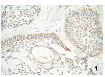
In AOT specimens, immunohistochemical stain-ing tended to show a focal distribution for integrins Figure 1. Diffuse labeling for integrin α2β1 in the central and peripheral cells of the follicles in solid ameloblastoma (strepta-vidin–biotin, magnification ×400).
Figure 2. Focal labeling for integrin α3β1 in the follicular type of ameloblastoma (streptavidin–biotin, magnification ×400).
Figure 3. Strong immunoreactivity to integrin α5β1 in cells undergoing squamous metaplasia in acanthomatous ameloblas-tomas (streptavidin–biotin, magnification ×400).
Figure 4. Diffuse immunohistochemical labeling for integrin
α2β1 and α3β1 (Figure 5) and a diffuse distribution for integrin α5β1 (Figure 6). In solid areas and cords, staining was observed at intercellular contacts, con-ferring a reticular pattern. In duct-like structures, granular or linear expression was observed at the luminal pole of cells in some areas and a tendency
toward bipolar expression in others. In these tumors, the immunostaining intensity did not differ signifi-cantly among the integrins studied (Tables 1 and 2). No immunohistochemical expression of integrin α3β1 was observed in tooth germs. Expression of integrin α2β1 was only detected in the late bell 280 International Journal of Surgical Pathology/ Vol. 16, No. 3, July 2008
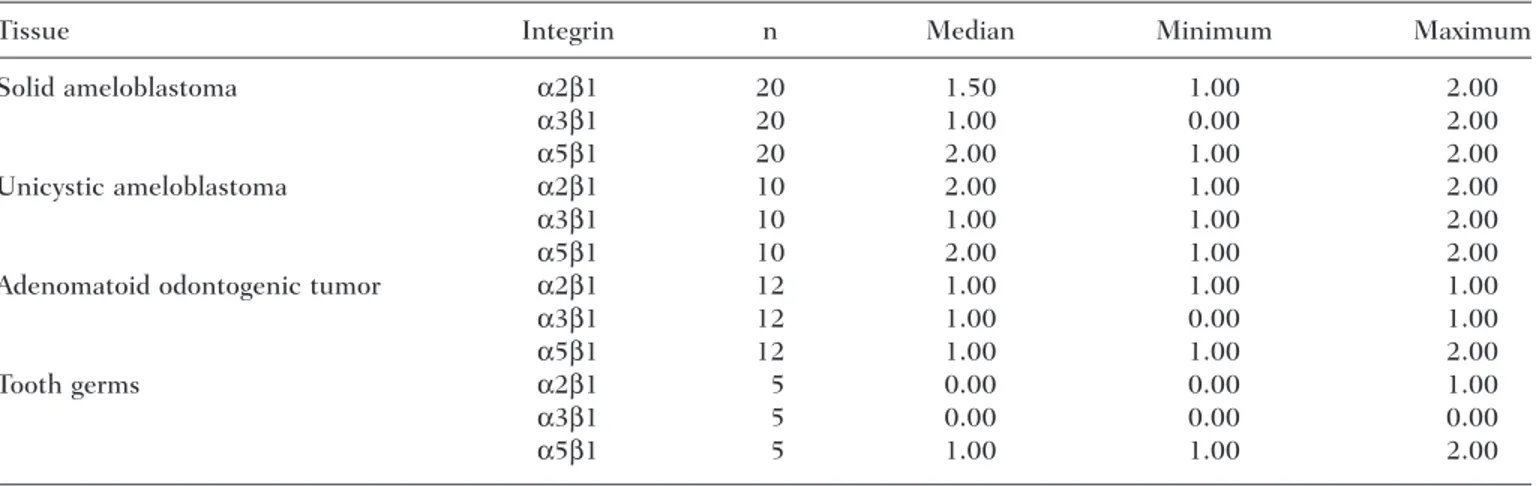
Table 1. Distribution and Variation in the Intensity of the Immunohistochemical
Expression of Integrins in the Cases Studied
Tissue Integrin n Median Minimum Maximum
Solid ameloblastoma α2β1 20 1.50 1.00 2.00
α3β1 20 1.00 0.00 2.00
α5β1 20 2.00 1.00 2.00
Unicystic ameloblastoma α2β1 10 2.00 1.00 2.00
α3β1 10 1.00 1.00 2.00
α5β1 10 2.00 1.00 2.00
Adenomatoid odontogenic tumor α2β1 12 1.00 1.00 1.00
α3β1 12 1.00 0.00 1.00
α5β1 12 1.00 1.00 2.00
Tooth germs α2β1 5 0.00 0.00 1.00
α3β1 5 0.00 0.00 0.00
α5β1 5 1.00 1.00 2.00
Table 2. Nonparametric Analysis of the Differences Observed Among the Integrins Studied
Tissue Integrin n Mean of Ranks Sum of Ranks Kruskal–Wallis KW Pvalue
Solid ameloblastoma α2β1 20 31.75 635.0 4.216 .1215
α3β1 20 25.05 501.0
α5β1 20 34.70 694.0
Unicystic ameloblastoma α2β1 10 17.50a,b 175.0 8.136 .0171
α3β1 10 10.00a 100.0
α5β1 10 19.00b 190.0
Adenomatoid odontogenic tumor α2β1 12 18.00 216.0 4.595 .1005
α3β1 12 16.58 199.0
α5β1 12 20.92 251.0
Tooth germs α2β1 5 6.30a 31.50 11.089 .0039
α3β1 5 5.00a 25.00
α5β1 5 12.70b 63.50
Note: KW and Pvalues for the Kruskal–Wallis test. Letters “a” and “b” express the results obtained with Dunn’s posttest, considering
P=.05.
Table 3. Immunoexpression of Integrins in the Different Types of Ameloblastoma
Integrin Ameloblastoma n Mean of Ranks Sum of Ranks Mann–Whitney U Pvalue
α2β1 Solid 20 14.5 290.0 80.0 .3840
Unicystic 10 17.5 175.0
α3β1 Solid 20 15.8 316.0 94.0 .8049
Unicystic 10 14.9 149.0
α5β1 Solid 20 14.5 290.0 80.0 .3818
Unicystic 10 17.5 175.0
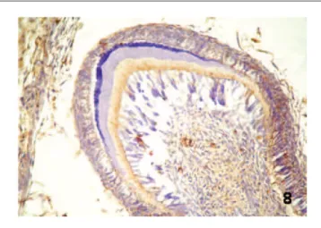
stage, with the observation of granular staining in the dentin layer being formed. Expression of inte-grin α5β1 was detected in all tooth germs. In the dental lamina stage, focal and granular staining was observed in some central epithelial cells (Figure 7). In the bud stage, granular and focal immunostaining was detected in the central cells of the epithelial component and in some condensed ectomesenchy-mal cells. In the cap stage, granular and diffuse staining was noted in the central cells of the enamel organ, at the interface with the basement membrane of the inner epithelium, and in some condensed ectomesenchymal cells. In the late bell stage, a gran-ular and diffuse expression pattern was observed in the cells of the inner epithelium (calcification area), stellate reticulum, outer epithelium, dentin layer,
and dental papilla (Figure 8). Comparison of the immunostaining intensity in this group showed a significant difference between integrins α2β1 and α3β1 and integrin α5β1, which exhibited a more intense staining (Tables 1 and 2).
Because no significant difference in the inten-sity of the immunohistochemical expression of the integrins studied was observed between solid and unicystic ameloblastomas (Table 3), these two clinical– pathological types were pooled into a single group for comparison with AOTs and tooth germs, which revealed a significant difference for all the inte-grins studied. A difference was observed between ameloblastomas and AOTs and between ameloblas-tomas and tooth germs for integrin α2β1, which showed a stronger immunostaining in ameloblastomas; Figure 5. Focal immunoreactivity for integrin α3β1 in the
inter-cellular contacts of adenomatoid odontogenic tumor (streptavidin– biotin, magnification ×400).
Figure 6. Diffuse labeling for integrin α5β1 in adenomatoid odontogenic tumor (streptavidin–biotin, magnification ×400).
Figure 8. Expression pattern for integrin α5β1 in late bell stage human tooth germs (streptavidin–biotin, magnification ×400).
between ameloblastomas and tooth germs and between AOTs and tooth germs for integrin α3β1, whose expression was stronger in tumors; and between ameloblastomas and AOTs for integrin α5β1, which exhibited a stronger staining in ameloblastomas (Tables 4 and 5).
Discussion
The binding of cells to ECM proteins mediated by integrins can lead to changes in the cytoplasmic domains of these proteins, which are associated with different postbinding events such as cell differentia-tion, migradifferentia-tion, proliferadifferentia-tion, and gene induction.17
Various inductive interactions between the epithe-lium and the ectomesenchyme have been shown to occur both during odontogenesis and in odonto-genic tumors,1and studies have demonstrated a cor-relation between the expression of integrins and the biological behavior of some tumors.12-14 In view of these findings, we investigated the expression of
integrins α2β1, α3β1, and α5β1, known ligands for ECM proteins (collagen, laminin, and fibronectin), in ameloblastomas and AOTs, two odontogenic tumors presenting different biological behaviors,4,6 and in tooth germs in different stages of odontogen-esis. Among ameloblastomas, integrin expression was compared between the solid and unicystic types because they are clinical types of the same tumor but with different biological behaviors.6 In view of the diversity and functional versatility of inte-grins15,16 and the lack of studies on these receptors in odontogenic tumors, immunohistochemical stain-ing for these molecules was analyzed in the present study in terms of location, distribution, expression pattern, and intensity to better understand the role of integrins in these tumors.
Focal distribution of integrin α3β1, an impor-tant receptor for laminin,18was observed in the two clinical–radiographic types of ameloblastoma (solid and unicystic). This finding agrees with the study of Souza et al,19 who demonstrated discontinuous 282 International Journal of Surgical Pathology/ Vol. 16, No. 3, July 2008
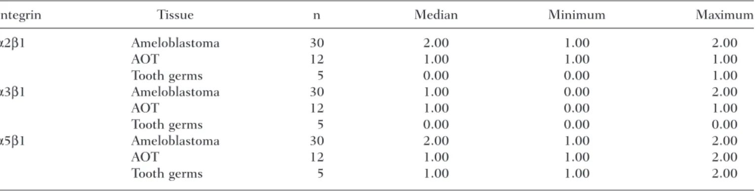
Table 4. Distribution and Variation in the Intensity of the Immunohistochemical Expression of Integrins
in Ameloblastomas, Adenomatoid Odontogenic Tumors (AOTs), and Tooth Germs
Integrin Tissue n Median Minimum Maximum
α2β1 Ameloblastoma 30 2.00 1.00 2.00
AOT 12 1.00 1.00 1.00
Tooth germs 5 0.00 0.00 1.00
α3β1 Ameloblastoma 30 1.00 0.00 2.00
AOT 12 1.00 0.00 1.00
Tooth germs 5 0.00 0.00 0.00
α5β1 Ameloblastoma 30 2.00 1.00 2.00
AOT 12 1.00 1.00 2.00
Tooth germs 5 1.00 1.00 2.00
Table 5. Nonparametric Analysis of the Differences Observed in Immunoexpression of Integrins in
Ameloblastomas, Adenomatoid Odontogenic Tumors (AOT), and Tooth Germs
Integrin Tissue n Mean of Ranks Sum of Ranks Kruskal–Wallis KW Pvalue
α2β1 Ameloblastoma 30 29.68a 890.0 21.648 <.0001
AOT 12 17.50b 210.0
Tooth germs 5 5.50b,c 27.5
α3β1 Ameloblastoma 30 28.18a 845.0 20.281 <.0001
AOT 12 21.87a 262.0
Tooth germs 5 4.00b 20.0
α5β1 Ameloblastoma 30 27.67a 830.0 8.661 .0132
AOT 12 15.92b 191.0
Tooth Germs 5 21.40a,b 107.0
laminin staining in the basement membrane of ameloblastomas. Integrin α3β1 plays an important role in the maintenance and integrity of the base-ment membrane,20and focal expression of this inte-grin by neoplastic ameloblastoma cells may therefore result in basement membrane defects or failure in the anchorage of these cells to the base-ment membrane, thus facilitating cell migration. However, the possibility that ameloblastoma cells use another integrin, such as integrin α6β4, for binding to laminin cannot be ruled out.
In unicystic ameloblastomas, a significant differ-ence in immunostaining intensity was observed between integrins α5β1 and α3β1, with the intensity being stronger for the former. However, no significant difference in intensity among the integrins studied was observed for solid ameloblastomas. Integrin α5β1 plays an important role in cell growth, migration, and tumorigenicity, and its expression inactivates cell pro-liferation genes such as c-fos, c-jun, and junB, decel-erating cell growth and reducing tumorigenicity.21 Taking together the staining intensity results obtained for the two clinical types of ameloblastoma and the focal expression of integrin α3β1, which, as discussed earlier, facilitates cell migration and tumor invasive-ness, we may suggest a compensatory mechanism of this invasiveness in unicystic ameloblastomas involv-ing a stronger expression of integrin α5β1, which was not observed in solid ameloblastomas. This fact might explain the more favorable biological behavior of uni-cystic ameloblastomas compared with the solid type. However, this hypothesis cannot be completely con-firmed in the present study because no significant dif-ference in the intensity of integrin staining was observed between solid and unicystic ameloblastomas (Table 3).
Some variations in the location and intensity of immunohistochemical staining were observed among the different histological types of solid ameloblas-tomas, which reflect the various functions attributed to these molecules.15,16
In the acanthomatous type, strong expression of integrin α5β1 was observed in cells undergoing squamous metaplasia, which were located in the center of tumor islands where the amount of ECM for anchorage of these cells is lower than at the periphery. Excessive production of this integrin in such a way that these molecules are not bound to ECM has been suggested as a mechanism of tumor suppression and promotion of apoptosis induced by the p16 gene.22 Studies on ameloblastomas have
shown the absence of staining for cyclin D1 and pro-tein p27, in addition to the presence of apoptotic bodies in cells undergoing squamous metaplasia.23,24 Thus, the presence of intensive staining for integrin α5β1 in the central cells of acanthomatous ameloblastomas observed in this study suggests a mechanism of terminal differentiation or apoptosis of these cells. Supporting this hypothesis, a signifi-cantly lower recurrence rate has been reported for acanthomatous ameloblastomas.6
In the present study, strong staining for all the integrins analyzed was observed in basal cell ameloblastomas. Studies using cell proliferation markers have demonstrated a higher proliferative rate in this histological type,23,25,26 as well as poor reactivity to apoptosis markers.24Furthermore, stud-ies have shown an integrin-mediated signaling pat-tern in cell cycle promotion and that the interaction of these molecules with growth factors regulates var-ious steps during the G1 to S phase transition of the cell cycle.27Our findings regarding integrins in basal cell ameloblastomas suggest an important role of these molecules in the survival and cell proliferation of this type of tumor.
In duct-like structures of AOTs, expression of the integrins studied showed a linear or granular pattern at the luminal pole of cells or even a bipolar pattern. The presence of these integrins in cells that surround duct-like structures might be related to the regulation of cell polarization in these structures because the participation of β1 integrins in this reg-ulation has been demonstrated in cell cultures.28In addition, expression of these integrins at the luminal pole of cells that surround these duct-like structures might be necessary for the activation of ECM pro-tein synthesis or for anchorage of these cells to ECM proteins whose secretion by AOT cells, associ-ated with their retention in the lumen of duct-like structures, has been demonstrated.29
fibronectin in the events of morphogenesis and dif-ferentiation during odontogenesis, as described in other studies.31,32 Furthermore, the wide distribution of integrin α5β1 during the various stages of odonto-genesis may facilitate cell migration in a fibronectin-rich matrix.
The intensity of staining for integrin α2β1 was significantly stronger in ameloblastomas than in AOTs or tooth germs (P<.0001). Integrin α2β1 acts as a receptor for collagen in platelets and epithelial cells and also activates collagen synthesis and induces the expression of collagenases and matrix metalloproteinases (MMPs) 1 and 13.33 MMP-1 binds to domain 1 (domain A) of the α2 subunit of integrin α2β1, and the association integrin–MMP locates the activity of ECM degradation in the same domain that is responsible for the plasma membrane adhesion, thus promoting cell migration.34 MMP-2 secretion is increased in human cervical tumor cells through a mechanism mediated by integrin α2β1.35 Furthermore, keratinocytes use this integrin for migration to collagen I,36 an ECM protein widely distributed in the stroma of ameloblastomas.9 A stronger expression of integrin α2β1 in ameloblas-tomas might therefore be associated with a greater cell migration in the collagen-rich matrix, or even with the formation of a complex between this inte-grin and MMPs whose presence and activity has been demonstrated in ameloblastomas,37a fact sug-gesting an important role of this integrin in the local invasiveness of these tumors.
The intensity of α3β1 integrin staining did not differ significantly between ameloblastomas and AOTs, whereas this integrin was not detected in tooth germs. Integrin α3β1 has been suggested to mediate the migration of tumor cells.38 Giannelli et al39demonstrated that this integrin is not present in normal hepatic parenchyma but is abundantly expressed in primary and metastatic hepatic carci-nomas, a fact that renders this integrin to be a potential marker for the development of hepatic car-cinomas and metastases. Similar results were obtained in the present study in which this integrin was detected in neoplastic odontogenic epithelium but not in tooth germs.
The intensity of α5β1 integrin staining was sig-nificantly stronger (P<.05) in ameloblastomas than in AOTs. Integrin α5β1 is widely expressed in tis-sues involved in the active deposition of pericellular fibronectin and in matrix remodeling.21 Extensive expression of fibronectin has been demonstrated during odontogenesis31,32 and in ameloblastomas.9
Thus, tooth germ cells and ameloblastomas use inte-grin α5β1 for migration and binding to a matrix rich in fibronectin. In addition, binding of integrin α5β1 to fibronectin increases the expression and secretion of MMPs involved in tumor invasion.21 Therefore, the extensive expression of fibronectin in ameloblas-tomas demonstrated in another study9 and the stronger expression of integrin α5β1 in these tumors than in AOTs suggest the participation of this inte-grin in the local invasiveness of ameloblastomas, probably mediated by a mechanism of induction of MMP synthesis.
In the present study, analyzing the immunohisto-chemical expression of integrins α2β1, α3β1, and α5β1 in ameloblastomas and AOTs (odontogenic tumors) and during the different stages of odontoge-nesis, important findings were obtained when evalu-ating the intensity of integrin staining, with the expression of these molecules tending to be stronger in ameloblastomas. The lack of detection of integrin α3β1 in tooth germs and its similar staining intensity in the odontogenic tumors studied suggest that this integrin might be used as a possible marker of neo-plastic transformation in odontogenic tissues. In addi-tion, blockade of integrins α2β1 and α5β1, which are more strongly expressed in ameloblastomas, may become an important strategy in the treatment or control of the local invasiveness of this tumor.
References
1. Neville BW, Damm DD, Allen CM, Bouquot, JE. Oral and Maxillofacial Pathology. 2nd ed. Philadelphia, PA: Saunders; 2002.
2. Melrose RJ. Benign epithelial odontogenic tumors. Semin Diagn Pathol. 1999;16:271-287.
3. Santos JN, Pereira Pinto L, de Figueredo CRLV, de Souza LB. Odontogenic tumors: analysis of 127 cases. Pesqui Odontol Bras. 2001;15:308-313.
4. Philipsen HP, Reichart PA. Unicystic ameloblastoma. A review of 193 cases from the literature. Oral Oncol. 1998; 34:317-325.
5. Barnes L, Eveson JW, Reichart P, Sidransky D. World Health Organization Classification of Tumours. Pathology and Genetics of Head and Neck Tumours. Lyon, France: IARC Press; 2005.
6. Reichart PA, Philipsen HP, Sonner S. Ameloblastoma: biological profile of 3677 cases. Oral Oncol. 1995;31B: 86-99.
Surg Oral Med Oral Pathol Oral Radiol Endod. 1997;48: 672-675.
8. Heikinheimo K, Morgan PR, Happonen RP, Stenman G, Virtanen I. Distribution of extracellular matrix pro-teins in odontogenic tumours and developing teeth. Virchows Arch B Cell Pathol. 1991;61:101-109.
9. Medeiros AMC. Expressão da fibronectina, tenascina e colágeno I em ameloblastomas e no tumor odontogênico adenomatóide [Tese, Doutorado em Patologia Oral]. Natal-RN, Brasil: Departamento de Odontologia, Universidade Federal do Rio Grande do Norte; 2001. 10. Yokosaki Y, Monis H, Chen J, Sheppard D. Differential
effects of the integrins α9β1, αvβ3, and αvβ6 on cell proliferative response to tenascin. Roles of the βsubunit extracellular and cytoplasmic domains. J Biol Chem. 1996;271:24144-24150.
11. Miles AJ, Knutson JR, Skubitz AP, Furcht LT, Mccarthy JB, Fields GB. A peptide model of basement membrane collagen α1 (IV) 531-543 binds the α3β1 integrin. J Biol Chem. 1995;270:29047-29050.
12. Van Waes C, Surh DM, Chen Z, et al. Increase in suprabasilar integrin adhesion molecule expression in human epidermal neoplasms accompanies increased proliferation occurring with immortalization and tumor progression. Cancer Res. 1995;55:5434-5444.
13. Franchi A, Santoro R, Paglierani M, Bondi R. Comparison of integrin α chain expression in benign and malignant salivary gland tumours. Oral Surg Oral Med Oral Pathol Oral Radiol Endod. 1997;83:588-595. 14. Benassi MS, Ragazzini P, Gamberi G, et al. Adhesion
molecules in high-grade soft tissue sarcomas: correlation to clinical outcome. Eur J Cancer. 1998;34:496-502. 15. Hynes RO. Integrins: a family of cell surface receptors.
Cell. 1987;48:549-554.
16. Hynes RO. Integrins: versatility, modulation, and signal-ing in cell adhesion. Cell. 1992;69:11-25.
17. Thomas GJ, Jones J, Speight PM. Integrins and oral cancer. Oral Oncol. 1997;33:381-388.
18. Kreidberg JA. Functions of α3β1 integrin. Curr Opin Cell Biol. 2000;12:548-555.
19. Souza LB, Sousa CVC, Pereira Pinto L. Estudo mor-fológico e imuno-histoquímico da membrana basal em diversos padrões histológicos do ameloblastoma. Rev Pós Grad. 2001;8:40-45.
20. Di Persio CM, Hodivala-Dilke KM, Jaenish R, Kreidberg JA, Hynes RO. α3β1 Integrin is required for normal development of the epidermal basement membrane. J Cell Biol. 1997;137:729-742.
21. Labat-Robert J. Fibronectin in malignancy. Effect of aging. Semin Cancer Biol. 2002;12:187-195.
22. Damsky CH, Ilic D. Integrin signaling: it’s where the action is. Curr Opin Cell Biol. 2002;14:594-602. 23. Kumamoto H, Kimi K, Ooya K. Detection of cell
cycle-related factors in ameloblastomas. J Oral Pathol Med. 2001;30:309-315.
24. Kumamoto H, Kimi K, Ooya K. Immunohistochemical analysis of apoptosis-related factors (Fas, Fas ligand, caspase-3 and single-stranded DNA) in ameloblastomas. J Oral Pathol Med. 2001;30:596-602.
25. Sandra F, Mitsuyasu T, Nakamura N, Shiratsuchi Y, Ohishi M. Immunohistochemical evaluation of PCNA and Ki-67 in ameloblastoma. Oral Oncol. 2001;37:193-198.
26. Sandra F, Nakamura N, Kanematsu T, Hirata M, Ohishi M. The role of MDM2 in the proliferative activity of ameloblastoma. Oral Oncol. 2002;38:153-157.
27. Coppolino MG, Deddhar S. Bi-directional signal trans-duction by integrin receptors. Int J Biochem Cell Biol. 2000;32:171-188.
28. Okajian GK, Schwimmer R. Regulation of epithelial cell surface polarity reversal by β1 integrins. J Cell Sci. 1994;107:561-576.
29. Murata M, Cheng J, Horino K, Hara K, Shimokawa H, Saku T. Enamel proteins and extracellular matrix mole-cules are co-localized in the pseudocystic stromal space of adenomatoid odontogenic tumor. J Oral Pathol Med. 2000;29:483-490.
30. Heikinheimo K, Salo T. Expression of basement mem-brane type IV collagen and type IV collagenases (MMP-2 and MMP-9) in human fetal teeth. J Dent Res. 1995; 74:1226-1234.
31. Thesleff I, Partanen AM, Vainio S. Epithelial-mesenchy-mal interactions in tooth morphogenesis: the roles of extracellular matrix, growth factors, and cell surface receptors. J Craniofac Genet Dev Biol. 1991;11:229-237. 32. Sawada T, Nanci A. Spatial distribution of enamel pro-teins and fibronectin at early stages of rat incisor tooth formation. Arch Oral Biol. 1995;40:1029-1038. 33. Nykvist P, Tu H, Ivaska J, Kapyla J, Pihlajaniemi T,
Heino J. Distinct recognition of collagen subtypes by
α1β1 and α2β1 integrins. J Biol Chem. 2000;275: 8255-8261.
34. Brown EJ. Integrin-associated proteins. Curr Opin Cell Biol. 2002; 14:603-607.
35. Mitra A, Chakrabarti J, Banerji A, Chatterjee A. Binding of α2 monoclonal antibody to human cervical tumor cell (SiHa) surface α2β1 integrin modulates MMP-2 activ-ity. Gynecol Oncol. 2004;94:33-39.
36. O’Toole EA. Extracellular matrix and keratinocyte migration. Clin Exp Dermatol. 2001;26:525-530. 37. Pinheiro JJ, Freitas VM, Moretti AI, Jorge AG, Jaeger
RG. Local invasiveness of ameloblastoma. Role played by matrix metalloproteinases and proliferative activity. Histopathology. 2004;45:65-72.
38. Hemler ME, Rutishauser U. Cell-to-cell contact and extracellular matrix. Curr Opin Cell Biol. 2000;12: 539-541.