SUPPURATIVE SIALADENITIS IN MANDIBULAR GLAND OF A DOG - ULTRASONOGRAPHIC ASPECT
Viviam Rocco Babicsak1 Vanesa Kutz Arruda2 Maria Claudia Lopes Silva2 Maria Lucia Gomes Lourenço3
Noeme Sousa Rocha3 Luiz Carlos Vulcano4 Maria Jaqueline Mamprim4
ABSTRACT
Sialoadenitis is a rare inflammatory disease of salivary glands, which may be caused by infection, trauma or auto-immune disease. Ultrasonography is an imaging method considered extremely advantageous in the identification and assessment of glandular diseases. In this report we describe the cervical sonographic findings in a dog diagnosed with suppurative sialadenitis in bilateral mandibular gland.
Keywords: ultrasonography, suppurative sialadenitis, dog.
SIALOADENITE SUPURATIVA EM GLÂNDULA MANDIBULAR DE UM CÃO – ASPECTO ULTRASSONOGRÁFICO
RESUMO
A sialoadenite é uma rara afecção inflamatória das glândulas salivares, que pode ter como causas infecção, trauma ou doença imunomediada. A ultrassonografia é considerada um método de imagem extremamente vantajoso na identificação e avaliação de doenças glandulares. Nesse relato descrevemos os achados ultrassonográficos cervicais em um cão diagnosticado com sialoadenite supurativa em glândula mandibular bilateral.
Palavras-chave: ultrassonografia, sialoadenite supurativa, cão.
SIALOADENITIS SUPURATIVA EN GLÁNDULA MANDIBULAR DE UN PERRO - ASPECTO ECOGRÁFICO
RESUMEN
La sialoadenitis es una enfermedad inflamatoria rara de las glándulas salivares, que puede ser provocada por infección, trauma e enfermedad autoinmune. El ultrasonido es un método imagenológico considerado valioso para la identificación y evaluación de las enfermedades glandulares. En este reporte se describen los hallazgos ecográficos cervicales en un perro con diagnóstico de sialoadenitis supurativa bilateral en las glándulas mandibulares.
Palabras clave: ecografía, sialoadenitis supurativa, perro.
1 Pós-graduanda do Departamento de Reprodução Animal e Radiologia Veterinária da Faculdade de Medicina Veterinária e
Zootecnia.
2
Residente do Departamento de Clínica Veterinária da Faculdade de Medicina Veterinária e Zootecnia.
3
Docente do Departamento de Clínica Veterinária da Faculdade de Medicina Veterinária e Zootecnia
4 Docente do Departamento de Reprodução Animal e Radiologia Veterinária da Faculdade de Medicina Veterinária e
INTRODUCTION
Sialoadenitis, despite being a rare disease in small animals, is the most common lesion that affects the salivary glands. This inflammatory condition can be caused by several factors, including trauma, infection and immune-mediated disease (1). The acute suppurative process commonly affects the parotid salivary gland, whereas the mandibular involvement is a rare condition. Generally, in human, suppurative sialoadenitis is caused by ascendant migration of agents from septic focus in oral cavity, such as chronic tonsillitis and dental sepsis, in patients with reduced salivary flow (2).
The patients affected by sialoadenitis show an enlargement of the salivary glands, which can be unilateral (3) or bilateral (4). Hypersialism and pain are also common clinical manifestations (4). Ultrasonography is an important imaging modality for evaluation of the salivary glands based on their accessibility, reliability and cost, besides having the advantage of not exposing the patient to radiation and not requiring anesthesia (5). In this case report we describe the cervical sonographic findings in a dog diagnosed with suppurative sialadenitis in bilateral mandibular gland.
CASE REPORT
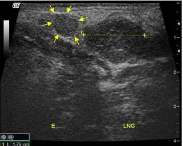
A 7-year-old male mixed breed dog showing apathy, anorexia and an enlargement of bilateral submandibular structures, which first began in the right portion, was seen at a veterinary hospital and submitted to a cervical ultrasonography in order to aid in the diagnosis determination. An enlargement (more pronounced in the right frame) and a decrease in the echogenicity of the mandibular lymph nodes were observed in the ultrasonographic examination, however, no significant changes in their contour and echotexture, which were regular and homogeneous, respectively, were identified (Figure 1). The mandibular salivary glands (Figure 1), which measured approximately 1.76 cm (left) and 2.46 cm (right) long, showed a slightly heterogeneous echotexture, a reduced echogenicity and a hypervascularization in color Doppler imaging.
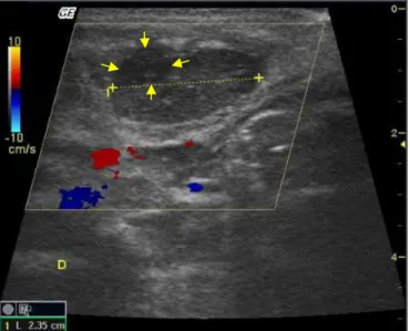
In the medial region of the right mandibular salivary gland was observed a hypoechogenic area with partially defined limits, measuring approximately 0.65 cm diameter, suggestive of abscess (Figure 2).
Figure 2. Transversal sonographic illustration demonstrating the hypoechogenic area (between arrows), suggestive of abscess, with partially defined limits, measuring approximately 0.65 cm diameter, in the medial region of the right mandibular salivary gland (between calipers).
Ultrasonography also allowed the verification of a hypoechoic linear structure, measuring 0.14 cm height, originating from the ventral portion of the bilateral mandibular salivary gland and extending to mandibular bone area, compatible with the dilated right mandibular salivary duct (Figure 3).
Figure 3. Transversal sonographic image showing the dilated right mandibular salivary duct (between arrows), a hypoechogenic linear structure, which measured 0.14 cm height, originated from the ventral region of the salivary gland (between calipers). The vascularization of the salivary gland can also be noted by color Doppler imaging.
DISCUSSION AND CONCLUSION
Sonographically, normal salivary glands are presented as slightly hypoechoic and homogeneous structures, however, in cases of acute sialoadenitis, the sonographic findings may be variable depending on the stage of evolution. Initially, the affected salivary glands only show a volumetric increase and a well defined limit. In a later stage, a decreased echogenicity and a heterogeneous echotexture can be visualized in the salivary glands, as observed in the case reported, indicating the existence of an edematous process in these structures (6).
Dilated salivary ducts can also be observed in ultrasonographic images, as identified in this case report, which can be filled with a purulent content in cases of suppurative sialadenitis. In salivary glands affected by this condition, hypoechogenic or anechogenic areas with slightly irregular and poorly defined limits can be identified within them, suggesting the existence of abscesses (7). In the animal reported a hypoechogenic area with similar sonographic appearance to the one mentioned above was observed in the right gland. Due to the sonographic aspect and the cytological alterations, an abscess was potentially suggested.
Other sonographic changes as hypervascularization of the salivary glands in color Doppler imaging and regional lymphadenopathy, both found in the animal reported, may also be observed in cases of sialoadenitis (6). In lymphadenopathy, the lymph nodes may be enlarged, rounded shaped and with a decreased echogenicity (8). In the patient reported, an enlargement of mandibular lymph nodes was verified since they presented a width greater than 0.5 cm (8). The measurements of the right and left lymph nodes were approximately 1.15 cm and 0.84 cm, respectively. The right structure presented a larger size, possibly due to an initial involvement of it since a right cervical enlargement was firstly observed.
Ultrasound can also aid in the diagnosis confirmation since, under ultrasonographic guidance, it is possible to collect material for cytological examination by fine needle aspiration or for histopathological examination by biopsy. This imaging modality allows the selection of the most representative area to collection be done. Ultrasound also enables the visualization of the tissue to be aspirated, and therefore, especially with the use of Color Doppler, it is used to ensure that there are no intervening vascular structures in the area selected to the collection procedure, in order to promote patient safety (9). In the animal reported, the material collection was safely performed, without any complication after the procedure.
Ultrasonography is an imaging modality of great importance for the diagnosis of salivary gland diseases such as sialoadenitis, as it enables the evaluation of glandular and extra-glandular structures and allows a safe collection of material, aiding therefore, in the determination of the definitive diagnosis.
REFERENCES
1. Spangler W, Culbertson MR. Salivary gland disease in dogs and cats: 245 cases (1985- 1988). J Am Vet Med Assoc. 1991;198:465-9.
2. Bradley PJ. Microbiology and management of sialadenitis. Curr Infect Dis Rep. 2002;4:217-24.
3. Simison WG. Sialadenitis associated with periorbital disease in a dog. J Am Vet Med Assoc. 1993;202:1983-5.
5. Zenk J, Iro H, Klintworth N, Lell M. Diagnostic imaging in sialadenitis. Oral Maxillofac Surg Clin North Am. 2009;21:275-92.
6. Brown J, Grees H, Zenk J. Diagnostic and imaging methods. In: Nahlieli O, Iro H, McGurk M. Modern management preserving the salivary glands. Herzeliya, Israel: Isradon; 2007. p.29-67.
7. Traxler M, Schurawitzki H, Ulm C, Solar P, Blahout R, Piehslinger E, et al. Sonography of nonneoplastic disorders of the salivary glands. Int J Oral Maxillofac Surg. 1992;21:360-3.
8. Zwingenberger A, Wisner E. Neck. In: Penninck D, d’Anjou MA. Atlas of small animal ultrasonography. Iowa: Blackwell Publishing; 2008. p.91-117.
9. Nyland TG, Mattoon JS, Herrgesell EJ, Wisner ER. Ultrasound-guided biopsy. In: Small animal diagnostic ultrasound. 2ª ed. Philadelphia: WB Saunders; 2002. p.30-48.