R E S E A R C H
Open Access
Annexin-A1 peptide down-regulates the
leukocyte recruitment and up-regulates
interleukin-10 release into lung after intestinal
ischemia-reperfusion in mice
Bruna Candido Guido
1†, Marianna Zanatelli
1†, Wothan Tavares-de-Lima
2, Sonia Maria Oliani
1*and Amílcar Sabino Damazo
3*Abstract
Background:Intestinal ischemia/reperfusion (IR) injury is a serious and triggering event in the development of remote organ dysfunction, from which the lung is the main target. This condition is characterized by intense neutrophil recruitment, increased microvascular permeability. Intestinal IR is also responsible for induction of adult respiratory distress syndrome, the most serious and life-threatening form of acute lung injury. The purpose of this study was to investigate the effect of annexin-A1 protein as an endogenous regulator of the organ remote injury induced by intestinal ischemia/reperfusion. Male C57bl/6 mice were subjected to intestinal ischemia, induced by 45 min occlusion of the superior mesenteric artery, followed by reperfusion.
Results:The intestinal ischemia/reperfusion evoked a high intensity lung inflammation as indicated by the number of neutrophils as compared to control group. Treatment with annexin-A1 peptidomimetic Ac2-26, reduced the number of neutrophils in the lung tissue and increased its number in the blood vessels, which suggests a regulatory effect of the peptide Ac2-26 in the neutrophil migration. Moreover, the peptide Ac2-26 treatment was associated with higher levels of plasma IL-10.
Conclusion:Our data suggest that the annexin-A1 peptidomimetic Ac2-26 treatment has a regulatory and protective effect in the intestinal ischemia/reperfusion by attenuation of the leukocyte migration to the lung and induction of the anti-inflammatory cytokine IL-10 release into the plasma. The anti-inflammatory action of annexin-A1 and its peptidomimetic described here may serve as a basis for future therapeutic approach in mitigating inflammatory processes due to intestinal ischemia/reperfusion.
Keywords:Annexin-A1, Lung, Macrophage, Neutrophil, Interleukin-10 (IL-10)
Background
Intestinal ischemia/reperfusion (I/R) injury is a serious and triggering event in the development of remote organ dysfunction, of which the lung is the main target. Indeed, intestinal I/R is a well recognized event involved in acute
lung injury (ALI) induction [1]. This condition is character-ized by intense neutrophil recruitment, increased micro-vascular permeability and is responsible for induction of adult respiratory distress syndrome (ARDS), the most serious and life-threatening form of acute lung injury [2].
Local injury associated with intestinal I/R are responsible
for the release into the blood stream of IL-1β,
TNF-α, prostanoids, oxygen and nitrogen-derived free
radicals. All of these mediators play pivotal role in the systemic inflammation [3]. Experimental and clinical evidences suggest that systemic inflammation contributes * Correspondence:smoliani@ibilce.unesp.br;asdamazo@yahoo.com.br
†
Equal contributors
1
Department of Biology; Institute of Biosciences, Letras e Ciências Exatas (IBILCE), São Paulo State University (UNESP), São José do Rio Preto, SP 15054-000, Brazil
3Department of Basic Science in Health; Faculty of Medicine (FM), Federal
University of Mato Grosso (UFMT), Mato Grosso, MT 78060-900, Brazil Full list of author information is available at the end of the article
to the induction of pulmonary microvascular permeability, neutrophil influx and lung function deterioration [4,5].
Moreover, neutrophil migration from blood into lung compartment is a significant event for acute lung injury induction [6]. However, endogenous mechanisms under-lying neutrophil trafficking after intestinal I/R are still unclear. Thus, it is important to employ new approaches in order to understand activation and sequestration of neutro-phils in the intestinal ischemic events [7].
The anti-inflammatory protein annexin-A1 (ANXA1) is a potent mediator of inflammation resolution and a 37-kDa
member of the annexin family of calcium and
phospholipid-binding proteins, expressed constitutively in many cells, including neutrophil gelatinase granules [8]. Exogenous AnxA1 or its N-terminal peptidomimetic (Ac2-26) administration has been shown to elicit protective
anti-inflammatory actions via both in vitro and in vivo
anti-neutrophil migration mechanisms [8-12]. Moreover, AnxA1 has been shown to have cardio protective effects against myocardial ischemia and reperfusion injury in rats and mice, at least in part due to its inhibitory actions on neutrophils [10,13]. However, the intracellular mechanisms involved in these actions have not been fully elucidated [14]. Because of the difficulties in producing a biologically active recombinant ANXA1 protein, its N-terminal peptide has been commonly used for in vivo andin vitro studies. The peptide Ac2-26 shares many of the anti-inflammatory activities of ANXA1 and as the name suggests, comprises the acetylated N-terminal sequence of ANXA1 [12,13]. Thus, we hypothesize that organ remote injury induced by intestinal I/R (notably acute lung injury) could be influenced by endogenous control by AnxA1.
Material and methods Animals
Male C57bl/6 mice (20–25 g of body weight),
main-tained on a standard chow pellet diet with tap water
ad libitum were used for all experiments. Animals were housed at a density of five animals per cage in a room with controlled lighting (lights on from 8:00 a.m. to 8:00 p.m.)
and temperature (21–23°C). Experiments were performed
according to Committee of Ethics in Animal Research, FAMERP, SP, Brazil (CEEA; Protocol 585107) and in conformity with the directives of the European Union.
Intestinal ischemia/reperfusion model
Laparotomy was carried out in mice (n = 10) under
anesthesia with ketamineW
[dopalen, Vetbrands, Brazil, 18.6 mg/Kg, intramuscular (i.m.)] and xylasineW(anasedan, Vetbrands, Brazil, 2.3 mg/Kg, i.m.). The superior mesen-teric artery was exposed through a midline abdominal inci-sion and occluded using a microsurgical clip [15]. After 45 min of arterial occlusion, the clip was removed and intestinal perfusion was re-established. The animals were
sacrificed 2 h and 24 h later by exsanguination, via the abdominal aorta, under deep anesthesia. The sham oper-ated group (n = 10) consisted of mice submitted to the same surgical procedures including mesenteric artery dissection but not submitted to the arterial occlusion. An additional group of non-manipulated mice (n = 10) was added to obtain normal values of the variables studied.
Peptide Ac2-26 treatment
Groups of mice (n = 10) were pretreated with the pep-tide Ac2-26 (Ac-AMVSEFLKQAWFIENEEQEYVQTVK; Invitrogen, USA) 1 mg/kg ip.1 h before the intestinal IR. As a negative control, mice were treated with Ac2-26 (i.p.), or vehicle (PBS) alone.
Blood and bronchoalveolar cell counts
At 2 h and 24 h post-reperfusion, mice were sacrificed as indicated above and blood samples (100μL) obtained from the abdominal artery were diluted 1:10 in Turk’s solution (0.1% crystal violet in 3% acetic acid) for cell count using a plastic syringe (1 mL). In a parallel set of experiments, brochoalveolar lavage (BAL) was performed according to Riffo-Vasquez et al. [16]. In brief, after semi-excision of the trachea, a plastic cannula was inserted and the lung was washed with 1 mL of saline solution (0.9%) containing 6 mM sodium citrate. This operation was repeated twice. BAL was centrifuged (600 g for 10 min, 4°C), and the
cell-free supernatants were frozen at −80°C for subsequent
cytokine analysis. An aliquot of cell-free supernatant was used to analyze the protein concentrations in BAL fluid using Bradford assay kit (Sigma, St. Louis, USA). Cell pellets were re-suspended with PBS, and an aliquot (190μL) was diluted 20:1 in Turk’s solution (3% crystal vio-let in 20% acetic acid) for cell counts. Total and differential counting was obtained using a Neubauer chamber utilizing a x40 objective upon light microscope Axioskop II mot plus (Zeiss, GR). BAL cells were distinguished as polymorpho-nuclear (PMN), monocyte/macrophage and lymphocytes, whereas peripheral blood cells were classified as PMN, peripheral blood mononuclear cells (PBMN) and lympho-cytes. All analyses were done by two blinded investigators.
Pulmonary microvascular leakage
of 620 nm (Bio-Tek Instruments, USA) using standard dilution of Evans blue in formamide (0.3–100 mg/mL). The dry/wet ratio of each lung sample was determined (index of edema) and used in the final calculation of Evans blue ex-travasation which was expressed as mg Evans blue/100 g of dry weight. The expression of the results as a function of dry weight of tissue avoided under-evaluation of changes due to edema.
Lung myeloperoxidase (MPO) activity
MPO was measured as an index of the presence of neu-trophils. Lung tissue samples were obtained from mice killed after 2 or 24 h of intestinal reperfusion (n = 5 per group). The lungs were perfused via the pulmonary artery with pH 7.0 phosphate buffered saline (PBS) containing 5 IU/mL heparin. Briefly, to normalize the pulmonary MPO activity among the group, whole lung was homogenized with 3 mL/g PBS containing 0.5% of hexadecyltrimethylammonium bromide and 5 mM EDTA, pH 6.0. The homogenized samples were soni-cated (Vibra Cell, Sonics Materials, USA) for 1 minute and were then centrifuged at 37,000 g for 15 min. Sam-ples of lung homogenates (20 mL) were incubated for
15 min with H2O2 and ortodianisidine; the reaction
was stopped by the addition of 1% NaNO3. Absorbance
was determined at 460 nm using a microplate reader (Bio-Tek Instruments, USA).
Histopathological analysis
In a new set of experiments, after euthanasia pulmonary ar-tery was perfused (20 mL of PBS) in a retrograde direction in order to remove the intravascular blood from the lung. The lungs were inflated with air, to avoid alveolar collapse, and fixed in 4% paraformaldehyde, 0.5% glutaraldehyde and 0.1 M sodium phosphate buffer (pH 7.4) for 2 h at 4°C. The lungs were then fragmented, washed, dehydrated in ethanol and embedded in LR Gold resin (London Resin, UK). Sections were cut (1μm thick) (Leica RM2265, Leica, GR), mounted on slides, and stained with toluidine blue. Quantification of leukocytes in tissue samples was performed with a high-power objective (x40) on Zeiss-Axioskop 2 light microscope (Carl Zeiss, Jena, Germany), and measures of the area of analysis was done with the soft-ware Axiovision (Zeiss, GR). Data was reported as cells/ mm2(analyzing at least 10 distinct sections per mouse). All analyses were done by two blinded investigators.
Interleukin 10 and tumoral necrosis factor (TNF)-α
quantification
Aliquots of blood fluids were centrifuged at 4,000 g for
10 min. Anti-inflammatory cytokine IL-10 and TNF-α
concentrations were measured using specific enzyme-linked immunosorbent assay kits purchased from R&D System (Abingdon, UK).
Annexin-A1 expression by immunohistochemistry and western blotting
Lung immunohistochemistry to detect AnxA1 protein was
performed [17]. Sections were blocked with 10%
bovine serum albumin in PBS (PBSA) followed by over-night incubation at 4°C with the polyclonal rabbit anti-AnxA1 antibody (1:200 in 1% PBSA; Invitrogen, USA). As a negative control of the reaction, some sections of lungs were incubated with non-immune rabbit serum (1:200 working dilution; Sigma-Aldrich) instead of the primary antibody. A goat rabbit IgG (Fc fragment-specific) anti-body conjugated to 5 nm colloidal gold (1:100; British BioCell International, UK) was then used as secondary anti-body and incubated for 1 h at room temperature. Silver enhancing solution (British BioCell International) was used to augment gold particle staining and sections were counterstained with haematoxylin. Densitometry was conducted using the Axioskop II microscope (Zeiss, Germany) and Axiovision (Zeiss) software was used to determine protein intensity of the sample (arbitrary 0–255 U scale).
To analyze AnxA1 immunoreactivity in the tissue, lung fragments were homogenized in EDTA free protease inhibi-tor (Roche, UK). Protein concentration was determined by
the Bradford assay [18]. Equal protein amounts (30 μg)
were diluted with sample buffer and electrophoresed in a 10% polyacrylamide gel in running buffer (0.3% Tris base, 1.44% glycine, 0.1% SDS in distilled water). Proteins were transferred onto Hybond-C extra nitrocellulose membranes with a transfer buffer (0.3% Tris base, 1.44% glycine, 20% methanol in distilled water). Membrane was initially blocked with 5% non-fat milk solution in TBS containing 0.1% Tween 20, followed by incubation with an antibody anti-AnxA1 to detect both cleaved (33 kDa) and intact pro-tein (37 kDa) (1:1000; Invitrogen, USA). The samples were incubated with HRP-linked anti-mouse secondary antibody (1:2000; Amersham Biosciences, USA) and the signal was amplified with ECL kit (Western blotting detection reagent; Amersham Biosciences, USA) and visualized with a photo-graphic film (Hyperbond, Amersham Biosciences). This ex-periment was performed five times with the animals of the experimental groups.
Statistical analysis
In all cases, data are reported as mean ± SEM of five mice per group. Statistical differences between groups were de-termined by one way ANOVA followed by the Bonferroni test. Values of P <0.05 were considered significant.
Results
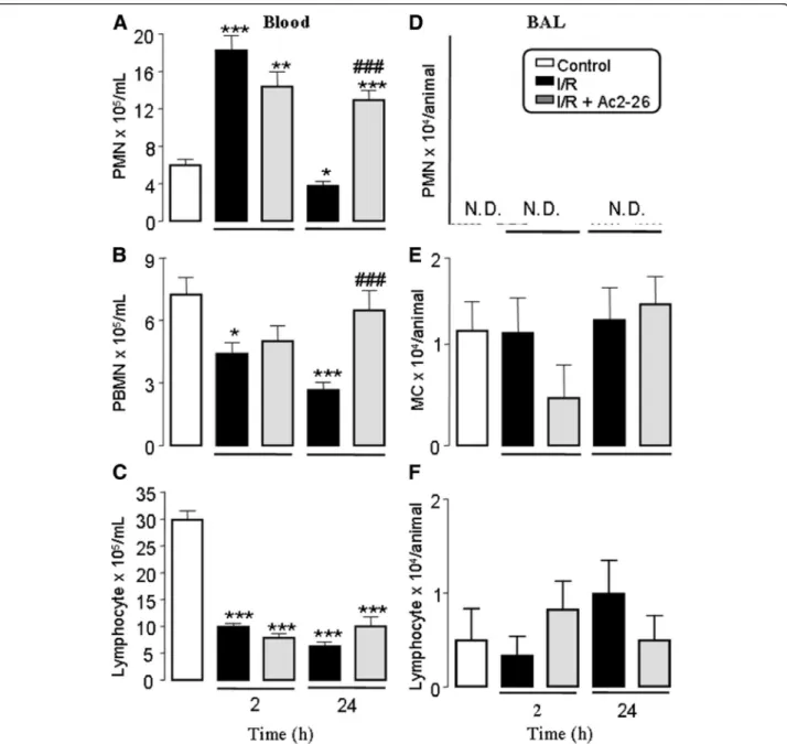
later, the number of these cells was similar to those in the control group (naive group: 6.1 ± 0.4; sham group:
6.0 ± 0.2; negative control group saline: 6.1 ± 0.2;
negative control group Ac2-26: 5.9 ± 0.4 × 10⁵/mL). On the other hand, PBMN and lymphocytes numbers were reduced (2 and 24 h) after reperfusion compared to control group (Figure 1 panels A-C).
Pretreatment with peptide Ac2-26 had no effect on the increases in the values of peripheral blood leukocytes 2 h
after reperfusion. In contrast, 24 h after reperfusion, Ac2-26 treatment prevented the reduction of circulating PBMN and PMN numbers as found in non-treated groups of mice upon intestinal I/R (Figure 1A and 1B). On the other hand, circulating lymphocytes number was not af-fected by pretreatment with peptide Ac2-26 (Figure 1C).
In order to verify the effects of intestinal I/R on the cell recruitment into bronchoalveolar compartment, we quanti-fied the cells in BAL, 2 h and 24 h after reperfusion. As
shown in Figure 1 PMN were not detected (Figure 1D), whereas monocyte/macrophages and lymphocytes number were not altered (Figure 1E and 1F).
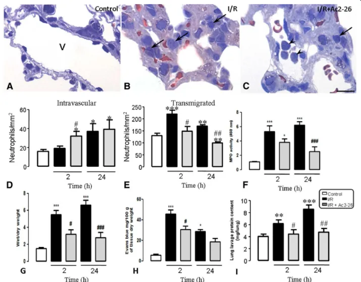
Edema and leukocytes infiltration in the lung after intestinal I/R induction and Ac2-26 treatment in mice As depicted in Figure 2 (A-C), neutrophil trafficking from the blood into the lung tissue was affected by intestinal I/R and Ac-2-26 treatment. Figure 2 (panels D and E) shows that neutrophils were present in the lung vessels and connective tissue of the control group
(naive group: 18.1 ± 0.5/135.0 ± 10.0; sham group:
18.5 ± 0.6/138.0 ± 15.0; negative control group saline: 19.5 ± 0.7/139.0 ± 12.0; negative control group Ac2-26:
17.6 ± 0.6/130.0 ± 8.0 neutrophils/mm2). After 2 h of
reperfusion, the neutrophils number in the vascular vessels was not affected, but a higher number of transmigrated cells were observed. However, 24 h post reperfusion, the number of intravascular neutrophils was increased tremendously.
Pre-treatment with Ac2-26 prevented neutrophil
migration into the lung tissue, as indicated by the
increased number of these cells in the intravascular compartment 2 h after reperfusion. Besides, transmigra-tion of neutrophils, 2 and 24 h after reperfusion was also reduced by prior treatment of mice with Ac-2-26 in comparison to the non-treated group (Figure 2D and E).
Similar to the histological findings, the intestinal I/R induces a large increase in MPO content in lung homoge-nates (5.5 ± 0.5 and 6.6 ± 0.6 optical density for MPO, respectively 2 and 24 after intestinal I/R) in comparison with animals from control group (naive group: 1.8 ± 0.5; sham group: 1.5 ± 0.4; negative control group + saline:
1.6 ± 0.6; negative control group + Ac2-26: 1.2 ± 0.4)
(Figure 2F). This increase was significantly attenuated by the pretreatment of peptide Ac2-26 (3.2 ± 0.5 and 2.8 ± 0.6, respectively 2 and 24 after intestinal I/R) (Figure 2F).
To analyze the pulmonary injury in response to intes-tinal I/R, we evaluate the Evans blue extravasation
(microvascular leakage) and the wet/dry weight
(pulmonary edema) technique. The pulmonary injury
was significantly higher (5.3 ± 0.8/45.4 ± 4.3 and
6.2 ± 0.5/28.5 ± 2.1, respectively Evans blue measurement and wet/dry weight at 2 and 24 h after intestinal I/R) in comparison with control group (naive group: 1.2 ± 0.2/ 5.6 ± 1.0; sham group: 1.0 ± 0.3/5.0 ± 1.2; negative control group + saline: 1.2 ± 0.4/6.2 ± 1.6; negative control group
+ Ac2-26: 1.0 ± 0.5/4.9 ± 1.0) (Figure 2G-H). The
pretreatment of peptide Ac2-26 significantly reduced pulmonary microvascular leakage 2 and 24 h after intes-tinal I/R (3.8 ± 0.5/30.6 ± 2.5 ± 0.7/3.3 and 18.5 ± 3.0, respectively) (Figure 2G-H).
Intestinal I/R mice also had significantly elevated lung plasma leak into alveolar cavity, detected by protein contents in the bronchoalveolar lavage (6.2 ± 0.6 and 8.4 ± 0.6 mg/lungs, respectively 2 and 24 h after intestinal I/R) compared to control mice (naive group: 3.9 ± 0.2; sham group: 4.0 ± 0.4; negative control group + saline: 4.2 ± 0.3; negative control group + Ac2-26: 4.3 ± 0.5) (Figure 2I). Peptide Ac2-26 treatment significantly attenu-ated the plasma leak in the lung alveolar cavity at 2 and 24 h post intestinal I/R (4.2 ± 0.4 and 4.7 ± 0.6 mg/lungs, respectively) (Figure 2I).
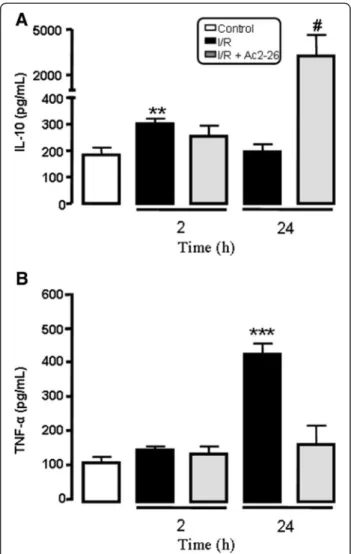
Effects of Ac-2-26 treatment on the IL-10 and TNF-αand
release after intestinal I/R
Figure 3 shows increased plasma level of IL-10 when mice were subjected to 2 h of reperfusion, but after 24 h of reperfusion, the level of this cytokine was similar to the
control group. The plasma level of TNF-α, a
pro-inflammatory cytokine, was elevated after intestinal I/R. Peptide Ac2-26 treatment significantly augmented in-crease (p < 0.05) in the level of IL-10 while it attenuated increase of TNF-α (p < 0.05) at 24 h of reperfusion when compared to levels found in non treated control mice (Figure 3).
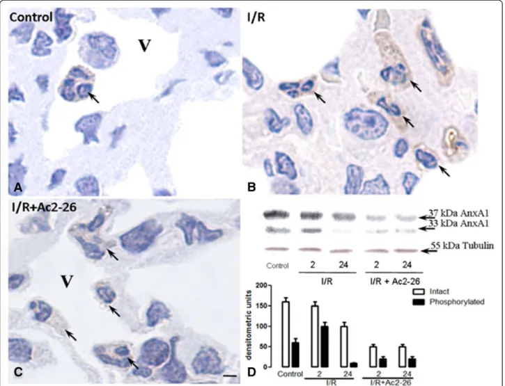
Effect of intestinal I/R on the annexin-A1 expression in the lung tissue
Considering the observed pharmacological effects of peptide Ac2-26 treatment, we decided to investigate the expression of endogenous AnxA1 protein in the leuko-cytes recruited into the lung due to intestinal I/R. The primary antibody used detected both the intact and cleaved AnxA1. However, it was not possible to distin-guish between the two by immunohistochemistry. As can be observed (Figure 4), control group of mice displayed basal AnxA1 immunostaining in the cytosol and the membrane of the leukocytes (Figure 4A). After 24 h of intestinal I/R, PBMN, alveolar monocyte/ macrophage and migrated neutrophils were highly stained for AnxA1 (Figure 4B and Table 1), but
connect-ive tissue monocyte/macrophage and intravascular
neutrophils showed a reduction in AnxA1 expression after 2 h of reperfusion (Table 1). After peptide Ac2-26 treat-ment, a reduction was observed in the AnxA1 endogenous expression in the intravascular (PBMN and PMN) and con-nective tissue (monocyte/macrophage and neutrophils) leu-kocytes after 24 h of intestinal I/R (Figure 4C and Table 1).
Finally, Western blotting analyzes indicated increa-sed endogenous AnxA1 expression after intestinal I/R (Figure 4D). However, after the peptide treatment, a reduction in the lung tissue protein content was ob-served, as the 37 kDa band was less intense (Figure 4D). Interestingly, the AnxA1 cleaved at 33 kDa was highly noticeable at 2 h post reperfusion (Figure 4D), whereas after peptide treatment, this post-translational modifica-tion was hardly present in all time-points (Figure 4D).
Discussion
Intestinal I/R is a risk factor for acute lung injury induc-tion, a lung disease where activated neutrophils play a part [2-4]. In this study, we analyzed the kinetics of leukocyte migration to the lung after intestinal I/R and we assessed AnxA1 expression. We also evaluated the role of Ac2-26, the annexin-1 N-terminal peptidomimetic, on the profile of lung tissue cells and peripheral blood leukocytes.
Our data showed that the period of reperfusion is a crucial factor in the changes to blood cell mobilization. In fact, the early increase of blood neutrophils number 2 h after reperfusion was reverted 24 h later. Thus, it is plausible to suggest that systemic inflammation caused by intestinal I/R caused the increment of blood leukocytes,
which migrated to target organ causing acute lung injury. Indeed we [1,15] and others [2-4] have demonstrated an increased influx of neutrophils into the lung and increased microvascular permeability in rodents subjected to intes-tinal I/R. Here, we observed that intesintes-tinal I/R intensified influx of neutrophil into the lung tissue, as evidenced by histological studies. Moreover, previous studies have demonstrated an increased lung myeloperoxidase activity due to intestinal I/R, indicating neutrophils activity at this inflammatory site [15]. These data agree with the concept that intestinal I/R induces remote organ injury, notably in the lung, where endothelial barrier plays a pivotal role in the organ injury [19]. Notwithstanding neutrophils are the principal cells that mediate acute lung injury after intestinal I/R. Mononuclear cells might also contribute to lung changes caused by gut trauma [5], causing immu-nodepression. Accordingly, innate immune response triggered by intestinal trauma has been associated to induction of lung failure [20]. It is noteworthy to state that our current data revealed a time-dependent decrease of blood monocytes and lymphocytes after intestinal I/R, a fact that was accompanied by their concomitant increase in the lung tissue, indicating, therefore, that these cells were activated by intestinal trauma.
Regarding the involvement of neutrophils in the intes-tinal I/R-induced remote organ inflammation, some treatments for this condition have been developed. Most of which include neutrophil depletion and direct inhib-ition of neutrophil activators [5]. However, leukocyte or pro-inflammatory mediator blocking may cause several adverse side effects, because they could also affect activation of the resolution phase of the inflammatory response [14]. Being so, we hypothesize that AnxA1 as a component of endogenous control of inflammatory response could constitute a new approach to control the magnitude of acute lung injury due to intestinal I/R. In fact, our data demonstrated that the AnxA1 mimetic Ac2-26 compound regulated the neutrophil trafficking from the blood vessels into the lung after intestinal I/R, as observed at 24 h time-point. In this scenario, Ac2-26
played a pivotal role in the control of neutrophil influx induced by intestinal I/R as found in other models such as heart ischemia [10,13,21,22]. To test this hypothesis, we assessed the number of intravascular neutrophil and those transmigrated into lung tissue after Ac2-26 treat-ment upon intestinal I/R. Intravascular neutrophils increased 2 h after Ac2-26 treatment followed by intes-tinal I/R and remained unaltered 24 h later. Such neutrophil mobilization dynamic was accompanied by a reduced transmigration of neutrophils into lung tissue 2 h and 24 h after reperfusion. Overall, our data suggest that the peptidomimetic Ac2-26 treatment regulates topographic distribution of neutrophils in order to control acute lung injury induced by intestinal I/R. AnxA1 and its peptidomimetic Ac2-26 regulate the leukocyte extravasation/activation through interaction with their receptor, the formyl-peptide receptor (FPR) [13]. Intravital-microscopy studies have demonstrated that AnxA1 does not inhibit the leukocytes recruitment or rolling/adhesion to the post-capillary venules, but affects the cell migration to the inflammatory sites [10,13]. Several studies have demonstrated that the inter-action between AnxA1/Ac2-26 and FPR induces a regu-lation of L-selectin and integrin CD11b expression in neutrophils and monocytes [13,23-25].
AnxA1 action has also been studied in other experi-mental models of I/R. In the myocardial injury induced by I/R of the left anterior descending coronary artery, the treatment with peptide Ac2-26 or the human recom-binant (hr)AnxA1 inhibited leukocyte migration and heart tissue damage [10,13,21,22]. Other models, like renal and cerebral I/R, also demonstrated the protective effect of AnxA1 treatment [26-28]. Moreover, some studies have demonstrated the inhibitory action of AnxA1 in the leukocyte migration to the intestine and cremaster tissue induced by intestinal I/R [28,29].
For a better understanding of other AnxA1 protect-ive effect during the intestinal I/R, we analyzed anti-inflammatory cytokine IL-10 and the pro-anti-inflammatory
cytokine TNF-α release. Our data indicated that the
Table 1 AnxA1 Immunohistochemistry analysis in lung leukocytes after intestinal ischemia-reperfusion
Control Intestinal I/R Intestinal I/R + Ac2-26
2 h 24 h 2 h 24 h
Intravascular PBMN 49,7 ± 7,2 55.9 ± 4.5 83,7 ± 3,1*** 65.4 ± 5.3 60,5 ± 5,2#
Alveolar monocyte/macrophage 43,2 ± 5,5 43.1 ± 4.4 66,7 ± 3,7* 44.4 ± 2.7 46,3 ± 7,3
Connective tissue monocyte/macrophage 60,2 ± 1,7 53.8 ± 2.1* 64,9 ± 1,9 45.6 ± 1.8***# 55,8 ±1,9#
Intravascular neutrophils 88,5 ± 4,2 71.3 ± 2.2*** 83,6 ± 2,7 72.6 ± 2.5*** 65,9 ± 3,0***###
Alveolar neutrophils —— 59.0 ± 3.8 80,6 ± 3,7 47.4 ± 3.4 68,1 ± 3,5
Connective tissue neutrophils 80,9 ± 2,6 71.2 ± 2.3 95,8 ± 2,9*** 69.3 ± 2.4* 70,3 ± 1,8*###
peptide Ac2-26 treatment increased IL-10 levels and de-crease that of TNF-αin the plasma after 24 h of intes-tinal I/R. Several works have indicated the induction of IL-10 release after AnxA1 treatment [29-31], and reduc-tion of IL-10 levels in AnxA1 deficient mice [23]. On one hand AnxA1 or peptide Ac2-26 treatment, in the
case of LPS-induced endotoxemia, inhibited TNF-α
re-lease [31,32], whereas in the absence of AnxA1, in-creased levels of the cytokine was observed [23]. In addition, antibodies against AnxA1 or an FPR antagon-ist, BOC-1, caused a reduction in levels of IL-10 in the plasma after intestinal I/R [29]. Also, it is important to highlight that, since endogenous AnxA1 promotes con-stitutive activation of ERK and innate immune stimula-tors such as CpG DNA up-regulate IL-10 production in macrophages by activating the extracellular signal-regulated kinase (ERK) pathways, it is conceivable that
ERK signaling pathway is involved in the
effect of IL-10 up-regulation via AnxA1 administration [33]. All these data indicate the importance of AnxA1 in the induction of the anti-inflammatory cytokine IL-10
release and downregulation of TNF-αlevel, which
high-lights the importance of this protein in the regulation of the inflammatory process. All this process might be or-chestrated through AnxA1 receptor, FPR/ALX [29].
Finally, we also analyzed endogenous AnxA1 protein expression by immunohistochemistry and Western blotting techniques in the lung tissue after intestinal I/R. Some studies have described endogenous expression of AnxA1 in the airways, in particular alveolar macro-phages and lung endothelial cells [23,31]. Our data indicated reduced AnxA1 expression after peptide Ac2-26 treatment. In contrast to our results, previous studies have shown increase in the endogenous AnxA1 after peptide Ac2-26 treatment in leukocytes [13]. This discrepancy can be explained by the high levels of clea-ved AnxA1 (33 kDa) obserclea-ved after intestinal I/R by Western blotting technique. One of the possible post-translational modifications described for these proteins is phosphorylation, which leads to protein translocation to the membrane and release during the inflammatory process [34-37]. Future studies will therefore address the secreted levels of AnxA1 after intestinal I/R.
Conclusion
In conclusion, our data indicated that AnxA1
peptidomimetic Ac2-26 treatment has both regulatory and protective effect during lung inflammation induced by intestinal I/R. The main mechanisms observed here were the reduction of leukocyte migration into the lung and the induction of the anti-inflammatory cytokine IL-10 release in the blood. The anti-inflammatory effects of AnxA1 as reported herein suggests that the protein
could be relevant to understanding mechanisms
underlying the pharmacological interventions to control inflammatory events related to intestinal I/R.
Abbreviations
ALI:Acute lung injury; I/R: Ischemia/reperfusion; ARDS: Adult respiratory distress syndrome; IL-1β: Interleukin-1β; TNF-α: Tumor necrosis
factor-α; AnxA1: Annexin A1; BAL: Bronchoalveolar lavage; PBMC: Peripheral blood monocytes; PBS: Phosphate-buffered saline; PBSA: Bovine serum albumin in phosphate-buffered saline; PMN: Polymorphonuclear; IL-10: Interleukin-10.
Competing interests
The authors declare that they have no competing interests.
Authors’contributions
BCG: carried out the experiments. MZ: carried out the experiments. WTL: conceived the study, and participated in its design, coordination and helped in writing of the manuscript. SMO: participated in the study design and participated in sequence alignment and in writing of the manuscript. ASD: participated in the study design, carried out the experiments and participated in sequence alignment and in writing of the manuscript. All authors read and approved the final manuscript.
Acknowledgements
This work was supported by Fundação de Amparo à Pesquisa do Estado de São Paulo (FAPESP, 05/56855-8 to A.S.D. and studentship 2007/01874-3 to B. C.G.) and Conselho Nacional de Desenvolvimento Científico e Tecnológico (CNPq, studentship number 100.042/2008-2 to M.Z.). S.M.O is supported by grants from CNPq (grant numbers 302768/2010-6).
Author details
1Department of Biology; Institute of Biosciences, Letras e Ciências Exatas
(IBILCE), São Paulo State University (UNESP), São José do Rio Preto, SP 15054-000, Brazil.2Department of Pharmacology, Institute of Biomedical
Sciences (ICB), University of São Paulo (USP), São Paulo 05508-900, Brazil.
3Department of Basic Science in Health; Faculty of Medicine (FM), Federal
University of Mato Grosso (UFMT), Mato Grosso, MT 78060-900, Brazil.
Received: 30 May 2012 Accepted: 1 March 2013 Published: 13 March 2013
References
1. Victoni T, Coelho FR, Soares AL, de Freitas A, Secher T, Guabiraba R, Erard F, de Oliveira-Filho RM, Vargaftig BB, Lauvaux G, Kamal MA, Ryffel B, Moser R, Tavares-de-Lima W:Local and remote tissue injury upon intestinal ischemia and reperfusion depends on the TLR/MyD88 signaling pathway.Med Microbiol Immunol2010,199(1):35–42.
2. Gropper MA, Wiener-Kronish J:The epithelium in acute lung injury/acute respiratory distress syndrome.Curr Opin Crit Care2008,14(1):11–15.
3. Bhatia M, Moochhala S:Role of inflammatory mediators in the pathophysiology of acute respiratory distress syndrome.J Pathol2004,
202(2):145–156.
4. Ishii H, Ishibashi M, Takayama M, Nishida T, Yoshida M:The role of cytokine-induced neutrophil chemoattractant-1 in neutrophil-mediated remote lung injury after intestinal ischaemia/reperfusion in rats.
Respirology2000,5(4):325–331.
5. Yang YJ, Chen SH, Ge XR:Role of interleukin-18 in the development of acute pulmonary injury induced by intestinal ischemia/reperfusion and its possible mechanism.J Gastroenterol Hepatol2007,22(2):253–260.
6. Moraes LB, Murakami AH, Fontes B, Poggetti RS, van Rooijen N, Younes RN, Heimbecker AM, Birolini D:Gut ischemia/reperfusion induced acute lung injury is an alveolar macrophage dependent event.J Trauma2008,
64(5):1196–1200.
7. Carden D, Xiao F, Moak C, Willis BH, Robinson-Jackson S, Alexander S:
Neutrophil elastase promotes lung microvascular injury and proteolysis of endothelial cadherins.Am J Physiol1998,275(2 Pt 2):H385–H392.
9. Facio FN Jr, Sena AA, Araújo LP, Mendes GE, Castro I, Luz MA, Yu L, Oliani SM, Burdmann EA:Annexin 1 mimetic peptide protects against renal ischemia/reperfusion injury in rats.J Mol Med (Berl)2011,89(1):51–63.
10. D’Amico M, Di Filippo C, La M, Solito E, McLean PG, Flower RJ, Oliani SM,
Perretti M:Lipocortin 1 reduces myocardial ischemia-reperfusion injury by affecting local leukocyte recruitment.FASEB J2000,14(13):1867–1869.
11. Gavins FN, Yona S, Kamal AM, Flower RJ, Perretti M:Leukocyte antiadhesive actions of annexin 1: ALXR- and FPR-related anti-inflammatory mechanisms.Blood2003,101(10):4140–4147.
12. Perretti M:Lipocortin-derived peptides.Biochem Pharmacol1994,47(6): 931–938.
13. Gavins FN, Kamal AM, D’Amico M, Oliani SM, Perretti M:Formyl-peptide receptor is not involved in the protection afforded by annexin 1 in murine acute myocardial infarct.FASEB J2005,19(1):100–102. 14. Perretti M, D’Acquisto F:Annexin A1 and glucocorticoids as effectors of
the resolution of inflammation.Nat Rev Immunol2009,9(1):62–70.
15. Cavriani G, Oliveira-Filho RM, Trezena AG, da Silva ZL, Domingos HV, de Arruda MJ, Jancar S, de Tavares Lima W:Lung microvascular permeability and neutrophil recruitment are differently regulated by nitric oxide in a rat model of intestinal ischemia-reperfusion.Eur J Pharmacol2004,494(2–3):241–249.
16. Riffo-Vasquez Y, de Ligeiro Oliveira AP, Page CP, Spina D, Tavares-de-Lima W:Role of sex hormones in allergic inflammation in mice.Clin Exp Allergy
2007,37(3):459–470.
17. Damazo AS, Yona S, Flower RJ, Perretti M, Oliani SM:Spatial and temporal profiles for anti-inflammatory gene expression in leukocytes during a resolving model of peritonitis.J Immunol2006,176(7):4410–4418.
18. Laemmli UK:Cleavage of structural proteins during the assembly of the head of bacteriophage T4.Nature1970,227(5259):680–685.
19. Fleming SD, Anderson J, Wilson F, Shea-Donohue T, Tsokos GC:C5 is required for CD49d expression on neutrophils and VCAM expression on vascular endothelial cells following mesenteric ischemia/reperfusion.Clin Immunol2003,106(1):55–64.
20. Uchida K, Mishima S, Ohta S, Yukioka T:Inhibition of inducible nitric oxide synthase ameliorates lung injury in rats after gut ischemia-reperfusion.J Trauma2007,63(3):603–607.
21. La M, D’Amico M, Bandiera S, Di Filippo C, Oliani SM, Gavins FN, Flower RJ,
Perretti M:Annexin 1 peptides protect against experimental myocardial ischemia-reperfusion: analysis of their mechanism of action.FASEB J
2001,15(12):2247–2256.
22. Facio FN Jr, Sena AA, Araújo LP, Mendes GE, Castro I, Luz MA, Yu L, Oliani SM, Burdmann EA:Annexin 1 mimetic peptide protects against renal ischemia/reperfusion injury in rats.J Mol Med2011,89(1):51–63.
23. Damazo AS, Yona S, D’Acquisto F, Flower RJ, Oliani SM, Perretti M:Critical
protective role for annexin 1 gene expression in the endotoxemic murine microcirculation.Am J Pathol2005,166(6):1607–1617.
24. Hannon R, Croxtall JD, Getting SJ, Roviezzo F, Yona S, Paul-Clark MJ, Gavins FN, Perretti M, Morris JF, Buckingham JC, Flower RJ:Aberrant inflammation and resistance to glucocorticoids in annexin 1−/−mouse.FASEB J2003,
17(2):253–255.
25. Hayhoe RP, Kamal AM, Solito E, Flower RJ, Cooper D, Perretti M:Annexin 1 and its bioactive peptide inhibit neutrophil-endothelium interactions under flow: indication of distinct receptor involvement.Blood2006,
107(5):2123–2130.
26. Relton JK, Strijbos PJ, O’Shaughnessy CT, Carey F, Forder RA, Tilders FJ,
Rothwell NJ:Lipocortin-1 is an endogenous inhibitor of ischemic damage in the rat brain.J Exp Med1991,174(2):305–310.
27. McKanna JA, Chuncharunee A, Munger KA, Breyer JA, Cohen S, Harris RC:
Localization of p35 (annexin I, lipocortin I) in normal adult rat kidney and during recovery from ischemia.J Cell Physiol1992,153(3):467–476.
28. Gavins FN, Dalli J, Flower RJ, Granger DN, Perretti M:Activation of the annexin 1 counter-regulatory circuit affords protection in the mouse brain microcirculation.FASEB J2007,21(8):1751–1758.
29. Souza DG, Fagundes CT, Amaral FA, Cisalpino D, Sousa LP, Vieira AT, Pinho V, Nicoli JR, Vieira LQ, Fierro IM, Teixeira MM:The required role of endogenously produced lipoxin A4 and annexin-1 for the production of IL-10 and inflammatory hyporesponsiveness in mice.J Immunol2007,
179(12):8533–8543.
30. Cuzzocrea S, Tailor A, Zingarelli B, Salzman AL, Flower RJ, Szabó C, Perretti M:Lipocortin 1 protects against splanchnic artery occlusion and reperfusion injury by affecting neutrophil migration.J Immunol1997,
159(10):5089–5097.
31. Parente L, Solito E:Annexin 1: more than an anti-phospholipase protein.
Inflamm Res2004,53(4):125–132.
32. de Coupade C, Ajuebor MN, Russo-Marie F, Perretti M, Solito E:Cytokine modulation of liver annexin 1 expression during experimental endotoxemia.Am J Pathol2001,159(4):1435–1443.
33. Ferlazzo V, D’Agostino P, Milano S, Caruso R, Feo S, Cillari E, Parente L: Anti-inflammatory effects of annexin-1: stimulation of IL-10 release and inhibition of nitric oxide synthesis.Int Immunopharmacol2003,
3(10–11):1363–1369.
34. Damazo AS, Paul-Clark MJ, Straus AH, Takahashi HK, Perretti M, Oliani SM:
Analysis of the annexin 1 expression in rat trachea.Annexin2004,
1(1):12–18.
35. Dorovkov MV, Ryazanov AG:Phosphorylation of annexin I by TRPM7 channel-kinase.J Biol Chem2004,279(49):50643–50646.
36. Damazo AS, Flower RJ, Solito E, Oliani SM:Annexin-A1 gene expression during liver development and post-translational modification after experimental endotoxemia.Inflamm Res2008,57(3):97–103.
37. Solito E, Christian HC, Festa M, Mulla A, Tierney T, Flower RJ, Buckingham JC:
Post-translational modification plays an crucial role in the translocation of annexin A1 from the cytoplasm to the cell surface.FASEB J2006,
20(9):1498–1500.
doi:10.1186/1476-9255-10-10
Cite this article as:Guidoet al.:Annexin-A1 peptide down-regulates the leukocyte recruitment and up-regulates interleukin-10 release into lung after intestinal ischemia-reperfusion in mice.Journal of Inflammation
201310:10.
Submit your next manuscript to BioMed Central and take full advantage of:
• Convenient online submission
• Thorough peer review
• No space constraints or color figure charges
• Immediate publication on acceptance
• Inclusion in PubMed, CAS, Scopus and Google Scholar
• Research which is freely available for redistribution