Research Article
Anti-Trichophyton
Activity of Protocatechuates and Their
Synergism with Fluconazole
Luciana Arantes Soares,
1Fernanda Patrícia Gullo,
1Janaina de Cássia Orlandi Sardi,
1Nayla de Souza Pitangui,
1Caroline Barcelos Costa-Orlandi,
1Fernanda Sangalli-Leite,
1Liliana Scorzoni,
1Luis Octávio Regasini,
2Maicon Segalla Petrônio,
2Patrícia Fernanda Souza,
2Dulce Helena Siqueira Silva,
2Maria José Soares Mendes-Giannini,
1and Ana Marisa Fusco-Almeida
1,31Laboratory of Clinical Mycology, Department of Clinical Analysis, Faculty of Pharmaceutical Sciences, UNESP,
Rodovia Araraquara-Ja´u, Km 1, 14801-902 Araraquara, SP, Brazil
2Institute of Chemistry, UNESP, Rua Professor Francisco Degni 55, 14800-900 Araraquara, SP, Brazil
3Department of Clinical Mycology, Faculty of Pharmaceutical Sciences, Universidade Estadual Paulista (UNESP),
Rodovia Araraquara-Ja´u, Km 1, 14801-902 Araraquara, SP, Brazil
Correspondence should be addressed to Ana Marisa Fusco-Almeida; ana.marisa@uol.com.br
Received 17 February 2014; Revised 23 May 2014; Accepted 1 June 2014; Published 18 June 2014
Academic Editor: Mohd Roslan Sulaiman
Copyright © 2014 Luciana Arantes Soares et al. This is an open access article distributed under the Creative Commons Attribution License, which permits unrestricted use, distribution, and reproduction in any medium, provided the original work is properly cited.
Dermatophytosis and superficial mycosis are a major global public health problem that affects 20–25% of the world’s population. The increase in fungal resistance to the commercially available antifungal agents, in conjunction with the limited spectrum of action of such drugs, emphasises the need to develop new antifungal agents. Natural products are attractive prototypes for antifungal agents due to their broad spectrum of biological activities. This study aimed to verify the antifungal activity of protocatechuic acid, 3,4-diacetoxybenzoic, and fourteen alkyl protocatechuates (3,4-dihydroxybenzoates) againstTrichophyton rubrumandTrichophyton mentagrophytesand to further assess their activities when combined with fluconazole. Susceptibility and synergism assays were conducted as described in M38-A2 (CLSI), with modifications. Three strains of Trichophyton rubrum and three strains of Trichophyton mentagrophyteswere used in this work. The pentyl, hexyl, heptyl, octyl, nonyl, and decyl protocatechuates showed great fungicidal effects, with minimum inhibitory concentration (MIC) values ranging from 0.97 to 7.8 mg/L. Heptyl showed a synergistic activity (FIC index= 0.49), reducing the MIC of fluconazole by fourfold. All substances tested were safe, especially the hexyl, heptyl, octyl, and nonyl compounds, all of which showed a high selectivity index, particularly in combination with fluconazole. These ester associations with fluconazole may represent a promising source of prototypes in the search for anti-Trichophytontherapeutic agents.
1. Introduction
Superficial fungal infections are a major global public health
problem that affects 20–25% of the population worldwide [1].
Among these diseases, dermatophytosis, or tinea, is one of the most frequent fungal infections. This infection is caused
by dermatophyte species that belong to the Trichophyton,
Microsporum,orEpidermophytongenera [2]. These
dermato-phytes commonly invade different keratinophilic regions of the body, causing tinea corporis, tinea cruris, tinea pedis,
tinea manus, tinea capitis, tinea barbae, and tinea unguium
[3]. Dermatophyte infections can lead to either mild or
severe symptoms, depending on the immunological response
of the host [4]. Several patient groups also seem to be
especially at risk of infection, including individuals with uncontrolled diabetes, AIDS, renal diseases, psoriasis, and types of immunosuppression, such as transplant recipients
and patients on long-term corticosteroid therapy [5].
There is an urgent need to find new sources of substances with antidermatophytic activity because the treatment of
Hindawi Publishing Corporation
Evidence-Based Complementary and Alternative Medicine Volume 2014, Article ID 957860, 9 pages
2 Evidence-Based Complementary and Alternative Medicine
dermatophytosis is long and expensive, particularly in the case of onychomycosis. Furthermore, the spectrum of the available drugs is limited, such drugs may induce adverse effects, and several reports of antifungal resistance have been
published [6–9]. For this reason, various antifungal agents
have been introduced into clinical practice, among them, amorolfine, ciclopirox, griseofulvin, terbinafine, itraconazole,
fluconazole, and more recently, voriconazole [10,11].
How-ever, efforts should be concentrated on the discovery and development of novel, safer, and effective antidermatophytic agents.
Protocatechuic acid (3,4-dihydroxybenzoic) is a phenolic compound produced by the secondary metabolism of plants. It is naturally present in almost all plant materials, including
food, fruits, and vegetables [12–14]. Together with its natural
and synthetic derivatives, it has been associated with a broad spectrum of biological actions and is known to have
antioxidant, proapoptotic [15], anti-inflammatory,
antiglyca-tive [16], and antimelanogenic [17] functions. However, the
major interest in protocatechuic acid and its derivatives is due to its antimicrobial properties. It has been reported that protocatechuic acids have activity against susceptible
and antibiotic-resistantCampylobacterspp. andHelicobacter
pylori [18, 19]. Furthermore, it has been shown that 𝑛
-octyl 3,4-dihydroxybenzoate has fungicidal activity against
Saccharomyces cerevisiae[20].
Thus, considering the broad spectrum of protocatechu-ates and the need for the discovery of new antifungal agents, this study aimed to investigate the antifungal activity of a
syn-thetic homologous series ofn-alkyl protocatechuates against
Trichophyton rubrumandTrichophyton mentagrophytes. This
study also aimed to investigate the chemical characteristics of the compounds responsible for the biological activity of dermatophytes, including the importance of free hydroxyl radicals and the size of the carbon side chain.
2. Methods
2.1. Compounds Synthesis. Synthetic compounds of
protocat-echuic acid were prepared as described by de Faria et al. [21],
with minor modifications. Briefly, a 3 mL solution ofN,N
-dicyclohexylcarbodiimide (DCC, 1 mmol) in𝑝-dioxane was
added to a cooled (5∘C) solution of 0.2 mmol protocatechuic
acid (1) (Sigma-Aldrich, St. Louis, MO, USA) and 20 mmol
of𝑛-alkyl alcohols in 6 mL of 𝑝-dioxane. The solution was
stirred for 48 h and the solvent was removed under reduced pressure. The residue was partitioned 3 times with EtOAc and filtered. The filtrate was washed successively with a saturated aqueous citric acid solution (3 times) and saturated aqueous
NaHCO3 (3 times), dried over anhydrous MgSO4, and
evaporated under reduced pressure. The crude products were purified over a silica gel column (0.06–0.20 mm, ACROS
Organics, USA) and eluted isocratically with CHCl3/MeOH
(98 : 2) to produce esters 2−15. Their structures were then
established by 1H and 13C NMR spectral analysis. For the
synthesis of compound 16, protocatechuic acid (20 mmol)
was dissolved in dried pyridine (5.0 mL) and anhydride acetic (5.0 mL) under a hydrogen atmosphere. The mixture was then
stirred for 48 h at room temperature, dried under reduced pressure, and purified by column chromatography with a
mixture of CHCl3/MeOH (85 : 15) to produce product16. The
NMR spectroscopic data for compound16were compatible
with it being 3,4-diacetoxybenzoic acid.
2.2. Microorganisms. To evaluate its antifungal activity, six
species of dermatophytes were tested: two clinical strains of
Trichophyton rubrum (Tr1 and Tr2), Trichophyton rubrum
ATCC MYA 3108, two clinical strains ofTrichophyton
menta-grophytes(Tm1 and Tm2), andTrichophyton mentagrophytes
ATCC 40131 (Tm3). The microorganisms were obtained from the collection of the Clinical Mycology Laboratory of the Department of Clinical Analyses at the School of Pharmaceutical Sciences of Universidade Estadual Paulista (UNESP). The strains were cultivated on Sabouraud dextrose
agar (Difco, BD Biosciences) and incubated at 28∘C for 7–
15 days. For all experiments, the strains were cultivated on Potato Dextrose Agar (Difco, BD Biosciences) and incubated
at 28∘C as described above or until sporulation.
2.3. Dilution of Test Substances. The dilution of the synthetic
compounds was performed with DMSO (Synth, Diadema,
Sao Paulo, Brazil) as described by Scorzoni et al. [22].
The concentrations of the compounds on 96-well plates (TPP, Trasadigen, Switzerland) ranged from 500 mg/L to 0.97 mg/L. The antifungal drugs were diluted according to
the CLSI M38-A2 document [23]. Stock solutions of
Flucona-zole (Sigma-Aldrich, St. Louis, MO, USA) were prepared, considering its power. Serial twofold dilutions were prepared according to the recommendations of Zhang and
collabora-tors [24], with some modifications.
2.4. Minimum Inhibitory Concentration (MIC). The
antifun-gal activity tests were performed using the broth microdilu-tion method as described in M38-A2, a document produced by the Clinical and Laboratory Standards Institute (CLSI,
2008) [23], with modifications. The medium used was RPMI
1640 with L-glutamine (Sigma-Aldrich, St. Louis, MO, USA) buffered to pH 7.0 with 0.165 M morpholinepropanesulfonic acid (MOPS) (Sigma-Aldrich, St. Louis, MO, USA), supple-mented with 2% glucose. The cell suspension was prepared in a 0.85% saline solution. The suspension of conidia was then transferred to small sterile test tubes where they remained for 40 minutes to separate the microconidia, which were lighter and therefore present in the supernatant. The separated microconidia were then counted with a hematocytometer, and their concentration was adjusted to obtain a final
con-centration ranging from 2.5×10³to 5×10³CFU/mL. These
suspensions were diluted in RPMI 1640 (Sigma-Aldrich, St. Louis, MO, USA) and inoculated on 96-well plates (TPP, Trasadigen, Switzerland) that had been previously prepared with the compounds diluted at concentrations from 250 to
0.48 mg/L. Positive (100𝜇L of RPMI medium with 100𝜇L
of inoculum) and negative (200𝜇L of RPMI) controls were
included in all experiments. The plates were incubated with
agitation at 35∘C for 7 days. The MIC reading was performed
Evidence-Based Complementary and Alternative Medicine 3
was defined as the concentration that produced a 50% inhibition of fungal growth.
2.5. Minimum Fungicide Concentration (MFC). A qualitative
analysis of the fungal viability was performed by transferring a portion of the wells to a plate with Sabouraud (Difco, BD
Biosciences) medium and incubating it at 35∘C for the time
determined for each fungal agent. The MFC was defined as the lowest extract concentration that did not allow the growth of any fungal colonies on the solid medium after
the incubation period [25]. A visual reading was performed
to confirm the death or growth inhibition provided by fluconazole and the sixteen semisynthetic substances derived from protocatechuic acid.
2.6. Synergistic Activity. The drug activity was assessed using
a checkerboard method derived from a standardised pro-cedure established by the National Committee for Clinical
Laboratory Standards (M38-A2) [23]. Briefly, the test was
performed on the same medium used for susceptibility
testing. Volumes of 50𝜇L of each drug in a concentration four
times the final concentration were dispensed in 96-well plates
(TPP, Trasadigen, Switzerland). To each well, 100𝜇L of the
fungal suspension was added to produce a final concentration
of 5.0×10³CFU/mL.
As a negative control, we used 200𝜇L of RPMI, while
as a positive control, we used 100𝜇L of RPMI medium with
100𝜇L of inoculum. The plates were incubated at 35∘C, and
the reading was completed after 168 hours. We conducted visual and spectrophotometric readings at 490 nm. To deter-mine the effect of combinatorial fractions, we calculated the fractional inhibitory concentration (FIC). The FIC was calculated by taking the MIC of the substance in combina-tion/MIC of the substance alone. The sum of the fractional inhibitory concentration (FIC) of each substance consists of the fractional inhibitory concentration index: (the MIC of drug A in combination/the MIC of drug A alone) + (the MIC of drug B in combination/the MIC of drug B alone).
A synergistic relationship was defined as FIC index≤0.5, an
additive relationship was defined as 0.5<FIC index≤1.0, an
indifferent relationship was defined as 1.0<FIC index≤4.0,
and an antagonistic relationship was defined as FIC index
>4.0 [26–28].
2.7. Cytotoxicity Assay. The cytotoxicity of sixteen
semisyn-thetic compounds derived from protocatechuic acid was assessed using a sulphorhodamine B assay in NOK (oral human keratinocyte) cell lines obtained from the American Type Culture Collection (Manassas, VA, USA). The strains were maintained in bottles appropriate for cell culture with keratinocyte serum free medium (Gibco, Life Technologies)
and incubated in standard conditions of 37∘C and 5% CO2.
Cell concentrations ranging from 2.5 to 5.0×10⁴ cells/mL
were used for the formation of cell monolayers. The con-centrations of pure substances were kept in contact with the cells for 24 hours. After the incubation period, the cells were treated with the sulphorhodamine B reagent (Sigma-Aldrich,
St. Louis, MO, USA) as previously described by Skehan et al.
[29], with some modifications.
2.8. Statistical Analysis. All experiments were performed
in triplicate. Statistical analysis was performed with a t
-test or one-way ANOVA with GraphPad Prism 5 software
(Version 5, USA).𝑃values<0.05 were considered statistically
significant.
3. Results
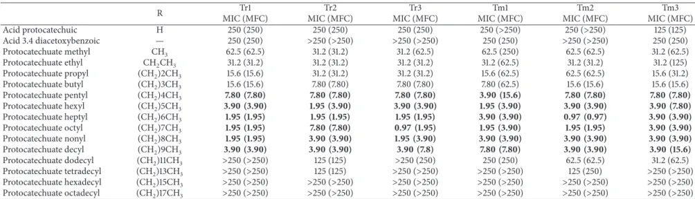
3.1. Antidermatophytic Activity. Protocatechuic acid, 3.4
diacetoxibenzoic acid, and fourteen-alkyl protocatechuate (3,4-dihydroxybenzoate) derivatives were evaluated against
6 strains of Trichophyton rubrum and Trichophyton
men-tagrophytes. The minimum inhibitory concentration (MIC)
ranged from≥250 to 1.95 mg/L. Pentyl, hexyl, heptyl, octyl,
nonyl, and decyl protocatechuate compounds showed the best MIC values, ranging from 1.95 to 7.8 mg/L for both species, with MFC values ranging from 1.95 to 15.6 mg/L
and 0.97 to 15.6 mg/L, for Trichophyton rubrum and
Tri-chophyton mentagrophytes,respectively (Table 1). In contrast,
protocatechuic acid and acid 3.4 diacetoxybenzoate showed
the highest values of MIC and MFC, from 125 to>250 mg/L,
for both species and were thus considered to have low antidermatophytic activity. The potentiation of antifungal activity was observed with an increase of methylation in the structure of protocatechuic acid. However, the addition
of nine methyl groups [(CH2) 9CH3] led to a progressive
reduction in the MIC values. The addition of methyl groups produced a reduction in the MIC and MFC values for strains of both species. The addition of a methyl group
(CH3) produced a range of MIC and MFC values of 31.25
to 62.50 mg/L for the isolates ofTrichophyton rubrum and
Trichophyton mentagrophytes. The only exception was for Tm1
because despite its MIC of 62.50 mg/L, its MFC remained at 250 mg/L. Ethyl and propyl showed MIC and MFC values ranging from 15.62 to 62.50 mg/L for both species of der-matophytes. The addition of four methyl groupings (pentyl protocatechuate) produced a strong antifungal activity, as the MIC values were low, ranging from 3.90 to 7.80 mg/L, while the MFC ranged from 3.90 to 15.60 mg/L. Therefore, dodecyl, tetradecyl, hexadecyl, and octadecyl showed low activities, with their MIC and MFC values ranging from 125
to>250 mg/L forTrichophyton rubrumstrains and from 31.25
and>250 mg/L forTrichophyton mentagrophytesstrains.
3.2. Synergistic Activity. The activity of the combination
of fluconazole with pentyl, hexyl, heptyl, octyl, and nonyl protocatechuates was evaluated and classified as antagonistic, indifferent, additive, and synergistic based on the FIC index,
as shown inTable 2. The FIC index was calculated based on
the results of the checkerboard test.
The combinations were tested in clinical isolates of
Trichophyton rubrum(Tr1) andTrichophyton mentagrophytes
(Tm1) and in the reference strain Trichophyton
4
E
vidence-B
as
ed
C
o
m
p
lemen
ta
ry
and
Al
te
rn
at
iv
e
M
ed
icine
Table 1: MIC and MFC values (mg/L) and quantitative analysis of fungal cellular viability of protocatechuic acid derivates againstTrichophytonspp.
R Tr1 Tr2 Tr3 Tm1 Tm2 Tm3
MIC (MFC) MIC (MFC) MIC (MFC) MIC (MFC) MIC (MFC) MIC (MFC)
Acid protocatechuic H 250 (250) 250 (250) 250 (250) 250 (>250) 250 (>250) 125 (125)
Acid 3.4 diacetoxybenzoic — 250 (250) >250 (>250) >250 (>250) 250 (250) >250 (>250) 250 (250)
Protocatechuate methyl CH3 62.5 (62.5) 31.2 (31.2) 31.2 (62.5) 62.5 (250) 62.5 (62.5) 31.2 (62.5)
Protocatechuate ethyl CH2CH3 31.2 (31.2) 31.2 (31.2) 31.2 (31.2) 31.2 (62.5) 31.2 (31.2) 31.2 (125)
Protocatechuate propyl (CH2)2CH3 15.6 (15.6) 31.2 (31.2) 31.2 (31.2) 15.6 (62.5) 62.5 (62.5) 15.6 (31.2)
Protocatechuate butyl (CH2)3CH3 15.6 (15.6) 7.80 (7.80) 7.80 (7.80) 7.80 (62.5) 15.6 (15.6) 15.6 (15.6)
Protocatechuate pentyl (CH2)4CH3 7.80 (7.80) 7.80 (7.80) 7.80 (7.80) 3.90 (15.6) 7.80 (7.80) 7.80 (7.80)
Protocatechuate hexyl (CH2)5CH3 3.90 (3.90) 1.95 (3.90) 3.90 (3.90) 1.95 (3.90) 3.90 (3.90) 3.90 (7.80)
Protocatechuate heptyl (CH2)6CH3 1.95 (1.95) 1.95 (1.95) 1.95 (1.95) 3.90 (3.90) 0.97 (0.97) 3.90 (3.90)
Protocatechuate octyl (CH2)7CH3 1.95 (1.95) 7.80 (7.80) 0.97 (1.95) 1.95 (3.90) 1.95 (1.95) 3.90 (3.90)
Protocatechuate nonyl (CH2)8CH3 1.95 (1.95) 3.90 (3.90) 1.95 (3.90) 3.90 (3.90) 3.90 (3.90) 3.90 (3.90)
Protocatechuate decyl (CH2)9CH3 3.90 (3.90) 3.90 (3.90) 3.90 (7.8) 7.80 (7.80) 3.90 (3.90) 3.90 (15.6)
Protocatechuate dodecyl (CH2)11CH3 >250 (>250) 125 (125) >250 (250) 250 (250) 62.5 (62.5) 31.2 (62.5)
Protocatechuate tetradecyl (CH2)13CH3 >250 (>250) 125 (125) >250 (>250) >250 (>250) 125 (250) >250 (>250) Protocatechuate hexadecyl (CH2)15CH3 >250 (>250) >250 (>250) >250 (>250) >250 (>250) >250 (>250) >250 (>250)
Protocatechuate octadecyl (CH2)17CH3 >250 (>250) >250 (>250) >250 (>250) >250 (>250) >250 (>250) >250 (>250)
MIC: minimal inhibitory concentration; MFC: minimal fungicidal concentration; Tr1 and Tr2:T. rubrumclinical isolates; Tr3:T. rubrumATCC MYA 3108; Tm1 and Tm2:T. interdigitaleclinical isolates; and Tm3:
E
vidence-B
as
ed
C
o
m
p
lemen
ta
ry
and
Al
te
rn
at
iv
e
M
edicine
5
Table 2: Activity of fluconazole combined with pentyl, hexyl, heptyl, octyl, and nonyl protocatechuates againstT. rubrumandT. mentagrophytes(mg/L).
MIC (mg/L) FIC index (association type)
Substances alone Substances combined
FLU 7 8 9 10 11 FLU +7 FLU +8 FLU +9 FLU +10 FLU +11 FLU +7 FLU +8 FLU +9 FLU +10 FLU +11
Tr1 2.0 7.8 3.9 1.9 1.9 1.9 0.12∗—7.80 2.00—0.48∗ 0.12∗—1.9 2.00—0.06∗∗ 2.00—0.03∗∗ 1.0 (A) 1.1 (I) 1.0 (A) 1.0 (A) 1.0 (A) Tm1 1.0 3.9 1.9 3.9 1.9 3.9 1.00—0.03∗∗ 1.00—0.03∗∗ 1.00—0.03∗∗ 1.00—0.03∗∗ 1.00—0.03∗∗ 1.0 (A) 1.0 (A) 1.0 (A) 1.0 (A) 1.0 (A)
Tm3 0.5 7.8 3.9 3.9 3.9 3.9 0.50—0.03 0.12—1.9∗ 0.12—0.97∗ 0.12—1.9∗ 0.50—0.03 1.0 (A) 0.72 (A) 0.48 (S) 0.72 (A) 1.0 (A)
MIC: minimal inhibitory concentration; FIC: fractional inhibitory concentration; S: synergistic effect; A: additive effect; I: indifferent effect Tr1: clinical strain ofT. rubrum; Tm1: clinical strain ofT. mentagrophytes;
6 Evidence-Based Complementary and Alternative Medicine
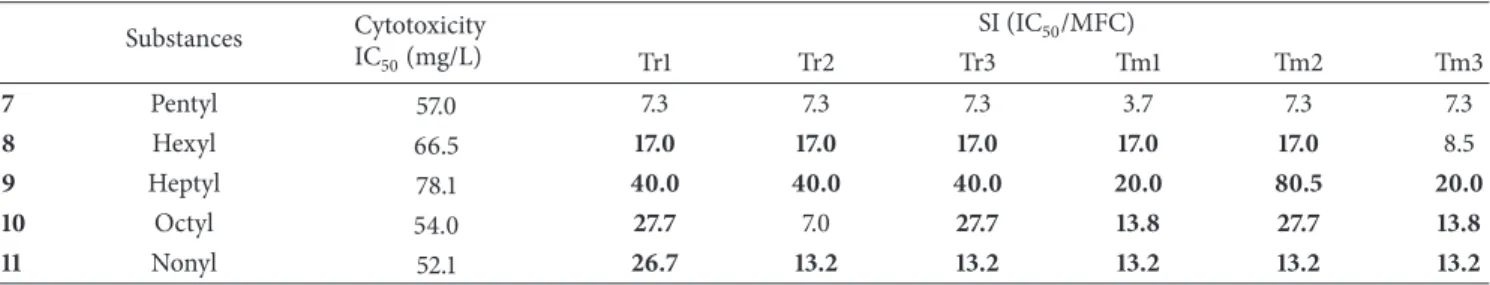
Table 3: Evaluation of IC50 and selectivity index of pentyl, hexyl, heptyl, octyl, and nonyl in NOK cells.
Substances Cytotoxicity
IC50(mg/L)
SI (IC50/MFC)
Tr1 Tr2 Tr3 Tm1 Tm2 Tm3
7 Pentyl 57.0 7.3 7.3 7.3 3.7 7.3 7.3
8 Hexyl 66.5 17.0 17.0 17.0 17.0 17.0 8.5
9 Heptyl 78.1 40.0 40.0 40.0 20.0 80.5 20.0
10 Octyl 54.0 27.7 7.0 27.7 13.8 27.7 13.8
11 Nonyl 52.1 26.7 13.2 13.2 13.2 13.2 13.2
For the clinical isolates ofTrichophyton rubrum(Tr1), the
MIC value of fluconazole was 2.0 mg/L. When associated with
the protocatechuate pentyl (compound7), a reduction in the
MIC of fluconazole of 16.66 times (0.12 mg/L) (𝑃 < 0.05)
and a conservation of the MIC of compound7 (7.8 mg/L)
were observed. The same occurred when fluconazole was
associated with protocatechuate heptyl (substance9) (𝑃 <
0.05). Thus, both associations were classified as additive,
with an FIC index of 1.0. The other acids tested showed
a conservation of the fluconazole MIC (2.0 mg/L) (𝑃 >
0.05) and a reduction in the MIC value of protocatechuates.
Compound8(hexyl protocatechuate) reduced the MIC value
by a factor of 8.125 (0.48 mg/L) (𝑃 < 0.05). Compound
10 (octyl protocatechuate) reduced the MIC by a factor of
31.66 (0.06 mg/L) (𝑃 < 0.01), and compound 11 (nonyl
protocatechuate) reduced the MIC by 63.33 times (0.03 mg/L) (𝑃 < 0.01). Thus, these combinations were classified as additive, with an FIC index of 1.0. The only exception was
compound8, which had an FIC index of 1.1 and was thus
classified as indifferent.
In the clinical isolate Tm1, the combination of fluconazole and all protocatechuates acids tested showed a conservation
of the fluconazole MIC (1.0 mg/L) (𝑃 > 0.05) and a
130-fold reduction in the MIC values of the protocatechuates
for substances 7, 9, and 11 (0.03 mg/L) (𝑃 < 0.01) and
in 63.33 times for compound8 and 10 (0.03 mg/L) (𝑃 <
0.01). All combinations were classified as additive for Tm1,
with an FIC index value of 1.0. However, for the strain ATCC 40131 (Tm3), a conservation in the fluconazole MIC
(0.5 mg/L) was observed when combined with compounds7
and11(𝑃 > 0.05). The combination of the activity between these compounds was classified as additive, with an FIC index equal to 1.0. A reduction in the MIC values of two-,
four-, and twofold, respectively, for substances8,9,and 10
(𝑃 < 0.05) was observed when combined with fluconazole. A fourfold reduction (0.12 mg/L) in the MIC value was observed
when associated with these compounds (𝑃 < 0.05). The
associations between fluconazole and compounds 8 and 10 were classified as additive and had an FIC index equal to
0.72, whereas the association with compound9instead had
a synergistic activity, with an FIC index equal to 0.48.
3.3. Cytotoxicity Assay. The compounds pentyl (7), hexyl (8),
heptyl (9), octyl (10), and nonyl (11) were evaluated in a
cytotoxicity assay in NOK cells. These substances showed
high values for IC50 after reaching the necessary
concen-tration to produce 50% lethality of 57 mg/L for cells treated
with compound7, 66.5 mg/L for cells treated with compound
8, 78.1 mg/L for cells treated with compound 9, 54 mg/L
for cells treated with compound10,and 52.1 mg/L for cells
treated with compound11. These results showed that these
compounds had a low toxicity in human oral keratinocytes (Table 3). The selectivity index (SI) was calculated for all
six strains ofTrichophyton rubrumand Trichophyton
men-tagrophytes. A selectivity index greater than 10 indicates a
substance with a higher selectivity for fungi. Considering all
six strains, compounds9and11had SI values greater than 10.
The SI values for compounds9and11ranged from 20.0 to
80.5 and 13.2 to 26.7, respectively.
4. Discussion
It is estimated that superficial mycoses affect approximately
25% of the world’s population [30]. Due to the high rate of
recurrence of superficial fungal infections and the increasing problem of antifungal resistance, especially against the azole family of drugs, new treatment alternatives with
fungici-dal activity are sorely needed [31–33]. In this context, the
anti-Trichophyton spp. activity of protocatechuic acid,
3,4-diacetoxibenzoic acid, and fourteen alkyl protocatechuates (3,4-dihydroxybenzoates) were evaluated on their own or in combination with fluconazole. In the sixteen compounds studied, significant antifungal activity was found. Remark-able results were observed in six compounds against the two species with pentyl, hexyl, heptyl, octyl, nonyl, and decyl compounds. These results showed that increasing the length of the side chain by up to 9 carbons enhanced both the hydrophobicity and thus the antifungal activity of the compounds. However, the addition of more than 9 carbons leads to a reduction in the antifungal activity. The same
protocatechuic acid esters were used by de Faria et al. [21].
However, in their work, the antioxidant activity of those com-pounds was evaluated. They also reported that the alkylation process increased the hydrophobicity of these compounds, resulting in an increased inhibition of the oxidative process.
Nihei and collaborators [20] evaluated the series of alkyl
3,4-dihydroxybenzoates (protocatechuates) and their fungicidal
activity against the yeast Saccharomyces cerevisiae. Nonyl
and octyl, 3,4-dihydroxybenzoates obtained the lowest values
of MIC. In addition, the anti-Saccharomyces activity was
Evidence-Based Complementary and Alternative Medicine 7
Faria and collaborators [21], but also in the antifungal activity
againstTrichophytonspp.
Due to the lack of new classes of drugs or different molecular targets, drug combinations can be considered a
strategy for therapy [34]. Several studies have proposed the
use of natural compounds in combination with drugs to establish a new strategy for the treatment and prevention
of certain diseases [35–38]. Different combinations of statins
and some antifungal drugs were tested against four
dermato-phyte species (Trichophyton mentagrophytes, Trichophyton
rubrum, Microsporum canis, and Microsporum gypseum).
Most of the synergistic activity was found with the com-bination of statins with terbinafine and the different azoles
[39]. Our results showed additive and synergistic activity of
the protocatechuic acid derivatives with fluconazole against
Trichophyton rubrumandTrichophyton mentagrophytes. We
observed a synergistic activity of fluconazole associated with
the heptyl derivative (9) when tested against Tm3, with a
reduction in the MIC value of fluconazole and heptyl by four-and eightfold, respectively; an additive activity was observed for Tm1 and Tm3 with this combination. For the isolate Tr1, we observed additive activity with the association of pentyl, heptyl, octyl, and nonyl with fluconazole. For this isolate,
the combination of a hexyl derivative (8) with this azole was
found to not produce a change in its antifungal activity, such that this association was considered indifferent. All other combinations produced additive activity against the isolates tested. The great advantage of using phenolic compounds in combination with conventional therapies is that they can increase the susceptibly of microorganisms compared to the usual drugs and therefore are associated with reduced toxicity
[38].
Palafox-Carlos and collaborators [40] reported an
antiox-idative synergistic effect between phenolic acids present in
mango (Mangifera indicaL.) and protocatechuic acid, gallic
acid, vanillic acid, and chlorogenic acid. The authors found that the association showed a synergistic effect and that the gallic and protocatechuic acids presented higher
antiox-idative capacities. Jayaraman and collaborators [38] studied
the combination of seven antibiotics and six phytochemical compounds, including protocatechuic acid, against isolates
ofPseudomonas aeruginosaand found that the combination
of protocatechuic acid and sulfamethoxazol showed syner-gistic activity against all bacterial isolates, with a fractional inhibitory concentration index of 0.25 to 0.5.
The efficacy of fluconazole can be improved using
combi-nation therapy [34]. Aala and collaborators [41] found good
activity between allicin in combination with ketoconazole or fluconazole demonstrating a synergistic or additive
inter-action against dermatophytes. Galg´oczy and coauthors [42]
evaluated thein vitro antifungal activity against strains of
dermatophytes, combining the protein (PAF) ofPenicillium
chrysogenum with fluconazole (FCZ). PAF and FCZ acted
synergistically and/or additively on all of the tested fungi
exceptMicrosporum gypseum, for which no interactions were
detected [42].
When the cytotoxicity of the substances was evaluated,
high IC50values were found. The IC50indicates the
concen-tration of the compound that is necessary to kill 50% of the
cell line. The selective index was also calculated using the ratio
of the IC50and MFC. For all of the isolates of the two species
tested, most IS values of the substances were higher than 10, and the best results were found for the heptyl and nonyl derivatives. The IS values indicate that the concentrations tested caused injury to the fungal cells but no toxicity to the
human cells [43–45].
Compounds not recognised as antifungal agents may cause potent inhibition of growth when used in combination with fluconazole. Our results suggest that the combination of substances can act with synergistic or additive activity in the treatment of dermatomycosis. More studies related to these combinations should be performed to identify a possible
mechanism of action and to provide a verification of theirin
vivoactivity. The evaluation of drug combinations defines a
strategy for the discovery of new therapies, leading to a future clinical evaluation. In addition to the potent antidermato-phytic capabilities, the protocatechuate derivatives presented a low toxicity to keratinocytes, demonstrating that the topical use of these novel compounds may represent a promising new option for the treatment of superficial mycoses.
5. Conclusion
In summary, new therapeutic trials are important to unravel biological data and thus result in the discovery of new combination drug. Overall, our results may contribute to a database of information on test susceptibility and synergism
of dermatophytes in vitro, targeting the development and
optimization of antifungal drugs. In the present work the esters of protocatechuic acid as well as their associations with commercial drug could be a promising therapeutic approach. More studies are needed to determine the mechanism of action of these substances providing the molecular basis of understanding.
Conflict of Interests
The authors report no conflict of interests regarding the publication of this paper.
Acknowledgments
The authors thank CAPES (Coordination for the Improve-ment of Higher Level) and PADC (Support Program for Scientific, Faculty of Pharmaceutical Sciences, UNESP) for their financial support.
References
[1] S. Asticcioli, A. di Silverio, L. Sacco, I. Fusi, L. Vincenti, and E. Romero, “Dermatophyte infections in patients attending a tertiary care hospital in Northern Italy,”New Microbiologica, vol. 31, no. 4, pp. 543–548, 2008.
8 Evidence-Based Complementary and Alternative Medicine
[3] H. Degreef, “Clinical forms of dermatophytosis (ringworm infection),”Mycopathologia, vol. 166, no. 5-6, pp. 257–265, 2008. [4] S. R. Almeida, “Immunology of dermatophytosis,”
Myco-pathologia, vol. 166, no. 5-6, pp. 277–283, 2008.
[5] G. Pi´erard, “Onychomycosis and other superficial fungal infec-tions of the foot in the elderly: a pan-European survey,” Dermatology, vol. 202, no. 3, pp. 220–224, 2001.
[6] A. K. Gupta, C. W. Lynde, G. J. Lauzon et al., “Cutaneous adverse effects associated with terbinafine therapy: 10 case reports and a review of the literature,”The British Journal of Dermatology, vol. 138, no. 3, pp. 529–532, 1998.
[7] J. L. S. Carazo, L. O. Losada, and V. P. Sanjuan, “Current treatment of superficial mycosis,” Revista Iberoamericana de Micologia, vol. 16, no. 1, pp. S26–S30, 1999.
[8] N. M. Martinez-Rossi, N. T. Peres, and A. Rossi, “Antifungal resistance mechanisms in dermatophytes,”Mycopathologia, vol. 166, no. 5-6, pp. 369–383, 2008.
[9] D. A. Santos and J. S. Hamdan, “In vitroactivities of four anti-fungal drugs againstTrichophyton rubrumisolates exhibiting resistance to fluconazole,”Mycoses, vol. 50, no. 4, pp. 286–289, 2007.
[10] C. B. Costa-Orlandi, G. M. Magalh˜aes, M. B. Oliveira, E. L. S. Taylor, C. R. S. Marques, and M. A. de Resende-Stoianoff, “Prevalence of dermatomycosis in a Brazilian tertiary care hospital,”Mycopathologia, vol. 174, no. 5-6, pp. 489–497, 2012. [11] G. S. Njateng, D. Gatsing, R. S. Mouokeu, P. K. Lunga, and J.
Kuiate, “In vitroandin vivoantidermatophytic activity of the dichloromethane-methanol (1:1 v/v) extract from the stem bark ofPolyscias fulvaHiern (Araliaceae),”BMC Complementary and Alternative Medicine, vol. 13, article 95, 2013.
[12] A. Kanaujia, R. Duggar, S. T. Pannakal et al., “Insulinomimetic activity of two new gallotannins from the fruits ofCapparis moonii,”Bioorganic and Medicinal Chemistry, vol. 18, no. 11, pp. 3940–3945, 2010.
[13] R. Masella, C. Santangelo, M. D'Archivio, G. LiVolti, C. Giovan-nini, and F. Galvano, “Protocatechuic acid and human disease prevention: biological activities and molecular mechanisms,” Current Medicinal Chemistry, vol. 19, no. 18, pp. 2901–2917, 2012. [14] R. Merkl, I. Hr´adkov´a, V. Filip, and J. ˇSmidrkal, “Antimicrobial and antioxidant properties of phenolic acids alkyl esters,”Czech Journal of Food Sciences, vol. 28, no. 4, pp. 275–279, 2010. [15] M. C. Yin, C. C. Lin, H. C. Wu, S. M. Tsao, and C. K. Hsu,
“Apoptotic effects of protocatechuic acid in human breast, lung, liver, cervix, and prostate cancer cells: potential mechanisms of action,”Journal of Agricultural and Food Chemistry, vol. 57, no. 14, pp. 6468–6473, 2009.
[16] C. Y. Lin, C. S. Huang, C. Y. Huang, and M. C. Yin, “Anticoagulatory, antiinflammatory, and antioxidative effects of protocatechuic acid in diabetic mice,”Journal of Agricultural and Food Chemistry, vol. 57, no. 15, pp. 6661–6667, 2009. [17] T. H. Chou, H. Y. Ding, R. J. Lin, J. Y. Liang, and C. H. Liang,
“Inhibition of melanogenesis and oxidation by protocatechuic acid from Origanum vulgare (oregano),” Journal of Natural Products, vol. 73, no. 11, pp. 1767–1774, 2010.
[18] M. C. Yin and C. Y. Chao, “Anti-Campylobacter, anti-aerobic, and anti-oxidative effects of roselle calyx extract and proto-catechuic acid in ground beef,”International Journal of Food Microbiology, vol. 127, no. 1-2, pp. 73–77, 2008.
[19] W. H. Liu, C. C. Hsu, and M. C. Yin, “In vitroanti-Helicobacter pylori activity of diallyl sulphides and protocatechuic acid,” Phytotherapy Research, vol. 22, no. 1, pp. 53–57, 2008.
[20] K. Nihei, A. Nihei, and I. Kubo, “Rational design of antimicro-bial agents: antifungal activity of alk(en)yl dihydroxybenzoates and dihydroxyphenyl alkanoates,” Bioorganic and Medicinal Chemistry Letters, vol. 13, no. 22, pp. 3993–3996, 2003. [21] C. M. de Faria, A. C. Nazar´e, M. S. Petrˆonio et al.,
“Proto-catechuic acid alkyl esters: hydrophobicity as a determinant factor for inhibition of NADPH oxidase,” Current Medicinal Chemistry, vol. 19, no. 28, pp. 4885–4893, 2012.
[22] L. Scorzoni, T. Benaducci, A. M. Fusco-Almeida, D. H. S. Silva, V. da Silva BolzaniI, and M. J. S. M. Gianinni, “The use of standard methodology for determination of antifungal activity of natural products against medical yeasts Candida sp and Cryptococcussp.,”Brazilian Journal of Microbiology, vol. 38, no. 3, pp. 391–397, 2007.
[23] CLSI, Reference Method for Broth Dilution Antifungal Sus-ceptibility Testing of Filamentous Fungi, Approved Standard, CLSI document M38-A2, Clinical and Laboratory Standards Institute, Wayne, Pa, USA, 2nd edition, 2008.
[24] J. Zhang, J. Chen, H. Q. Huang et al., “Comparison of a glucose consumption based method with the CLSI M38-A method for testing antifungal susceptibility of Trichophyton rubrumand Trichophyton mentagrophytes,”Chinese Medical Journal, vol. 123, no. 14, pp. 1909–1914, 2010.
[25] F. Barchiesi, C. Silvestri, D. Arzeni et al., “In vitrosusceptibility of dermatophytes to conventional and alternative antifungal agents,”Medical Mycology, vol. 47, no. 3, pp. 321–326, 2009. [26] G. M. Eliopoulos and C. T. Eliopoulos, “Abtibiotic
combina-tions: should they be tested?”Clinical Microbiology Reviews, vol. 1, no. 2, pp. 139–156, 1988.
[27] R. L. White, D. S. Burgess, M. Manduru, and J. A. Bosso, “Comparison of three differentin vitromethods of detecting synergy: time-kill, checkerboard, and E test,” Antimicrobial Agents and Chemotherapy, vol. 40, no. 8, pp. 1914–1918, 1996. [28] M. D. Johnson, C. MacDougall, L. Ostrosky-Zeichner, J. R.
Perfect, and J. H. Rex, “Combination antifungal therapy,” Antimicrobial Agents and Chemotherapy, vol. 48, no. 3, pp. 693– 715, 2004.
[29] P. Skehan, R. Storeng, D. Scudiero et al., “New colorimetric cytotoxicity assay for anticancer-drug screening,”Journal of the National Cancer Institute, vol. 82, no. 13, pp. 1107–1112, 1990. [30] B. Havlickova, V. A. Czaika, and M. Friedrich, “Epidemiological
trends in skin mycoses worldwide,”Mycoses, vol. 51, supplement 4, pp. 2–15, 2008.
[31] G. Tchernev, P. K. Penev, P. Nenoff et al., “Onychomycosis: mod-ern diagnostic and treatment approaches,”Wiener Medizinische Wochenschrift, vol. 163, no. 1-2, pp. 1–12, 2013.
[32] A. K. Gupta and Y. Kohli, “Evaluation of in vitroresistance in patients with onychomycosis who fail antifungal therapy,” Dermatology, vol. 207, no. 4, pp. 375–380, 2003.
[33] M. Ghannoum, N. Isham, A. Verma, S. Plaum, A. Fleischer Jr., and B. Hardas, “In vitro antifungal activity of naftifine hydrochloride against dermatophytes,” Antimicrobial Agents and Chemotherapy, vol. 57, no. 9, pp. 4369–4372, 2013. [34] E. H. Endo, D. A. Garcia Cortez, T. Ueda-Nakamura, C. V.
Nakamura, and B. P. Dias Filho, “Potent antifungal activity of extracts and pure compound isolated from pomegranate peels and synergism with fluconazole againstCandida albicans,” Research in Microbiology, vol. 161, no. 7, pp. 534–540, 2010. [35] D. F. Coutinho, I. H. Pashkuleva, C. M. Alves, A. P. Marques, N.
Evidence-Based Complementary and Alternative Medicine 9
[36] H. D. Coutinho, J. G. Costa, E. O. Lima, V. S. Falc˜ao-Silva, and J. P. Siqueira, “Herbal therapy associated with antibiotic therapy: potentiation of the antibiotic activity against methicillin— resistantStaphylococcus aureusbyTurnera ulmifoliaL,”BMC Complementary and Alternative Medicine, vol. 9, article 13, 2009. [37] H. Coutinho, E. Matias, K. Santos et al., “Enhancement of the norfloxacin antibiotic activity by gaseous contact with the essential oil ofCroton zehntneri,”Journal of Young Pharmacists, vol. 2, no. 4, pp. 362–364, 2010.
[38] P. Jayaraman, M. K. Sakharkar, C. S. Lim, T. H. Tang, and K. R. Sakharkar, “Activity and interactions of antibiotic and phytochemical combinations againstPseudomonas aeruginosa in vitro,”International Journal of Biological Sciences, vol. 6, no. 6, pp. 556–568, 2010.
[39] I. Nyilasi, S. Kocsub, K. Krizsan et al., “Susceptibility of clinically important dermatophytes against statins and different statin-antifungal combinations,”Medical Mycology, vol. 52, no. 2, pp. 140–148, 2014.
[40] H. Palafox-Carlos, J. Gil-Ch´avez, R. R. Sotelo-Mundo, J. Namiesnik, S. Gorinstein, and G. A. Gonz´alez-Aguilar, “Antiox-idant interactions between major phenolic compounds found in “Ataulfo” mango pulp: chlorogenic, gallic, protocatechuic and vanillic acids,”Molecules, vol. 17, no. 11, pp. 12657–12664, 2012. [41] F. Aala, U. Yusuf, A. Khodavandi, and F. Jamal, “In vitro
antifungal activity of allicin alone and in combination with two medications against six dermatophytic fungi,”African Journal of Microbiology Research, vol. 4, no. 5, pp. 380–385, 2010. [42] L. Galg´oczy, T. Papp, I. P´ocsi, N. Heged˝us, and C. V´agv¨olgyi,
“In vitroactivity ofPenicillium chrysogenumantifungal protein (PAF) and its combination with fluconazole against different dermatophytes,”Antonie van Leeuwenhoek, vol. 94, no. 3, pp. 463–470, 2008.
[43] C. B´ezivin, S. Tomasi, F. D. Lohezic-Le, and J. Boustie, “Cyto-toxic activity of some lichen extracts on murine and human cancer cell lines,”Phytomedicine, vol. 10, no. 6-7, pp. 499–503, 2003.
[44] M. Protopopova, C. Hanrahan, B. Nikonenko et al., “Identi-fication of a new antitubercular drug candidate, SQ109, from a combinatorial library of 1,2-ethylenediamines,” Journal of Antimicrobial Chemotherapy, vol. 56, no. 5, pp. 968–974, 2005. [45] F. P. Gullo, J. C. O. Sardi, V. A. F. F. M. Santos et al., “Antifungal
Submit your manuscripts at
http://www.hindawi.com
Stem Cells
International
Hindawi Publishing Corporation
http://www.hindawi.com Volume 2014
Hindawi Publishing Corporation
http://www.hindawi.com Volume 2014
MEDIATORS
INFLAMMATION
of
Hindawi Publishing Corporation
http://www.hindawi.com Volume 2014
Behavioural
Neurology
Endocrinology
International Journal ofHindawi Publishing Corporation
http://www.hindawi.com Volume 2014
Hindawi Publishing Corporation
http://www.hindawi.com Volume 2014
Disease Markers
Hindawi Publishing Corporation
http://www.hindawi.com Volume 2014
BioMed
Research International
Oncology
Journal of Hindawi Publishing Corporationhttp://www.hindawi.com Volume 2014
Hindawi Publishing Corporation
http://www.hindawi.com Volume 2014
Oxidative Medicine and Cellular Longevity
Hindawi Publishing Corporation
http://www.hindawi.com Volume 2014
PPAR Research
The Scientific
World Journal
Hindawi Publishing Corporationhttp://www.hindawi.com Volume 2014
Immunology Research
Hindawi Publishing Corporation
http://www.hindawi.com Volume 2014
Journal of
Obesity
Journal ofHindawi Publishing Corporation
http://www.hindawi.com Volume 2014
Hindawi Publishing Corporation
http://www.hindawi.com Volume 2014
Computational and Mathematical Methods in Medicine
Ophthalmology
Journal ofHindawi Publishing Corporation
http://www.hindawi.com Volume 2014
Diabetes Research
Journal ofHindawi Publishing Corporation
http://www.hindawi.com Volume 2014
Hindawi Publishing Corporation
http://www.hindawi.com Volume 2014
Research and Treatment
AIDS
Hindawi Publishing Corporation
http://www.hindawi.com Volume 2014
Gastroenterology Research and Practice
Hindawi Publishing Corporation
http://www.hindawi.com Volume 2014
Parkinson’s
Disease
Evidence-Based Complementary and Alternative Medicine
Volume 2014