J of Evolution of Med and Dent Sci/ eISSN- 2278-4802, pISSN- 2278-4748/ Vol. 3/ Issue 13/ Mar 31, 2014 Page 3278
GIANT CELL TUMOUR OF THE TENDON SHEATH: A CASE REPORT
Ravikumar A. S1, Amit Chaudhary2
HOW TO CITE THIS ARTICLE:
Ravikumar A. S, Amit Chaudhary. Giant Cell Tumour of the Tendon Sheath: A Case Report . Journal of Evolution of Medical and Dental Sciences 2014; Vol. 3, Issue 13, March 31; Page: 3278-3282,
DOI: 10.14260/jemds/2014/2280
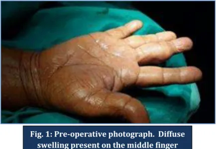
ABSTRACT: INTRODUCTION: The giant cell tumor of tendon sheath is among the more common soft tissue tumors of the hand. It is a slowly progressive, usually painless, rubbery mass predominating on the radial three digits of the hand and is typically identified adherent to the digital flexor tendon sheath of the hand. The histology is variable but the tumors consistently contain multinucleated giant cells and xanthoma cells. CASE PRESENTATION: A case report of a 57 years old female patient who came with a diffuse, painless swelling on the radial aspect of middle finger of the right hand at the Orthopedic OPD of our institution. MANAGEMENT & OUTCOME: A planned operative procedure was done. After clinical examination and other investigations, a careful excision of the mass was done by detaching it from the underlying tendon sheath. The tissue was sent for biopsy to two different labs for examination. The reports were confirmed as Giant Cell tumor of the tendon Sheath. DISCUSSION: A giant cell tumors of the tendon sheath (GCTTS) is an uncommon and usually benign lesion that arises from the tendon sheath though they are the second most common tumors of the hand. Recurrence rates are as high. A recent study found that following surgical excision, the only lesions that recurred were those that originally had multiple discrete tumors. Tumors composed of single masses did not recur following surgical excision. MRI may be helpful in determining the anatomic extent of the lesion.
KEYWORDS: Giant cell tumor, Tendon Sheath, Recurrence, Benign neoplasm, Excision.
INTRODUCTION: Giant cell tumor of tendon sheath (GCTTS) is clinically a slow growing soft tissue mass that develops over a period of months to years. It is the second commonest tumor of the hand Trauma, inflammation; metabolic disease and a neoplastic etiology are considered as etiological factors. It is a slowly progressive, usually painless, rubbery mass predominating on the radial three digits of the hand and is typically identified adherent to the digital flexor tendon sheath of the hand. It is notorious for its recurrence. Many factors are considered as causing recurrence, including proximity to the distal interphalangeal joints, presence of degenerative joint disease, pressure erosions in the radiographs, increased mitotic activity.1
J of Evolution of Med and Dent Sci/ eISSN- 2278-4802, pISSN- 2278-4748/ Vol. 3/ Issue 13/ Mar 31, 2014 Page 3279
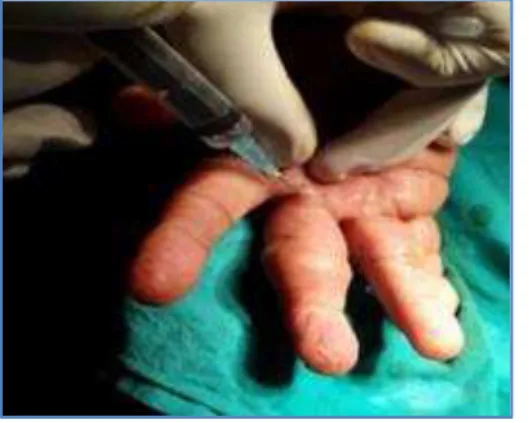
MANAGEMENT AND OUTCOME: A planned operative procedure was carried out. Local infiltration of Xylocaine 2% was given by the ring method at the base of the middle finger. After setting of anesthetic effect, a linear incision was made over the mass. A tourniquet was applied to control bleeding and clearing the surgical field. Special care was taken to excise the tumor in total, retaining the capsule, with margin of normal tissue. The operating field was searched for presence of satellite lesions or daughter cysts. The entire specimen was then subjected to histopathological examination at two different labs, and the margins were observed for clearance.
DISCUSSION: A giant cell tumor of the tendon sheath (GCTTS) is an uncommon and usually benign lesion that arises from the tendon sheath though they are the second most common tumors of the hand, with simple ganglion cysts being the most common. It is unclear whether these lesions represent neoplasm or simply reactive masses.1 Clinically these masses typically present in the hand (although they are found elsewhere also) with localized swelling with or without pain. They are slow growing. Typically, they present in 3rd -5th decades and have a slight female predilection with a M: F ratio of 1.5-2.1:1.2 They have been divided macroscopically into localized or diffuse forms, and appear as rubbery multinodular masses that are well circumscribed. They have an enveloping fibrous capsule, and the cut surface is variably coloured depending on the relative proportions of fibrous tissue, hemosiderin and pigmented foam cells.3
As is true for most soft-tissue tumors, the etiology of giant cell tumors of the tendon sheath is unknown. Pathogenetic theories have included trauma, disturbed lipid metabolism, osteoclastic proliferation, infection, vascular disturbances, immune mechanisms, inflammation, neoplasia, and metabolic disturbances.Probably the most widely accepted theory, as Abimelac et al proposed, is that of a reactive or regenerative hyperplasia associated with an inflammatory process.4, 5 Histochemical evidence shows that the mononuclear cells and giant cells present in these lesions resemble osteoclasts, suggesting a bone marrow–derived monocyte/macrophage lineage for these tumors.
The tumor is histologically identical to pigmented villonodular synovitis (PVNS)6 and is composed of fibroblasts and multinucleated giant cells, foamy histiocytes and inflammatory cells on a background fibrous matrix. Giant cell tumors of the soft tissue are classified into 2 types: the common localized type and the rare diffuse type.7
The rare diffuse form is considered the soft tissue counterpart of diffuse pigmented villonodular synovitis (PVNS) and typically affects the lower extremities.Its anatomic distribution parallels that of PVNS, with lesions most commonly found around the knee, followed by the ankle and foot; however, the diffuse form occasionally affects the hand. Typically, these lesions, like those of PVNS, occur in young patients; 50% of cases are diagnosed in patients younger than 40 years. The diffuse form is often locally aggressive, and multiple recurrences after excision are common.
Recurrence of Giant cell tumor of the tendon sheath (GCTTS) is an unresolved issue, though it is basically a non-malignant condition. Reported rate of recurrence of GCTTS is 9–44% and some authors have reported recurrence rate as high as 45%.
J of Evolution of Med and Dent Sci/ eISSN- 2278-4802, pISSN- 2278-4748/ Vol. 3/ Issue 13/ Mar 31, 2014 Page 3280 nodules is considered as the most important factor deciding recurrence pattern. MRI may be helpful in determining the anatomic extent of the lesion. Adequate surgical exposure, meticulous dissection and use of magnification are necessary to reduce recurrence and should remain the mainstay of surgical management.
REFERENCES:
1. Rao AS, Vigorita VJ. Pigmented villonodular synovitis (Giant-Cell tumor of the tendon sheath and synovial membrane): a review of 81 cases. J Bone Joint Surg. 1984; 66A:76–94. [PubMed] 2. Ly JQ, Carlson CL, Lagatta LM et-al. Giant cell tumor of the peroneus tendon sheath. AJR Am J
Roentgenol. 2003; 180 (5): 1442. AJR Am J Roentgenol (full text) - Pubmed citation
3. Enzinger FM, Weiss SH. Benign tumors and tumor like lesions of synovial tissue. In: Enzinger FM, Weiss SW, eds. Soft Tissue Tumors. St Louis, Mo: Mosby; 1995:735-55.
4. Choudhury M, Jain R, Nangia A. Localized tenosynovial giant cell tumor of tendon sheath. A case report. Acta Cytol. May-Jun 2000; 44(3):463-6. [Medline].
5. Abimelec P, Cambiaghi S, Thioly D. Subungual giant cell tumor of the tendon sheath. Cutis. Oct 1996; 58(4):273-5. [Medline].
6. Jaffe HL, Lichtenstein HL, Elsutro CJ. Pigmented villonodular synovitis, bursitis, and tenosynovitis. Arch Pathol. 1941; 31:731-65.
7. Abdul-Karim FW, el-Naggar AK, Joyce MJ. Diffuse and localized tenosynovial giant cell tumor and pigmented villonodular synovitis: a clinicopathologic and flow cytometric DNA analysis. Hum Pathol. Jul 1992; 23(7):729-35. [Medline]
8. Lowyck H, Smet L. Recurrence rate of giant cell tumors of the tendon sheath. Eur J Plast Surg. 2006; 28:385–388. doi: 10.1007/s00238-005-0791-6. [Cross Ref]
9. Agarwal PK, Gupta M, Srivastava A. Cytomorphology of giant cell tumor of tendon sheath. A report of two cases. Acta Cytol. Mar-Apr 1997; 41(2):587-9. [Medline].
J of Evolution of Med and Dent Sci/ eISSN- 2278-4802, pISSN- 2278-4748/ Vol. 3/ Issue 13/ Mar 31, 2014 Page 3281
Fig. 2: The base of the finger was infiltrated with local anesthesia (Xylocaine2%)
Fig. 3: A deeper dissection was made to assess the basal attachment of the mass
J of Evolution of Med and Dent Sci/ eISSN- 2278-4802, pISSN- 2278-4748/ Vol. 3/ Issue 13/ Mar 31, 2014 Page 3282 AUTHORS:
1. Ravikumar A.S. 2. Amith Chaudhary
PARTICULARS OF CONTRIBUTORS:
1. Assistant Professor, Department of Orthopaedics, Sri Siddhartha Medical College and Hospital, Tumkur, Karnataka.
2. Resident, Department of Orthopaedic Surgery, Sri Siddhartha Medical College and Hospital, Tumkur, Karnataka.
NAME ADDRESS EMAIL ID OF THE CORRESPONDING AUTHOR:
Dr. Ravikumar A. S, Assistant Professor,
Department of Orthopaedics,
Sri Siddhartha Medical College & Hospital, Tumkur-572107, Karnataka.
E-mail: [email protected]
Date of Submission: 04/03/2014. Date of Peer Review: 05/03/2014. Date of Acceptance: 14/03/2014. Date of Publishing: 25/03/2014.