1Laboratório de Neurociências, Núcleo de Pesquisas Tecnológicas/Universidade de Mogi das Cruzes (NPT/ UMC), Mogi das Cru z e s S P, Brazil;2Laboratório de Neurologia Experimental, Universidade Federal de São Paulo/Escola Paulista de Medicina (UNIFESP/EPM), São Paulo SP, Brazil. FAEP, FAPESP, CNPq and CAPES supported this study.
Received 30 May 2005, received in final form 27 July 2005. Accepted 19 August 2005.
D r. Fulvio Alexandre Scorza - Rua Botucatu 862 - Ed. Leal Prado, Disciplina de Neurologia Experimental / UNIFESP/EPM - 04023-900 São Paulo SP - Brasil. E-mail: scorza.nexp@epm.br
LOVASTATIN REDUCES NEURONAL CELL DEATH
IN HIPPOCAMPAL CA1 SUBFIELD AFTER
PILOCARPINE-INDUCED
STATUS EPILEPTICUS
Preliminary results
Pauline Rangel
1, Roberta Monterazzo Cysneiros
2, Ricardo Mario Arida
1,2,
Marly de Albuquerque
1, Diego Basile Colugnati
1,2, Carla Alessandra Scorza
2,
Esper Abrão Cavalheiro
2, Fulvio Alexandre Scorza
1,2ABSTRACT -Objective:To further characterize the capacity of lovastatin to prevent hippocampal neuronal loss after pilocarpine-induced status epilepticus (SE) Method:Adult male Wistar rats were divided into four gro u p s : (A) control rats, received neither pilocarpine nor lovastatin (n=5); (B) control rats, received just lovastatin (n=5); (C) rats that received just pilocarpine (n=5); (D) rats that received pilocarpine and lovastatin (n=5). After pilo-carpine injection (350mg/kg, i.p.), only rats that displayed continuous, convulsive seizure activity were included in our study. Seizure activity was monitored behaviorally and terminated with an injection of diazepam (10 mg/kg, i.p.) after 4 h of convulsive SE. The rats treated with lovastatin received two doses of 20mg/kg via an oesophag-ic probe immediately and 24 hours after SE induction. Seven days after pilocarpine-induced SE, all the animals w e reperfused and their brains were processed for histological analysis through Nissl method. Results:The cell counts in the Nissl-stained sections perf o rmed within the hippocampal formation showed a significant cell loss in rats that received pilocarpine and presented SE (CA1= 26.8 ± 13.67; CA3= 38.1 ± 7.2; hilus= 43.8 ± 3.95) when c o m p a red with control group animals (Group A: CA1= 53.2 ± 9.63; CA3= 63.5 ± 13.35; hilus= 59.08 ± 10.24; Gro u p B: CA1= 74.3 ± 8.16; CA3= 70.1 ± 3.83; hilus= 70.6 ± 5.10). The average neuronal cell number of CA1 subfield of rats that present SE and received lovastatin (44.4 ± 17.88) was statically significant increased when compare d with animals that just presented SE. Conclusion:Lovastatin exert a neuro p rotective role in the attenuation of brain damage after SE.
KEY WORDS: epilepsy, pilocarpine, lovastatin, hippocampus.
Lovastatina reduz a lesão celular na região CA1 do hipocampo após o status epilepticusi n d u z i d o pela pilocarpina: resultados preliminares
RESUMO - Objetivo:Capacidade da lovastatina em prevenir a perda de neurônios hipocampais após o status epilepticus (SE) induzido pela pilocarpina. Método:Ratos adultos Wistar foram divididos em 4 grupos: (A) ratos c o n t roles que não receberam pilocarpina nem lovastatina (n=5); (B) ratos controles que receberam somente lovas-tatina (n=5); (C) ratos que receberam somente pilocarpina (n=5); (D) ratos que receberam pilocarpina e lovastati-na (n=5). Após a administração de pilocarpilovastati-na (350mg/kg, i.p.), somente ratos que evoluíram para o status epilep-ticus foram incluídos em nosso estudo. A atividade epiléptica foi interrompida com uma injeção de diazepam (10 mg/kg, i.p.) após 4h do início do SE. Os ratos tratados com lovastatina receberam duas doses de 20mg/kg via esofágica, imediatamente e 24 h após a indução do SE. Sete dias após o SE induzido pela pilocarpina, todos os animais foram perfundidos e seus cére b ros processados para análise histológica através do método de Nissl.
Resultados:A contagem celular da formação hipocampal mostrou uma significante perda celular nos animais que receberam pilocarpina e apresentaram SE (CA1= 26,8 ± 13,67; CA3= 38,1 ± 7,2; hilus= 43,8 ± 3,95) quando comparados com animais pertencentes ao grupo controle (Grupo A: CA1= 53,2 ± 9,63; CA3= 63,5 ± 13,35; hilus= 59,08 ± 10,24; Grupo B: CA1= 74,3 ± 8,16; CA3= 70,1 ± 3,83; hilus= 70,6 ± 5,10). O número de células neuro n a i s na região CA1 do hipocampo de ratos que apresentaram SE e receberam lovastatina (44,4 ± 17,88) foi estatisti-camente maior quando comparado com animais que somente apresentaram SE. Conclusão:A lovastatina exerc e papel neuro p rotetor na atenuação do dano cerebral após o SE.
Status epilepticus(SE), a neurological emerg e n-cy characterized by abnormally prolonged seizure s , remains an important clinical pro b l e m1. Animal m o
d-els of SE have been used to produce chronic epilep-s y, thuepilep-s epilep-suggeepilep-sting that SE itepilep-self iepilep-s epileptogenic2.
The systemic administration of a potent agonist p i l o-carpine to rats leads to a pattern of repetitive lim-bic seizures and SE, which can last for up 12 h o u r s3 , 4.
Morphological analysis of hippocampal form a t i o n after pilocarpine-induced SE shows an extensive loss of neurons within the hilar area of the denta-te gyru s4 , 5, as well a loss of selective populations of
interneurons in areas CA1 and CA3 and in the hi-l u s4. Furt h e rm o re, SE-associated cell loss is a critical
step that activates a cascade of events leading to mossy fiber sprouting,de novore c u rrent excitat i o n of granule cells, culminating in spontaneous re-current seizures (SRSs)4-6.
Lovastatin, a fungal antibiotic used in hyperc h o-l e s t e roo-lemia treatment, is a competitive inhibitor of HMG-CoA (3-hydro x y - 3 - m e t h y l g l u t a ryl coenzime A reductase), the major re g u l a t o ryenzyme of d e n o v oc h o l e s t e rol synthesis7. Recent data reveal that
statins reduce vascular inflammatory re s p o n s e s8,
p romote angiogenesis9, modulate cytokine pro d u
c-t i o n1 0and decrease oxidative stre s s1 1. Furt h e rm o re ,
recent studies have shown that statins reduces the extent of brain damage after ischemic insult1 2.
Based on this, the purpose of our study was to f u rther characterize the capacity of lovastatin to prevent hippocampal neuronal loss after pilocar-pine-inducedstatus epilepticus.
METHOD
Adult male Wistar rats (n=20), weighting 200-250g w e re housed under standard controlled conditions (7:00 AM/7:00 P.M. light/dark cycle; 20-22oC; 45-55%
humidi-ty) with food and waterad libitumw e re used in our stu-d y. The pro c e stu-d u res involving the animals anstu-d their care at the Neuroscience Laboratory at University of Mogi das Cruzes respected the Institution’s guidelines, which comply with national and international rules and polic i e s . Rats were divided randomly into four groups: (A) cont ro l rats, received neither pilocarpine nor lovastatin (n=5); (B) control rats, received just lovastatin (n=5); (C) rats that received just pilocarpine (n=5); (D) rats that re c e i v e d pilocarpine and lovastatin (n=5).
SE were induced according to the pro c e d u re des-cribed pre v i o u s l y4. In brief, 30 minutes after
methylsco-polamine injection (1mg/kg, s.c - used to reduce periph-eral effects of pilocarpine), pilocarpine (350mg/kg, i.p.) was administered to rats. Only rats that displayed conti-nuous, convulsive seizure activity after pilocarpine tre a
t-ment were included in these studies. Seizure activity w a s m o n i t o red behaviorally and terminated with an i.p. i n-jection of diazepam (10 mg/kg; Roche, Brazil) after 4 h of convulsive SE. Lovastatin was provided by Millenium P h a r-macy (São Paulo, Brazil) and was diluted using sterile s a-line 0,9%. The rats treated with lovastatin received two doses of 20mg/kg via an oesophagic probe immediatel y and 24 hours after SE induction.
Seven days after pilocarpine-induced SE, the animals w e re perfused and the brains were processed for histo-logical analysis. Sections cut 20µm thick were stained a c c o rding to Nissl method. Cell counts in the Nissl-stained sections, were perf o rmed within the hippocampal pyra-midal cell layer and the dentate hilus, as previously re-p o rted by Mouritzen-Dam1 3. For each animal, both the
right and left hemispheres of three diff e rent sections w e re counted to provide a total of five individuals valu-es per animal. Only cells with evident nucleus and nucle-olus were included in the counts. The mean value observ e d in control rats was considered 100% of normal cell popu-lation. Statistical analysis was perf o rmed by one-way analysis of variance (ANOVA), followed by post-hoc Dunnett´s test; p values of p<0.05 were considered stati-cally significant.
RESULTS
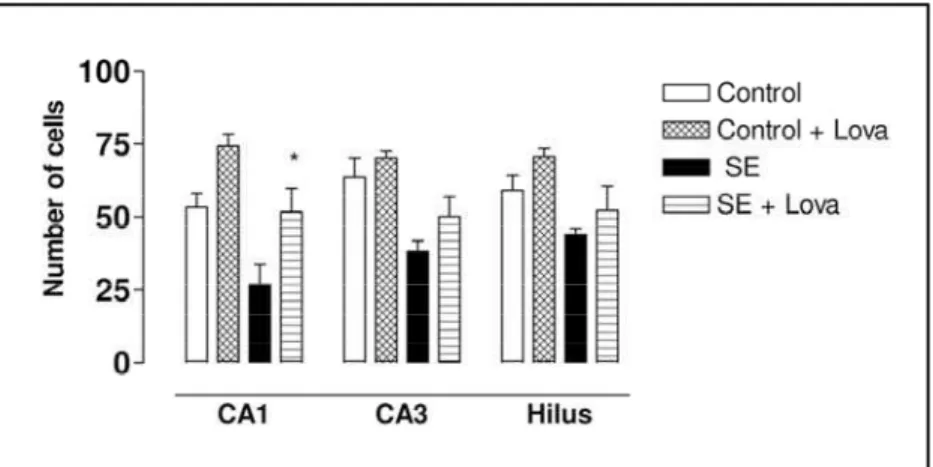
Pilocarpine treatment induced the following behavioral changes: akinesia, facial automatisms, and limbic seizures consisting of forelimb clonus with rearing, salivation, and masticatory jaw move-ments and falling. This type of behavior built up p ro g ressively into motor limbic seizures that re-c u rred repeatedly and rapidly and developed into SE. After SE, animals were comatose or unre s p o n-sive to their environment and akinetic; behavior re t u rned toward normal over a 3 to 5-day period. We next analyzed the qualitative morphological changes in the hippocampus associated with lovas-tatin treatment and SE. In the control animals (Fig 1A, 1B), the Nissl staining of hippocampal form a t i o n and dentate gyrus showed integrity of all cells lay-ers. In contrast, animals that received just pilocarpine and presented SE (Fig 1C) we observed a significant cell loss in hippocampal subfields CA1, CA3 and hilus of dentate gyrus. Interesting, the structural analy-sis of the brains from animals that presented SE and received lovastatin (Fig 1D) was similar when com-p a red with animals in the control gro u com-p .
Fig 2. Neuronal cell count. Data re p re s e n t average neuronal cell counts in contro l rats, rats that received just pilocarpina and rats that received pilocarpine and lovas tatin. Data were analyzed by ANOVA, fol -lowed by post hoc Dunnett´s test. Note the reduced cell loss in CA1 subfield of rats that received pilocarpine and lovastatin when compared with rats that just re c e i v e d pilocarpina. Data expressed as mean ± S.E. *p<0.05.
and presented SE (CA1= 26.8 ± 13.67; CA3= 38.1 ± 7.2; hilus= 43.8 ± 3.95) when compared with con-t rol group animals (Group A: CA1= 53.2 ± 9.63; CA3= 63.5 ± 13.35; hilus= 59.08 ± 10.24; Group B: CA1= 74.3 ± 8.16; CA3= 70.1 ± 3.83; hilus= 70.6 ± 5.10). The average neuronal cell number of CA1 subfield of rats that present SE and received lovas-tatin (44.4 ± 17.88) was statistically significant in-c reased when in-compared with animals that just pre-sented SE. It is therefore of interest that the cells in CA3 subfield and hilus of rats that present SE and received lovastatin (CA3= 50.14 ± 15.33; hilus= 52.20 ± 18.71) tended to be relatively protect when c o m p a redwith animals that just received pilocar-pine and presented SE.
DISCUSSION
This study evaluated the effects of lovastatin administration in rats after pilocarpine-induced S E . We demonstrated that lovastatin treatment was able to prevent hippocampal neuronal loss in CA1 subfield after an epileptic insult. In these lines, our results are in agreement with a recent study that re-p o rted an effective neuro re-p rotective action of sta-tins after an acute brain injury14.
Reactive oxygen species (ROS) are a part of nor-mal human metabolism; however, when pro d u c e d in excess, ROS can cause tissue injury including lipid p e roxidation, DNA damage and enzyme inactiva-t i o n8. ROS is a common denominator among acute
n e u rological conditions8 , 1 5, including epilepsy1 6 , 1 7. In
the pilocarpine model, there is a involvement of exci-totoxic neuronal injury1 8and ROS production has
been considered to be a part of mechanisms involved with glutamatergic excitotoxicity in vitro1 5and in
v i v o1 6 , 1 7 , 1 9. More o v e r, it was demonstrated that
lovas-tatin treatment inhibits free radical injury2 0. Thus,
this antioxidant effect of statins could explain the n e u ro p rotective pro p e rties found in our study.
T h e re is one classical argument supporting a possible role of nitric oxide (NO) in convulsive phe-nomena: excitatory amino acids, such as N-methyl-D- aspartate (NMDA) and kainate, are known to be potent convulsants2 1, and the activation of N M D A
receptors is accompanied by the formation of NO2 2.
In fact, the role of NO in epileptogenesis has been examined in a number of studies, suggesting to be a proconvulsive endogenous substance2 3 , 2 4.
Fur-t h e rm o re, some sFur-tudies have revealed Fur-thaFur-t sFur-taFur-tins inhibit the production of NO in brain pare n c h ym a1 0,
indicating that statins, secondarily, may play the
role of an anticonvulsant substance, does not pro-moting glutamate-mediated neuro t o x i c i t y. Other possibility is that the inhibition of brain endothe-lial nitric oxide synthase (eNOS) leads to incre a s e d blood pre s s u re2 5, which, in turn, may affect the
ex-citability of central nervous system2 6; however, it
was demonstrated that statins are able to upre g u-late eNOS2 7, may be pivotal in enhance cerebral a rt
e-rial vasodilator responses, decreasing with this, the firing threshold.
F i n a l l y, anti-inflammatory effects of statins could also contribute to neuro p rotection after pilo-carpine-induced SE observed in our study. The pa-thological alterations that occur in the hippocam-pus following prolonged seizures begin within hours and cause changes that last throughout life. M o re o v e r, several studies has implicated a number of cytokines in seizure - related hippocampal pathol-o g y2 8 , 2 9. Intere s t i n g l y, Pahan and colleagues1 0h a v e
been shown that lovastatin reduces the induction of inflammatory mediators. Taken together, lovas-tatin treatment may provide an important appro-ach to suppression of the inflammatory re s p o n s-es after SE induced by pilocarpine.
Based on these facts, our pre l i m i n a ryresults s u p-p o rt p-previous evidence that statins reduces neuro-nal death after an acute brain insult. Future studi-es are needed to gain a better understanding of t h e-se and other possible mechanisms of lovastatin du-ring epileptogenesis
Acknowledgements -The authors would like to thank Hilda S. Reis for her help with histological techni-ques and Jaqueline Botelho her help in cell counts analy-sis. FA E P, FA P E S P, CNPq and CAPES supported this study.
REFERENCES
1. Lowenstein DH, A l l d redge BK. Current concepts: status epilepticus. N Engl J Med 1998;388:970-976.
2. Treiman DM. Therapy of status epilepticus in adults and children. Curr Opin Neurol. 2001;14:203-210.
3. Turski WA, Cavalheiro EA, Schwarz M, Czuczwar SJ, Kleinrok Z, Tu r s k i L. Limbic seizures produced by pilocarpine in rats: behavioural, elec-t roencephalographic and neuropaelec-thological selec-tudy. Behav Brain Res 1983;9:315-335.
4. C a v a l h e i ro EA. The pilocarpine model of epilepsy. Ital J Neurol Sci 1995;16:33-37.
5. Mello LEAM, Cavalheiro EA, Tan AM, et al. Circuit mechanisms of sei-zure in the pilocarpine model of chronic epilepsy: cell loss and mossy fiber sprouting. Epilepsia 1993;34:985-995.
6. Mathern GW, Betram EH, Babb TL, Pretorius JK, Kuhlman PA, Men-doza D. In contrast to kindled seizures, the frequency of spontaneous epilepsy in the limbic status model correlates with greater aberrant fas-cia dentata excitatory and inhibitory axon sprouting, and incre a s e d staining for NMDA, AMPA and GABA receptors. Neuroscience 1997; 77:1003-1019.
8 . Gutteridge JM, Halliwell B. Free radicals and antioxidants in the year 2000: a historical look to the future. Ann N Y Acad Sci 2000;899:136-147. 9. K u reishi Y, Luo Z, Shiojima I, et al. The HMG-CoA reductase inhibitor simvastatin activates the protein kinase Akt and promotes angiogene-sis in normocholesterolemic animals. Nat Med 2000;6:1004-1010. 10. Pahan K, Sheikh FG, Namboodiri AM, Singh I, Lovastatin and
phenyla-cetate inhibit the induction of nitric oxide synthase and cytokines in rat primary astrocytes, microglia, and macrophages. J Clin Invest1997; 100:2671-2679.
11. Aviram. M, Rosenblat M, Bisgaier CL, Newton RS. Atorvastatin and g e m f i b rozil metabolites, but not the parent drugs, are potent antioxi-dants against lipoprotein oxidation. A t h e ro s c l e rosis 1998;138:271-280. 12. S i roni L, Cinino M, Guerrini U, et al. Treatment with statins after induc-tion of focal ischemia in rats reduces the extent of brain damage. A r t e-rioscler Thromb Vasc Biol. 2003;23:322-327.
13. Mouritzen-Dam A. Hippocampal neurons loss in epilepsy and after experimental seizures. Acta Neurol Scand 1992;66:601-642. 14. Daimon A, Shigeyuki A, Takakazu K, Kurosawa H. Pravastatin, a
3-h y d roxy-3-met3-hylglutaryl coenzyme A reductase in3-hibitor, reduces de-layed neuronal death following transient forebrain ischemia in the adult rat hippocampus. Neurosci Lett 2004;362:122-126.
15. Bonfoco E, Krainc D, A n k a rc rona M, Nicotera P, Lipton AS. A p o p t o s i s and necrosis: two distinct events induced, re s p e c t i v e l y, by mild and intense insults with N-methyl-D-aspartate or nitric oxide/supero x i d e in cortical cell cultures. Proc Natl Acad Sci 1995;92:7162-7166. 16. B ruce AJ, Baudry M. Oxygen free radicals in rat limbic stru c t u res after
kainate-induced seizures. Free Radic Biol Med 1995;18:993-1002. 1 7 . Ueda Y, Yokoyama H, Niwa R, Konaka R, Ohya-Nishiguchi H, Kamada
H. Generation of lipid radicals in the hippocampal extracellular space during kainic acid-induced seizures in rats. Epilepsy Res 1997;26:329-333. 18. C a v a l h e i ro EA, Leite JP, Bortolotto ZA, Turski WA, Ikonomidou C,
Turski L. Long-term effects of pilocarpine in rats: structural damage of the brain triggers kindling and spontaneous re c u r rent seizures. Epilepsia 1991;32:778-782.
19. Schulz JB, Henshaw DR, Siwek D, et al. Involvement of free radicals in excitotoxicity in vivo. J Neurochem. 1995;64:2239-2247.
2 0 . Chen L, Haught WH., Yang B, Saldeen TGP, Parathasarathy S, Metha JL. P reservation of endogenous antioxidant activity and inhibition of lipid p e roxidation as common mechanisms of antiathero s c l e rotic effects of vita-min E, lovastatin and amlodipine. J Am Coll Cardiol 1997;30:569-575. 21. M e l d rum BS. Amino acid neurotransmitters in new approaches to
anti-convulsant drug action. Epilepsia 1984;22:140-149.
22. Garthwaite J. Glutamate, nitric oxide and cell-cell signalling in the nerv-ous system. Trends Neurosci 1991;14:60-67.
23. De Sarro GB, Donato Di Paola E, Sarro A, Vidal JM. Role of nitric oxide in the genesis of excitatory amino acid-induced seizures from the deep prepiriform cortex. Fundam Clin Pharmacol 1991;5:503-511. 24. Mulsch A, Busse R, Mondvintcev PL, et al. Nitric oxide promotes seizure
activity in kainate-treated rats. Neuroreport 1994;21:2325-2328. 25. Moncada S,. Palmer RMJ, Higgs AS. Nitric oxide: physiology,
pathophy-siology, and pharmacology. Pharmacol Rev 1991;43:109-142. 26. Fewell JE, Johnson P. Acute increases in blood pressure cause arousal
from sleep in lambs. Brain Res 1994;311:259-265.
27. Laufs U, Gertz K, Huang P, et al. Atorvastatin upregulates type III nitric oxide synthase in thrombocytes, decreases platelet activation, and pro-tects from cerebral ischemia in normocholesterolemic mice. Stro k e 2000;31:2442-2449.
28. Rothwell NJ. Annual review prize lecture cytokines - killers in the brain? J Physiol 1999;514:3-17