639
PROTECTIVE EFFECT OF NARINGENIN
ON GLUTAMATE-INDUCED NEUROTOXICITY
IN CULTURED HIPPOCAMPAL CELLS
Xiao-Hui Xu*, Cong-Min Ma, Yue-Zhen Han, Yan Li, Chao Liu,
Zhi-Hui Duan, Hui-Lin Wang, De-Quan Liu and Rui-Hua Liu
Department of Neurology, Luoyang Central Hospital Affiliated to Zhengzhou University, Luoyang, PR China
*Corresponding author: xhxu86@gmail.com
Abstract: Monosodium glutamate induces excitotoxicity in the central nervous system through hyperactiva-tion of both ionotropic and metabotropic glutamate receptors, which leads to neuronal cell death. In this study, we investigated the neuroprotective effects of naringenin on excitotoxicity induced by glutamate in primary hippocampal neurons of neonatal mice. The expression levels of apoptosis-inducing proteins and as well as ischemic factors were observed by Western blot analysis. Immunocytochemistry and morphometric analysis of hippocampal cells with or without glutamate and naringenin treatment were performed. We observed that naringenin regulated Erk1/2 and Akt phosphorylation and reduced the demise of dendrites due to glutamate exposure in cultured hippocampal neurons. Furthermore, naringenin induced the brain-derived neurotrophic factor and other neuroprotective cytokines, and markedly improved the survival rates of the neurons 24 h fol-lowing glutamate exposure. The observed results suggest that the naturally occurring bioflavonoid (naringenin) exerts neuroprotective effects via highly specific molecular targets in neurons.
Key words: excitotoxicity; glutamate; hippocampal neuronal cells; naringenin; neuroprotection
Received August 11, 2014; Revised October 14, 2014; Received October 29, 2014
INTRODUCTION
Oxygen/glucose deprivation (OGD), glutamate disturbance and inflammation comprise the mul-tifactorial pathophysiological process of cerebral ischemia and stroke (Harukuni and Bhardwaj, 2006). Glutamate-induced neurotoxicity has been implicated in severe brain disorders, including the Alzheimer’s disease, epilepsy, ischemic stroke and also in Parkinson’s disease (Fukui et al., 2009; Xu et al., 2011). Previous studies have reported
of glutamate receptors, namely α-amino-3-hydroxy-5-methyl-4-isoxazolepropionic acid (AMPA), N-methyl D-aspartate (NMDA) and kai-nate receptors (KARs). The main toxicity of MSG includes neurotoxicity, disorders of the endocrine glands associated with neurological activity, learn-ing difficulties, epileptic seizures, increases in glu-cose levels and increased incidences of metabolic diseases (Egbuonu et al., 2009). One of the ways to counteract these deleterious effects of neurotoxic-ity produced by excess MSG is to evaluate an effec-tive neuroproteceffec-tive compound of natural origin.
Naringenin, a naturally-occurring bioflavonoid that is found in fruit and vegetables such as grape-fruit, orange and tomato, is reported to have anti-inflammatory as well as antioxidant properties (Shi et al., 2009; Baumann et al., 1980). The antioxidant effect of naringenin is mainly attributed to the pres-ence of a 4’ hydroxyl group in the B ring, which effectively quenches free radicals (van Acker et al., 2000). It exhibits beneficial effects in oxidative stress-mediated diseases, such as airway inflammation and neuroinflammation possibly due to its anti-oxidative and anti-inflammatory properties (Yang et al., 2011; Assini et al., 2013). Since glutamate-induced excito-toxicity and neuronal death are currently thought to contribute to the progression of cerebral ischemia and the occurrence of strokes, it is plausible to hy-pothesize that naringenin might exhibit neuropro-tective potential. The validity of its potential neuro-protection has been evaluated in this study.
MATERIALS AND METHODS
Mouse hippocampal neuron isolation and culture
The primary cultures of hippocampal neurons were isolated from neonatal (C57Bl/6J) mice as
described earlier by Berbari et al. (2007), with slight modifications. The study was approved by the Institutional Ethical Animal Care Commit-tee of Zhengzhou University and was performed according to the institutional animal care com-mittee guidelines. The tissues from hippocam-pal regions were isolated from neonatal mice on postnatal day (P) 1. The isolated tissues were treated with papain (0.4 mg/mL) and DNase (50 µg/mL) in Leibovitz L-15 medium containing bo-vine serum albumin (0.2 mg/mL) for around 15 min at 37°C. Treated tissue samples were washed with NEUROBASAL-A medium supplemented with B-27 thrice and gently ground. Following centrifugation at 4°C for 5 min at 200 g, the cells were resuspended in NEUROBASAL-A/B-27 medium containing DNase. Prior to plating, the cells were passed through a cell strainer (100 µm mesh) and plated (1.0 x10⁵ cells/cm2) onto
poly-D-lysine precoated glass coverslips.
Experimental design
Western blot analysis
Equal concentrations of the isolated proteins were separated by SDS-PAGE and blotted to a nitrocellulose membrane. Phosphatase activity was inhibited by the addition of sodium fluoride (1 mM) to sample lysis buffer. The blots were blocked with blocking buffer (Tris-buffered sa-line with Tween 20 (TBST) containing 10% (w/v) non-fat dry milk) for 1 h at room temperature. Following treatment with blocking buffers, the blots were incubated overnight at 4°C with pri-mary antibodies against Erk, p-Erk, Akt, p-Akt, caspase 3 and calpain 1 (1:500). After the incuba-tion period, the blots were incubated with sec-ondary antibody (1:500) conjugated with alkaline phosphatase, incubated at room temperature for 1 h and later washed with TBST, where NBT/ BCIP was used to visualize the available immune reactive bands in the blots.
Morphometric analysis and immunocytochemistry study
The neurons were fixed on glass coverslips that were permeabilized with ice-cold methanol. The cells were then blocked with skimmed milk (10%) in phosphate-buffered saline (PBS) for around 30 min, followed by incubation with anti-MAP2 antibody (1:200 dilution) at 4°C for over-night. Samples were incubated with secondary antibody that was conjugated with Alexa Fluor 488 in a 1:200 dilution for 30 min at room tem-perature, after a washing with PBS. The cells were further stained with 4′ ,6-diamidino-2-phenylin-dole (DAPI) and mounted on a glass slide. Im-ages of the stained cells were investigated using a microscope that was equipped with a CCD color camera and DP2-BSW software for recording the images. As cell compactness affects dendrite length, the cell density images tend to
culate the mean dendrite length due to overlap, random field selection was employed.
Real-time PCR analysis
Using TRIZOL reagent, total RNA was isolated from the experimental cell groups as per the manufacturer’s instructions (Gibco-BRL, Grand Island, New York) and its purity was established by determining its absorbance at 260 and 280 nm. According to the manufacturer’s instruction, the superscript preamplification system was used with Superscript II RNase H-reverse transcriptase for synthesizing the first cDNA from total RNA. Further amplification of the cDNA (1 μl) tem-plate was performed by PCR reaction that con-tained supermix (18 μl) and each specific primer (10 pmol) making up the total volume of 20 μl. For PCR reaction, primer sequences of caspase-3 (forward 5′-TGTCATCTCGCTCTGGTACG-3′ and reverse 5′-AA ATGACCCCT TCAT-CACCA-3′) and calpain 1 (forward, 5′ -GT-GGTAGCCGCTGAAACTC C-3′; reverse, 5′-TGTTCCCGCTCTCATCTGC-3′) were used along with glyceraldehy3-phosphate de-hydrogenase (GAPDH), which was amplified and used (forward 5′ -AACTTTGGCATTGTG-GAAGG-3′ and reverse 5′ -GGAGACAACCTG-GTCCTCAG-3′) as an internal control. For the amplification of the cDNA, the PCR reaction for calpain 1 employed 30 cycles, caspase-3 35 cycles, and the internal control (GAPDH) 28 cycles. The obtained amplified products were separated us-ing agarose gel (1.5%) electrophoresis and were analyzed using a Bio-Rad Imaging Densitometer (Bio-Rad, Hercules, California).
Statistical analysis
The obtained results were subjected to statistical analysis and values were expressed as the mean ± SEM. A two-tailed t-test was used to compare
the differences between two groups and one-way analysis of variance (ANOvA) was used to com-pare differences that are seen among multiple groups. A post hoc test was also used to compare a few selected groups and P<0.05 was considered significant.
RESULTS
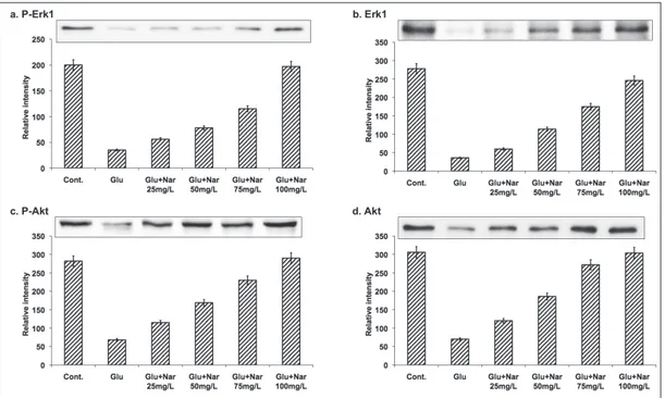
Naringenin protects glutamate-induced Erk1 phosphorylation
The neuroprotective effects of naringenin were assessed. Mouse hippocampal neurons were sub-jected to an excitotoxic concentration of gluta-mate in the presence of naringenin and the levels of Erk1 phosphorylation were assessed. The ini-tial trial was carried out using different concen-trations (25, 50, 75 and 100 mg/L) of naringenin against glutamate excitotoxicity. On exposure of cultured hippocampal neuron cells to glutamate (50 mM) for 30 min, a pronounced reduction of Erk1 phosphorylation was observed (Fig. 1). However, in glutamate-exposed cells treated with 100 mg/L naringenin, significant Erk1 phosphor-ylation (Fig.1) was observed, while lower concen-trations of naringenin were ineffective. Further, naringenin also caused a similar increase in the phosphorylation of Akt (Fig. 1). These data col-lectively demonstrate that naringenin at a con-centration of 100 mg/L showed the optimum protection of Erk1 during excitotoxic stress in cultured hippocampal neuronal cells.
Neuroprotective effects of naringenin
identifi-cation of the surviving neurons and determina-tion of dendrite length, the cells were immunos-tained (Fig. 2). Fig. 2 shows that 17% of neurons survived after glutamate exposure alone, whereas the survival rate increased to around 30% in those exposed to glutamate along with naringenin. Morphometric analysis (i.e., the total neuronal number and dendrite length) revealed that the mean dendrite length was longer for those treat-ed with naringenin than for those treattreat-ed with glutamatealone (Fig. 2) resulting in naringenin’s dendritic protection and also cell survival after glutamate toxicity. The observations suggest that naringenin could protect the dendrites from glu-tamate toxicity and as well improve cell viability.
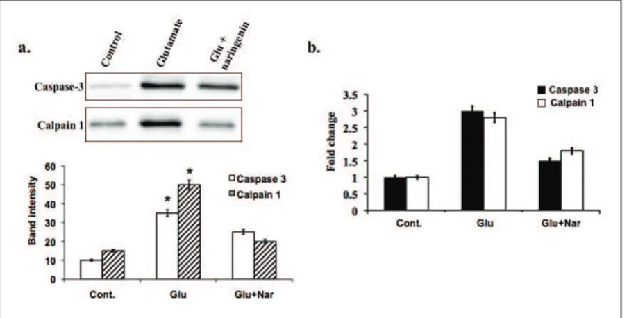
Evaluation of apoptotic signaling protein/mRNA level
The mRNA levels of caspase-3 and calpain 1 were assessed following exposure to glutamate
and naringenin. A 30-min exposure of cells to glutamate caused a robust increase in the lev-els of caspase-3 and calpain mRNA. (Fig. 3a). In neurons treated with 100 mg/L naringenin, the expression levels of caspase and calpain 1 decreased (Fig. 3b) following 6 h of glutamate exposure. These results suggest that naringenin could be used effectively to target the caspase-mediated apoptosis cascade.
DISCUSSION
Preventing hippocampal neuronal death could be a beneficial therapeutic approach against cognitive and memory impairments accompa-nying brain disorders. The bioflavonoid, nar-ingenin could be a potential therapeutic candi-date. Glutamate-induced oxidative stress causes apoptotic and necrotic neuronal death in HT22 cells (Fukui et al., 2009). Glutamate-mediated
toxicity is mainly induced via excitotoxicity me-diated by glutamate receptors and through ROS-mediated oxidative stress (Fukui et al., 2010; Pod-dar et al., 2010).
Overactivation of NMDA, along with other glutamate/glycine receptors, disturbs calcium homeostasis, which is the key mediator of glu-tamate-induced excitotoxic neuronal damage. Oxidative stress and excitotoxicity are the fore-most activities induced by brain ischemia, lead-ing to the damage of neuronal cells (Coyle and Puttfarcken, 1993). Namura et al. (2001) report-ed earlier a rreport-eduction in the phosphorylation of Erk1/2 in the hippocampus of the mice during a forebrain ischemia experiment. However, the underlying mechanism contributing to the cy-totoxic action of glutamate has not been clearly established. This present research demonstrates that naringenin eased the glutamate-induced re-duction in the level of Erk1/2 phosphorylation in hippocampal neuronal cells and protected the dendrites from refutation during a 30-min gluta-mate exposure. Almeida et al. (2005) previously reported that the brain-derived neurotrophic fac-tor increased the phosphorylation of Erk1/2 in
a hippocampal neuronal culture by preventing glutamate-induced apoptosis. Our study results are consistent with those of Almeida et al. (2005). Our results also exhibited the phosphorylation of the Akt-signaling pathway induced by narin-genin hippocampal neurons.
Further, the cell survival rate and dendrite length of the cultured hippocampal neuronal cells after 24 h of glutamate exposure was exam-ined for confirmation of neuroprotective effect induced by naringenin, though the molecular mechanism is still to be understood. It can be inferred that naringenin inhibits glutamate re-ceptors or the direct modulators, thereby reduc-ing the excitotoxicity in the hippocampal neu-ronal cells. Gao et al. (2006) demonstrated that concentrations (10-100 mM) of trans-resveratrol can inhibit postsynaptic glutamate receptors in hippocampal neurons, with NMDA receptors being more sensitive than AMPA receptors. Inhibition of Erk phosphatases could also lead to an increase in Erk phosphorylation in cells stimulated with glutamate and exposed to narin-genin (Levinthal and Defranco, 2005; Choi et al., 2006). From the analysis of the mRNA levels of
caspase-3, it could be concluded that naringenin inhibits glutamate-induced caspase-3 activation at an initial stage. Activation of caspase occurs in the execution phase, as it is an effector caspase that can destroy cells extensively.
The glutamate-induced oxidative toxicity in HT22 cells was reported to be mediated via a caspase-independent pathway involving calpain (Elphick et al., 2008; Zhang and Bhavnani, 2006). Calpains (Ca2+-dependent proteases) require an
increase in the cytosolic concentration of cal-cium via extracellular influx and an increase of this level has been reported in glutamate-treated HT22 cells (Herrera et al., 2007). Elevated levels of ROS and the activation of calpains through the induction of caspase-independent cell death have also been reported in association with some en-vironmental pollutants (Tofighi et al., 2011). Our data suggest that naringenin protects neurons by preventing apoptosis. These beneficial effects of naringenin could be possibly attributed its anti-oxidant and anti-inflammatory properties (Yang et al., 2011; Assini et al., 2013).
Conflict of interest disclosure: The authors declare that they have no conflict of interest.
REFERENCES
Almeida, R. D., Manadas, B. J., Melo, C. V., Gomes, J. R., Mendes, C.S. et al. (2005). Neuroprotection by BDNF against glutamate-induced apoptotic cell death is mediated by ERK and PI3-kinase pathways. Cell. Death. Differ. 12, 1329-1343.
Assini, J.M., Mulvihill, E.E., Sutherland, B.G., Telford, D.E., Saw-yez, C.G. et al. (2013). Naringenin prevents cholesterol-induced systemic inflammation, metabolic dysregula-tion, and atherosclerosis in Ldlr -/- mice. J. Lipid. Res.
54, 711-724.
Baumann, J., Wurm, G. and F. Bruchhausen (1980). Prostaglan-din synthetase inhibition by flavonoids and phenolic com-pounds in relation to their O2 scavenging.
Berbari, N. F., Bishop, G. A., Askwith, C. C., Lewis, J. S. and K. Mykytyn (2007). Hippocampal neurons possess primary cilia in culture. J. Neurosci. Res. 85, 1095-1100.
Choi, B. H., Hur, E. M., Lee, J. H., Jun, D. J. and K. T. Kim (2006). Protein kinase Cdelta mediated proteasomal degradation of MAP kinase phosphatase-1 contributes to glutamate-induced neuronal cell death. J. Cell. Sci.119, 1329-1340.
Coyle, J. T. and P. Puttfarcken (1993). Oxidative stress, glutamate, and neurodegenerative disorders. Science.262, 689-695.
Egbuonu, A. C., Obidoa, O., Ezeokonkwo, C. A., Ezeanyika, L. U.
and P. M. Ejikeme (2009). Hepatotoxic effects of low dose oral administration of monosodium glutamate in male albino rats. Afr. J. Biotech. 8, 3031-3035.
Elphick, L. M., Hawat, M., Toms, N. J., Meinander, A., Mikhailov, A. et al. (2008) Opposing roles for caspase and calpain death proteases in L glutamate-induced oxidative neuro-toxicity. Toxicol. Appl. Pharmacol. 232, 258-267.
Fukui, M., Choi, H. J. and B.T. Zhu (2010). Mechanism for the protective effect of resveratrol against oxidative stress-induced neuronal death. Free. Radic. Biol. Med. 49 800-813.
Fukui, M., Song, J. H., Choi, J., Choi, H.J. and B.T. Zhu (2009). Mechanism of glutamate- induced neurotoxicity in HT22 mouse hippocampal cells. Eur. J. Pharmacol. 617, 1-11.
Gao, Z.B., Chen, X.Q. and G.Y. Hu (2006). Inhibition of excitatory synaptic transmission by trans-resveratrol in rat hippocam-pus. Brain. Res. 1111, 41-47.
Harukuni, I. and A. Bhardwaj (2006). Mechanisms of brain injury after global cerebral ischemia. Neurol. Clin. 24, 1-21.
Herrera, F., Martin, V., Garcia-Santos, G., Rodriguez-Blanco, J., Antolin, I. and C. Rodriguez (2007). Melatonin prevents glutamate-induced oxytosis in the HT22 mouse hippo-campal cell line through an antioxidant effect specifically targeting mitochondria. J. Neurochem. 100, 736-746.
Hwang, I. K., Yoo, K. Y., Kim, D.S., Jeong, Y. K., Kim, J. D. et al.
(2004). Neuroprotective effects of grape seed extract on neuronal injury by inhibiting DNA damage in the gerbil hippocampus after transient forebrain ischemia. Life. Sci. 75, 1989-01.
Levinthal, D. J. and D. B. Defranco (2005). Reversible oxidation of ERK-directed protein phosphatases drives oxidative toxic-ity in neurons. J. Biol. Chem. 280, 5875-5883.
Namura, S., Iihara, K., Takami, S., Nagata, I., Kikuchi, H. et al.
(2001). Intravenous administration of MEK inhibitor U0126 affords brain protection against forebrain ischemia and focal cerebral ischemia. Proc. Natl. Acad. Sci. U S A. 98, 11569-74.
Pavlovic, V., Pavlovic, D., Kocic, G., Sokolovic, D., Sarac, M. and Z. Jovic (2009). Ascorbic acid modulates monosodium gluta-mate induced cytotoxicity in rat thymus. Bratisl. Lek. Listy.
110, 205-209.
Poddar, R., Deb, I., Mukherjee, S. and S. Paul (2010). NR2B-NMDA receptor mediated modulation of the tyrosine phosphatase STEP regulates glutamate induced neuronal cell death. J. Neurochem.115, 1350-1362.
responsiveness and inhibits NF-kappaB activity in a murine model of asthma. Can. J. Physiol. Pharmacol.87, 729-735.
Stanciu, M., Wang, Y., Kentor, R., Burke, N., Watkins, S. et al. (2000). Persistent activation of ERK contributes to glutamate-induced oxidative toxicity in a neuronal cell line and primary cortical neuron cultures. J. Biol. Chem. 275, 12200-06.
Tofighi, R., Johansson, C., Goldoni, M., Ibrahim, W. N., Gogvadze, V. et al. (2011). Hippocampal neurons exposed to the envi-ronmental contaminants methyl mercury and poly chlori-nated biphenyls undergo cell death via parallel activation of calpains and lysosomal proteases. Neurotox. Res. 19, 183-194.
van Acker, F. A., Schouten, O., Haenen, GR., van der Vijgh, W. J.
and A. Bast (2000). Flavonoids can replace alpha-tocoph-erol as an antioxidant. FEBS Lett. 473,145-148.
Xu, J., Xilouri, M., Bruban, J., Shioi, J., Shao, Z. et al. (2011). Extra-cellular progranulin protects cortical neurons from toxic insults by activating survival signaling. Neurobiol. Aging.32, 2326.e5-16.
Yang, J., Li, Q., Zhou, X.D., Kolosov, V. P. and J. M. Perelman
(2011). Naringenin attenuates mucous hypersecretion by modulating reactive oxygen species production and inhib-iting NF-κB activity via EGFR-PI3K-Akt/ERK MAPKinase signaling in human airway epithelial cells. Mol. Cell. Bio-chem.351, 29-40.