Comparison of Equivalence between Two
Commercially Available S499-Phosphorylated
FMRP Antibodies in Mice
Conner D. Reynolds1, Gregory D. Smith2, Taylor S. Jefferson1, Joaquin N. Lugo1,2*
1Department of Psychology and Neuroscience, Baylor University, Waco, Texas, United States of America,
2Institute of Biomedical Sciences, Baylor University, Waco, Texas, United States of America
Abstract
Fragile X syndrome (FXS) develops from excessive trinucleotide CGG repeats in the 5’ -untranslated region at Xq27.3 of theFmr-1gene, which functionally silences its expression and prevents transcription of its protein. This disorder is the most prominent form of herita-ble intellectual deficiency, affecting roughly 1 in 5,000 males and 1 in 10,000 females globally. Antibody specificity and selectivity are essential for investigating changes in intra-cellular protein signaling and phosphorylation status of the Fragile X Mental Retardation Protein (FMRP). Currently, both PhosphoSolutions1
and abcam1produce commercially
available S499-phosphorylated FMRP specific antibodies. The antibody from PhosphoSo-lutions1
has been validated in previous studies; however, the antibody from abcam1
anti-body has yet to receive similar validation. This study aims to determine whether these two antibodies are true equivalents through western blot analysis of bothNS-Ptenknockout (KO) andFmr-1KO mice strains. We prepared hippocampal synaptosomal preparations and probed the samples using total FMRP, abcam1
phosphorylated FMRP, and Phospho-Solutions1
phosphorylated FMRP antibodies. We found that there was a significant increase in phosphorylated FMRP levels using the abcam1and PhosphoSolutions1
anti-bodies in theNS-PtenKO mice compared to wildtype mice. However, there was much more variability using the abcam1
antibody. Furthermore, there was a band present in the
Fmr-1KO for the phosphorylated FMRP site using the abcam1
antibody for western blot-ting but not for the PhosphoSolutions1
antibody. Our findings strongly suggest that the
anti-body from abcam1
is neither specific nor selective for its advertised targeted substrate, S499-phosphorylated FMRP.
Introduction
Since theFmr-1gene was discovered in 1991, its product, the Fragile X Mental Retardation Protein (FMRP), has been a topic of major discussion in the investigation of inherited intellec-tual deficiencies [1]. It is most frequently addressed in Fragile X Syndrome (FXS), the most OPEN ACCESS
Citation:Reynolds CD, Smith GD, Jefferson TS, Lugo JN (2015) Comparison of Equivalence between Two Commercially Available S499-Phosphorylated FMRP Antibodies in Mice. PLoS ONE 10(11): e0143134. doi:10.1371/journal.pone.0143134
Editor:Ellen R Goldman, Naval Research Laboratory, UNITED STATES
Received:August 5, 2015
Accepted:October 31, 2015
Published:November 18, 2015
Copyright:© 2015 Reynolds et al. This is an open access article distributed under the terms of the
Creative Commons Attribution License, which permits unrestricted use, distribution, and reproduction in any medium, provided the original author and source are credited.
Data Availability Statement:All relevant data have been uploaded to Figshare with the following DOI:
http://dx.doi.org/10.6084/m9.figshare.1582675.
Funding:Support was provided by the NIH NINDS: NS088776 [http://www.ninds.nih.gov/] and the Epilepsy Foundation [http://www.epilepsyfoundation. org/]. The funders had no role in study design, data collection and analysis, decision to publish, or preparation of the manuscript.
prominent form of heritable intellectual disability [2]. This disorder develops from excessive trinucleotide CGG repeats in the 5’-untranslated region at Xq27.3 of theFmr-1gene, function-ally silencing its expression and preventing transcription of its protein [1]. Fragile X syndrome is observed in approximately 1 in 5,000 males and 1 in 10,000 females globally [3,4]. Altered FMRP levels have also been observed in disorders unassociated with an expansion inFmr-1, such as schizophrenia, bipolar disorder, major depression, autism spectrum disorder, and epi-lepsy [5–7]. The involvement of FMRP in such a wide array of disorders results from its role in protein synthesis throughout the brain.
Primarily associating with polyribosomes, FMRP has been shown to negatively regulate pro-tein synthesis via post-transcriptional degradation of target mRNA [8]. Phosphorylation status determines where in the cell FMRP performs this function. Unphosphorylated FMRP tends to associate with actively translating polyribosomes, while phosphorylated FMRP tends to associ-ate with stalled polyribosomes. This protein is preferentially phosphorylassoci-ated at the highly con-served serine 499 (S499) residue, which subsequently triggers the phosphorylation of nearby serine residues [9]. Thus, in studies investigating the expression of FMRP, discerning its phos-phorylation status is imperative.
FMRP-mediated transcriptional repression has been well-characterized in the Phosphoino-sitide 3-kinase|serine/threonine kinase|mammalian target of rapamycin (PI3K-Akt-mTOR) intracellular signaling pathway. This pathway has essential roles in nutrition, energy metabo-lism, and cell growth throughout the entire body, with specific roles in neural plasticity, learn-ing, and memory in the brain [10–13]. FMRP is activated downstream of this pathway, subsequently initiating a negative feedback loop and preventing hyperactivation of this path-way. While it is hypothesized to result via FMRP-mediated suppression of the PI3K-enhancer, PIKE-S [14], a definitive mechanism of action still remains unclear.
Phosphatase and tensin homolog (PTEN) also functions as a critical negative regulator of PI3K-mediated conversion of Phosphatidylinositol 4,5-bisphosphate (PIP2) to Phosphatidyli-nositol (3,4,5)-trisphosphate (PIP3). Disruption of this protein causes hyperactive PI3K-Akt-mTOR signaling and has been associated with spontaneous seizures, disorganization of neural circuitry, macrocephaly, developmental delay, and autism spectrum disorders [15,16]. Previ-ous studies have also confirmed this disruption leads to a downstream increase of total FMRP and phosphorylated FMRP in the brain [17,18].
Protein signaling changes in the brain are most readily visualized using western blot analy-sis. This method uses primary antibodies that bind with target protein sequences to show rela-tive expression between groups. However, producing antibodies that recognize specific phosphorylated amino-acid residues is a difficult process, potentially leading to target substrate variability [reviewed in [19]]. PhosphoSolutions1
and abcam1
are two companies that have produced an antibody that recognizes phosphorylated FMRP. The efficacy of PhosphoSolu-tions1
antibody product has been validated in aFmr-1KO mice study [7]. However, the anti-body from abcam1has yet to receive any similar validation of efficacy. Therefore, the aim of this study was to compare expression levels observed using the PhosphoSolutions1
and abcam1antibody products and to determine whether they yield equivalent results when used in assays. Given the link between PTEN disruption and pFMRP hyperphosphorylation described above, theNS-PtenKO mice is an ideal animal model for comparing the equivalency of these antibodies. These antibodies will also be compared usingFmr-1KO mice in order to confirm that both antibodies are truly targeting S499-phosphorylated FMRP. The results below provide evidence that the antibody from abcam1
is not labeling S499-phosphorylated FMRP, while the antibody from PhosphoSolutions1
Methods and Methods
Ethics Statement
This study was carried out in strict accordance with the recommendations in the Guide for the Care and Use of Laboratory Animals of the National Institutes of Health. The protocol was approved by Baylor University Institutional Care and Use Committee (Animal Assurance Number A3948-01)
Animals
Two strains of mice were used in this investigation. Neuron subset-specificPtenconditional mice have been previously described in literature as GFAP-CreloxP/loxP[15,20].NS-Pten+/+ wild type (WT) andNS-PtenloxP/loxPknockout (KO) mice were produced for this experiment by breedingNS-PtenloxP/+heterozygous parents.Fmr-1+/+wild type (WT) andFmr-1-/- knock-out (KO) mice were produced for this experiment by breedingFmr-1-/-male and female mice and separate breeding ofFmr-1+/+male and female mice. We originally purchased this strain through The Jackson Laboratory (FVB.129P2-Pde6b+ Tyrc-chFmr1tm1Cgr/J). All mice were subsequently housed at an ambient temperature of 22°C, with a 14-hour light and 10-hour dark diurnal cycle. These mice were also givenad libitumaccess to food and water. All proce-dures involving mice were conducted in compliance with the National Institute of Health Guidelines for the Care and Use of Laboratory Animals.
Western Blotting
NS-PtenandFmr-1wildtype and knockout mice were sacrificed at approximately 8-weeks of age and hippocampi were rapidly dissected. The resulting samples were then rinsed in 1X phos-phate buffer solution, placed on dry ice, and stored at -80°C until used. Hippocampi were homogenized in ice-cold homogenization buffer (0.32M sucrose, 1mM EDTA, 5mM Hepes) containing protease inhibitor cocktail (Sigma, USA) and processed for western blotting as pre-viously described [21]. This procedure produced both crude synaptosomes and total homoge-nate samples. Only the crude synaptosomes were used in our total analyses of FMRP and phosphorylated FMRP expression. Equivalent protein concentrations were confirmed using the Bradford Protein Assay (Bio Rad, Hercules, CA, USA) and diluted in Laemmli loading buffer (4X: 0.25M Tris, pH 6.8, 6% SDS, 40% Glycerol, 0.04% Bromophenol Blue, 200mM Dithiothreitol). Following SDS-PAGE, proteins were transferred to Hybond-P polyvinyl difluoride membranes (GE Healthcare, Piscataway, NJ, USA). Membranes were then incubated for 1 hour at room temperature in blocking solution [5% non-fat milk diluted in 1X Tris Buff-ered Saline (50mM Tris-HCl, pH 7.4, 150mM NaCl) with 0.1% Tween (1X TTBS) and 1mM Na3VO4]. The membranes were then incubated overnight at 4°C with the following primary antibodies in 5% milk in TTBS: FMRP (1:1,000; Cell Signaling Technology, Catalog Number 4317, Boston, MA, USA); pFMRP (1:500 PhosphoSolutions1
, Catalog Number p1125-499, Aurora, CO, USA); pFMRP (1:500 abcam1
Optical Densitization
ProteinSimple AlphaView software was then used to measure the optical density of resulting immunoreactive bands. Measurements obtained from all bands of interest were normalized to actin levels within the same lane. All experimental points represent a single mouse each (n = 1). All groups were normalized to the average of the control group (NS-PtenWT) per blot. All values from WT mice represent biological replicates and were collected from WT
littermates.
Statistical Analysis
The western blotting data were analyzed using an independent samples t-test. All data were analyzed using either GraphPad Prism 6 software (La Jolla, CA) or SPSS 20.0 (IBM, USA). Val-ues are shown as mean ± S.E.M. for each group.
Results
NS-Pten
KO Mice Show Increased Total FMRP Expression
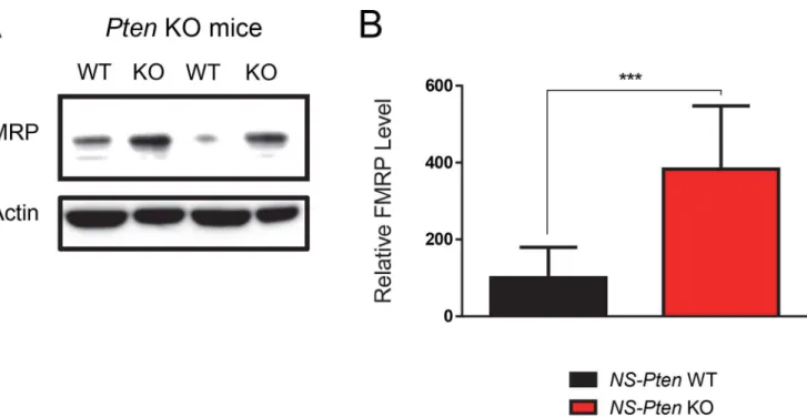
We first confirmed the results of a previous study by Lugo et al. (2014) that indicatedPten dis-ruption substantially increases total FMRP (Cell Signaling Technology, Catalog Number 4317, Boston, MA, USA) levels. As expected, differential expression of the protein between groups showed significant increase in total FMRP levels in theNS-PtenKO mice compared to WT micet(1,16) = 4.6,p<0.001 (n = 9) (Fig 1).
Fig 1.NS-PtenKO leads to increased expression of FMRP.Hippocampal tissue fromNS-Ptenwildtype (WT) and knockout (KO) mice were examined for
total FMRP levels using western blotting. (Fig 1A) The figure shows a representative blot from twoNS-PtenWT and KO samples. (Fig 1B) Graphs show the mean (±SEM) of WT and KO mice.***= p<0.001. n = 9 per group.
PhosphoSolutions
1’
& abcam
1’
s S499-Phosphorylated FMRP
Antibodies Both Indicate Increased Expression in
NS-Pten
KO Mice
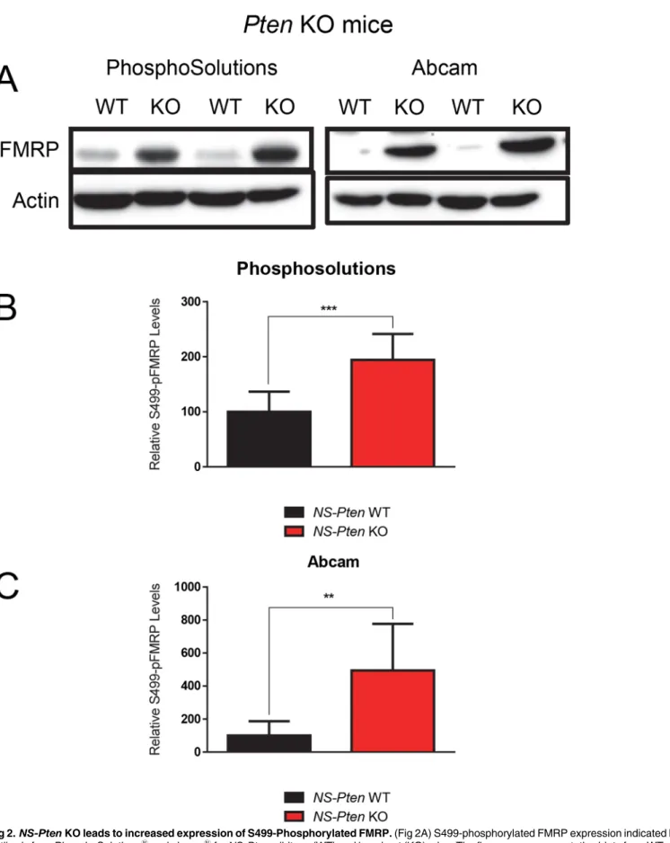
Previous studies have found that disruptions in the PI3K-Akt-mTOR pathway lead to increases in total phosphorylated FMRP in the brain [17,18]. Because the efficacy of PhosphoSolu-tions1
’antibody has been confirmed in previous studies [7,22], we used it as a control in examining the influence ofNS-Ptendeletion on pFMRP expression. We found a significant increase in total S499-phosphorylated FMRP levels in theNS-PtenKO mice compared to con-trolst(1,16) = 4.76,p<0.001 (n = 9) (Fig 2B). We then used the abcam1antibody to
deter-mine how its results compare to those of PhosphoSolutions1
. We found that it also showed a significant increase in total pFMRP levels in theNS-PtenKO mice compared to controls t(1,16) = 4.0,p<0.01 (n = 9) (Fig 2C). This confirms that both antibody products are
reason-ably able to detect total S499-phosphorylated FMRP expression.
PhosphoSolutions
1’
and abcam
1’
s S499-Phosphorylated FMRP
Antibodies Differ in Expression and Variance in
NS-Pten
KO Mice
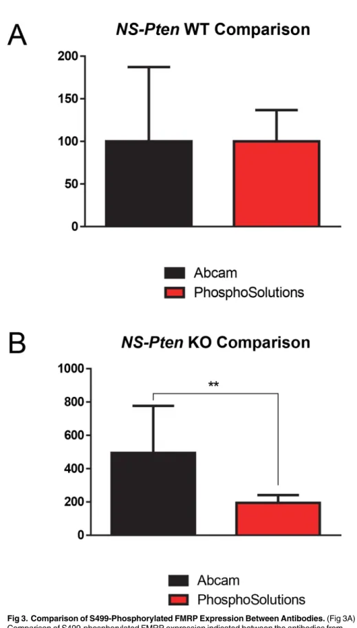
We next wanted to determine if there were any differences between the results obtained by PhosphoSolutions1and abcam1antibodies. Comparing the results from both antibodies in NS-PtenWT S499-phosphorylated FMRP expression, we found no significant differences t(1,16) = 0.01,p>0.99 (n = 9) (Fig 3A). However, there were significant differences in variance
between the two antibody productsF(8,8) = 5.67,p<.05, with the antibody from abcam1
showing greater variance than the antibody from PhosphoSolutions1
.
In comparing relative S499-phosphorylated FMRP expression inNS-PtenKO mice, we found significant differences between the two antibodiest(1,8.4) = 3.1,p<0.01 (n = 9) (Fig 3B),
with the abcam1
antibody indicating much greater expression than the PhosphoSolutions1
antibody. There were also significant differences in variance between the two antibody prod-uctsF(8,8) = 36.29,p<0.001, with the antibody from abcam1showing greater variance than
the antibody from PhosphoSolutions1.
Expression Levels Indicated By PhosphoSolutions
1and abcam
1Antibodies S499-Phosphorylated FMRP Antibodies Show No
Correlational Relationship in
NS-Pten
KO Mice
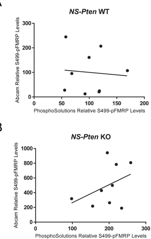
With the previous differences in indicated expression found between antibodies, we next wanted to determine whether there is a correlational relationship between the S499-phosphor-ylated FMRP expressions indicated by PhosphoSolutions1
and abcam1
antibody products. Theoretically, if these antibodies are measuring the same target substrate, then a perfect corre-lation should exist. InNS-PtenWT mice, correlational analysis between the expression levels between antibodies failed to show a significant relationshipr(16) = -0.09,p= 0.83 (Fig 4A). In NS-PtenKO mice, correlational analysis between the expression levels between antibodies also failed to show a significant relationshipr(16) = 0.41,p= 0.28 (Fig 4B). A lack of correlational relationships between the S499-phosphorylated FMRP expressions indicated between Phos-phoSolutions1
and abcam1
antibodies in bothNS-PtenWT and KO mice indicates that these antibodies may not be detecting changes in the same substrate.
abcam
1Phosphorylated FMRP Antibody Indicates Presence of Target
Protein in
Fmr-1
KO mice
In order to determine whether the PhosphoSolutions1
and abcam1
Fig 2.NS-PtenKO leads to increased expression of S499-Phosphorylated FMRP.(Fig 2A) S499-phosphorylated FMRP expression indicated by the antibody from PhosphoSolutions1
and abcam1
forNS-Ptenwildtype (WT) and knockout (KO) mice. The figures are representative blots from WT and KO
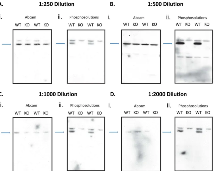
levels inFmr-1KO mice. Because these mice have a congenital absence of FMRP, neither anti-body should detect any presence of S499-phosphorylated FMRP. In order to rule out the non-specific binding effects resulting from too low or high antibody concentration, we used 4 different concentrations (Fig 5A–5D). This would help to eliminate the possibility that the appearance of a band in the knockout mice is due to too high a concentration of antibody. Visual analysis of S499-phosphorylated FMRP expression using the PhosphoSolutions1
anti-body shows an alternating pattern, indicating an absence of any expression in lanes withFmr-1 KO samples using a 1:250 dilution (Fig 5A), 1:500 dilution (Fig 5B), 1:1000 dilution (Fig 5C), or 1:2000 dilution (Fig 5D). In all dilutions there is a clear pattern that the PhosphoSolutions1
antibody correctly probes for phosphorylated FMRP, while abcam1
does not. Visual analysis of S499-phosphorylated FMRP expression using abcam1
antibody shows a constitutive expression in all lanes (Fig 5A–5D).
Discussion
Although there are no universally accepted guidelines for validating antibodies, Bordeaux et al. (2010) recommended several steps to ensure antibody selectivity and reproducibility [23]. First, these authors advise testing antibodies against a cell line with known target expression. By comparing experimental data obtained from the antibody in question with expected values, researchers are able to determine its target selectivity. Cell lines with target gene deletion are considered to be the“Gold Standard”in antibody verification because true antibodies should not detect any measurable expression of the gene product. Bordeaux et al. (2010) also recom-mend verifying reproducibility of the antibody results through use of multiple molecular techniques, such as western blotting, immunoprecipitation, immunohistochemistry, or immunofluorescence.
In the present study S499-phosphorylated FMRP antibodies from PhosphoSolutions1
and abcam1
were tested againstNS-PtenKO andFmr-1KO mouse hippocampal cell lines in west-ern blot analysis. InNS-PtenKO mice both antibodies correctly identified an increase in target protein expression compared to WT mice, though the abcam1
antibody indicated significantly greater protein expression than the antibody from PhosphoSolutions1
. The experimental data using the abcam1
antibody also showed much greater variance between data points, suggesting a possible lack of specificity. Their performances continued to differ when tested inFmr-1KO mice. In this cell line, only the antibody from abcam1
antibody identified the presence of S499-phosphorylated FMRP, which would be impossible considering that the congenital absence of genes coding for this protein. These results confirm that the abcam1
antibody is not selective for S499-phosphorylated FMRP.
One possible contributor to the variance in the results from theNS-PtenKO mice may be due to an incorrect concentration of the antibody. If the antibody concentration used was too high there could have been an increase in nonspecific binding, which would contribute to the variability seen in the phosphorylated FMRP levels inNS-PtenWT and KO mice. We included a series of experiments in the FMRP WT and KO mice with concentrations of abcam1
and PhosphoSolutions1
antibodies at 1:250, 1:500, 1:1000, and 1:2000 (Fig 5A–5D). A clear increase in nonspecific binding was observed in the abcam1
treated samples, since an addi-tional band appeared on top of the target band when a 1:250 concentration was used (Fig 5A). However, in each of the abcam1
dilutions there was a consistent band seen in the knockout mice that should not be present. We did not observe this band in the FMRP KO mice when we
(Fig 2C) S499-phosphorylated FMRP expression indicated by abcam1
’s antibodyNS-Ptenfor WT and KO mice. Graphs show the mean (±SEM). n = 9 per
group.**= p<0.01;***= p<0.001.
Fig 3. Comparison of S499-Phosphorylated FMRP Expression Between Antibodies.(Fig 3A) Comparison of S499-phosphorylated FMRP expression indicated between the antibodies from PhosphoSolutions1and abcam1inNS-PtenWT mice (Fig 3B) Comparison of S499-phosphorylated FMRP expression indicated between PhosphoSolutions1and abcam1antibodies inNS-PtenKO mice.
**= p<0.01. Graphs show the mean (±SEM). n = 9 per group.
Fig 4. Comparison of S499-Phosphorylated FMRP Expression Between Antibodies.(Fig 4A)
Correlation of S499-phosphorylated FMRP expression indicated between PhosphoSolutions1and abcam1 antibodies inNS-PtenWT mice. (Fig 4B) Correlation of S499-phosphorylated FMRP expression indicated between PhosphoSolutions1
and abcam1
antibodies inNS-PtenKO mice. The line represents the line of
best fit. n = 9 per graph.
used the Phosphosolutions1
antibody. Therefore, the variability seen in theNS-PtenKO sam-ples using the abcam1antibody was not due to nonspecific binding, but to lack of antibody specificity.
Although we did not choose to repeat this study using other molecular techniques, the reli-ability of the PhosphoSolutions1
antibody was reproduced in two different hippocampal cell lines in western blot analysis; one of which being the gold standard target gene knockout. Two previous studies have also verified this antibody by using other molecular techniques [7,22]. Therefore, our study supports existing evidence for the selectivity and reproducibility of the PhosphoSolutions1
antibody, as well as informs researchers to preferentially use this product over the antibody from abcam1when investigating phosphorylation changes in FMRP. Fig 5. Comparison of S499-Phosphorylated FMRP Expression Between Antibodies.We examined phosphorylated levels of FMRP inFmr-1wildtype
(WT) and knockout (KO) mice using 4 concentrations of the abcam1
and PhosphoSolutions1
antibody. The full blot of the samples is presented in each set of figures. There are 2 alternating lanes of WT and KO probed samples throughout all dilutions. In each dilution experiment the first figure used the abcam1 S499 phosphorylated antibody (i) and the second figure used the PhosphoSolutions1S499 phosphorylated antibody (ii). The blue arrows point to the location of the target site. (Fig 5A) WT and KO hippocampus tissue probed for antibodies using a 1:250 dilution, (Fig 5B) 1:500 dilution, (Fig 5C) 1:1000 dilution, (Fig 5D) 1:200 dilution
It is still unclear what the antibody from abcam1
might be measuring, but several possibili-ties exist. In a recent study by Bartley et al (2014), which validated commercially available S499-phosphorylated FMRP antibodies, it was found that one antibody also recognized unpho-sphorylated FMRP. These authors did not identify the company that produced this nonspecific antibody. If the antibody from abcam1is measuring FMRP indiscriminant of phosphorylation status, this may explain why it indicated greater S499-phosphorylated FMRP expression than the PhosphoSolutions1
antibody inNS-PtenKO mice. However, this would not account for its identification of protein inFmr-1KO mice.
It is also possible that this antibody is measuring proteins with homologous sequences to FMRP. Fragile-X Related proteins 1 and 2 (FXR1 and FXR2) are members of the same protein family as FMRP and are 70–80% homologous in the N-terminal region [24,25]. If the antibody from abcam1
is measuring the FXR1/2 proteins, in addition to the total FMRP, this may explain for the protein expression it indicated inFmr-1KO mice. This would also account for the large variability between its data points inNS-PtenKO mice.
There are several published studies that have used abcam1S499-phosphorylated FMRP antibody. Given the results of this study, it may be necessary for these researchers to reproduce their findings using the PhosphoSolutions1
antibody in order to ensure their validity.
Antibodies are some of the most frequently used tools in basic science and clinical investiga-tion, allowing researchers to probe specific aspects of molecular physiology. However, synthe-sizing these powerful tools is a highly intricate process that requires careful planning and precision. An even greater degree of difficulty is introduced when producing antibodies that recognize only specific phosphorylated amino-acid residues. Therefore, it is imperative to for researchers to validate the specificity and reproducibility of antibodies used in their investigations.
Acknowledgments
We would also like to acknowledge the Baylor University Molecular Biosciences Core for the use of equipment for this study.
Author Contributions
Conceived and designed the experiments: CDR GDS JNL. Performed the experiments: CDR GDS TSJ. Analyzed the data: CDR GDS TSJ JNL. Contributed reagents/materials/analysis tools: CDR GDS TSJ JNL. Wrote the paper: CDR JNL.
References
1. Verkerk AJ, Pieretti M, Sutcliffe JS, Fu YH, Kuhl DP, Pizzuti A, et al. Identification of a gene (FMR-1) containing a CGG repeat coincident with a breakpoint cluster region exhibiting length variation in fragile X syndrome. Cell. 1991; 65(5):905–14. PMID:1710175
2. Garber KB, Visootsak J, Warren ST. Fragile X syndrome. Eur J Hum Genet. 2008; 16(6):666–72. doi: 10.1038/ejhg.2008.61PMID:18398441
3. Hawkins M, Boyle J, Wright KE, Elles R, Ramsden SC, O'Grady A, et al. Preparation and validation of the first WHO international genetic reference panel for Fragile X syndrome. Eur J Hum Genet. 2011; 19 (1):10–7. doi:10.1038/ejhg.2010.135PMID:20736975
4. Hersh JH, Saul RA, Committee on G. Health supervision for children with fragile X syndrome. Pediat-rics. 2011; 127(5):994–1006. doi:10.1542/peds.2010-3500PMID:21518720
5. Fatemi SH, Folsom TD, Kneeland RE, Liesch SB. Metabotropic glutamate receptor 5 upregulation in children with autism is associated with underexpression of both Fragile X mental retardation protein and GABAA receptor beta 3 in adults with autism. Anatomical record. 2011; 294(10):1635–45.
7. Bernard PB, Castano AM, O'Leary H, Simpson K, Browning MD, Benke TA. Phosphorylation of FMRP and alterations of FMRP complex underlie enhanced mLTD in adult rats triggered by early life seizures. Neurobiol Dis. 2013.
8. Laggerbauer B, Ostareck D, Keidel EM, Ostareck-Lederer A, Fischer U. Evidence that fragile X mental retardation protein is a negative regulator of translation. Hum Mol Genet. 2001; 10(4):329–38. PMID: 11157796
9. Ceman S, O'Donnell WT, Reed M, Patton S, Pohl J, Warren ST. Phosphorylation influences the transla-tion state of FMRP-associated polyribosomes. Hum Mol Genet. 2003; 12(24):3295–305. PMID: 14570712
10. Wullschleger S, Loewith R, Hall MN. TOR signaling in growth and metabolism. Cell. 2006; 124(3):471– 84. PMID:16469695
11. Hoeffer CA, Klann E. mTOR signaling: at the crossroads of plasticity, memory and disease. Trends Neurosci. 2010; 33(2):67–75. doi:10.1016/j.tins.2009.11.003PMID:19963289
12. Owonikoko TK, Khuri FR. Targeting the PI3K/AKT/mTOR pathway: biomarkers of success and tribula-tion. American Society of Clinical Oncology educational book / ASCO American Society of Clinical Oncology Meeting. 2013.
13. Graber TE, McCamphill PK, Sossin WS. A recollection of mTOR signaling in learning and memory. Learn Mem. 2013; 20(10):518–30. doi:10.1101/lm.027664.112PMID:24042848
14. Sharma A, Hoeffer CA, Takayasu Y, Miyawaki T, McBride SM, Klann E, et al. Dysregulation of mTOR signaling in fragile X syndrome. J Neurosci. 2010; 30(2):694–702. doi:10.1523/JNEUROSCI.3696-09. 2010PMID:20071534
15. Backman SA, Stambolic V, Suzuki A, Haight J, Elia A, Pretorius J, et al. Deletion of Pten in mouse brain causes seizures, ataxia and defects in soma size resembling Lhermitte-Duclos disease. Nat Genet. 2001; 29(4):396–403. PMID:11726926
16. Zhou J, Parada LF. PTEN signaling in autism spectrum disorders. Curr Opin Neurobiol. 2012.
17. Lugo JN, Smith GD, Arbuckle EP, White J, Holley AJ, Floruta CM, et al. Deletion of PTEN produces autism-like behavioral deficits and alterations in synaptic proteins. Front Mol Neurosci. 2014; 7:27. doi: 10.3389/fnmol.2014.00027PMID:24795561
18. Lugo JN, Smith GD, Morrison JB, White J. Deletion of PTEN produces deficits in conditioned fear and increases fragile X mental retardation protein. Learning & Memory. 2013; 20(12):670–3.
19. Stoevesandt O, Taussig MJ. Phospho-specific antibodies by design. Nat Biotechnol. 2013; 31 (10):889–91. doi:10.1038/nbt.2712PMID:24104753
20. Kwon CH, Zhu X, Zhang J, Knoop LL, Tharp R, Smeyne RJ, et al. Pten regulates neuronal soma size: a mouse model of Lhermitte-Duclos disease. Nat Genet. 2001; 29(4):404–11. PMID:11726927
21. Lugo JN, Barnwell LF, Ren Y, Lee WL, Johnston LD, Kim R, et al. Altered phosphorylation and localiza-tion of the A-type channel, Kv4.2 in status epilepticus. J Neurochem. 2008; 106(4):1929–40. doi:10. 1111/j.1471-4159.2008.05508.xPMID:18513371
22. Bartley CM, O'Keefe RA, Bordey A. FMRP S499 is phosphorylated independent of mTORC1-S6K1 activity. PLoS One. 2014; 9(5):e96956. doi:10.1371/journal.pone.0096956PMID:24806451
23. Bordeaux J, Welsh A, Agarwal S, Killiam E, Baquero M, Hanna J, et al. Antibody validation. Biotechni-ques. 2010; 48(3):197–209. doi:10.2144/000113382PMID:20359301
24. Zhang Y, O'Connor JP, Siomi MC, Srinivasan S, Dutra A, Nussbaum RL, et al. The fragile X mental retardation syndrome protein interacts with novel homologs FXR1 and FXR2. EMBO J. 1995; 14 (21):5358–66. PMID:7489725