Overexpression of
Mitochondrial Phosphate
Transporter 3
Severely Hampers Plant
Development through Regulating
Mitochondrial Function in
Arabidopsis
Fengjuan Jia1, Xiaomin Wan1, Wei Zhu1¤, Dan Sun1, Chengchao Zheng1, Pei Liu2*, Jinguang Huang1*
1State Key Laboratory of Crop Biology, College of Life Sciences, Shandong Agricultural University, Taian, Shandong, P.R. China,2College of Food Science and Engineering, Shandong Agricultural University, Taian, Shandong, P.R. China
¤ Current address: Department of Ophthalmology and Visual Sciences, University of Iowa, Iowa City, Iowa, United States of America
*[email protected](PL);[email protected](JH)
Abstract
Mitochondria are abundant and important organelles present in nearly all eukaryotic cells, which maintain metabolic communication with the cytosol through mitochondrial carriers. The mitochondrial membrane localized phosphate transporter (MPT) plays vital roles in di-verse development and signaling processes, especially the ATP biosynthesis. Among the threeMPTgenes inArabidopsisgenome,AtMPT3was proven to be a major member, and its overexpression gave rise to multiple developmental defects including curly leaves with deep color, dwarfed stature, and reduced fertility. Transcript profiles revealed that genes in-volved in plant metabolism, cellular redox homeostasis, alternative respiration pathway, and leaf and flower development were obviously altered inAtMPT3overexpression (OEMPT3) plants. Moreover, OEMPT3 plants also accumulated higher ATP content, faster respiration rate and more reactive oxygen species (ROS) than wild type plants. Overall, our studies showed thatAtMPT3was indispensable forArabidopsisnormal growth and devel-opment, and provided new sights to investigate its possible regulation mechanisms.
Introduction
Mitochondria are important organelles that can integrate numerous metabolic pathways, in-cluding energy homeostasis, redox balance, cellular growth and stress adaptation [1,2]. The drastic expression fluctuation of mitochondrion associated genes would disturb mitochondrial function, which had significant effects on growth and development. For example, overexpres-sion of the unedited form of the subunit 9 of ATP synthase gene (35S::u-atp9) caused mito-chondrial dysfunction, dwarf morphology and male sterility [3]. Loss of the mitochondrial Mg2+transporterAtMGT5led to pollen abortion and male sterility [4]. TransgenicArabidopsis
OPEN ACCESS
Citation:Jia F, Wan X, Zhu W, Sun D, Zheng C, Liu P, et al. (2015) Overexpression ofMitochondrial Phosphate Transporter 3Severely Hampers Plant Development through Regulating Mitochondrial Function inArabidopsis. PLoS ONE 10(6): e0129717. doi:10.1371/journal.pone.0129717
Academic Editor:Wagner L. Araujo, Universidade Federal de Vicosa, BRAZIL
Received:November 22, 2014
Accepted:May 12, 2015
Published:June 15, 2015
Copyright:© 2015 Jia et al. This is an open access article distributed under the terms of theCreative Commons Attribution License, which permits unrestricted use, distribution, and reproduction in any medium, provided the original author and source are credited.
Data Availability Statement:All relevant data are within the paper and its Supporting Information files.
Funding:This work was supported by the National Natural Science Foundation (Grant No. 31370305, 31000121) in China.
Competing Interests:The authors have declared
plants overexpressing mtDNA-asscciatedAtWhy2showed defects in mitochondrial function, smaller and distorted leaves [5]. Double mutant of mitochondrial malate dehydrogenase (mMDH)mmdh1mmdh2showed growth retardation due to the lower net CO2assimilation rate [6]. All these studies indicate that finely tuned mitochondrion activities are indispensable for plant normal growth and development.
The transport of various metabolites, nucleotides and cofactors across the inner mitochon-drial membrane relies on the presence of a family of related proteins: the mitochonmitochon-drial carrier family. This superfamily contains a tripartite structure of 100 amino acid segments each con-sisting of two membrane-spanningα-helices separated by an extra-membrane hydrophilic loop [7]. These transporters operate as homodimers with a 12 transmembrane domain struc-ture and are responsible for the transport of a wide variety of metabolites between mitochon-drion and cytosol [8]. InArabidopsis, the mitochondrial carrier family contains as many as 58 members [9]. In recent years, several of these transporters have been cloned and investigated at the molecular and biochemical levels, including the adenine nucleotide transporter (ANT), the oxoglutarate-malate transporter (OMT), the uncoupling protein (UCP), the dicarboxylate transporter, the tricarboxylate transporter and the phosphate transporter [10–14]. However, the biological functions of most members in this family remain unknown. In eukaryotes, the mitochondrial phosphate transporters (MPTs), which belong to the mitochondrial carrier fam-ily, play crucial roles in respiration by uptaking orthophosphate (Pi) into the mitochondrial matrix, where the Pi is utilized for the conversion of ADP to ATP [15–17]. In human, disorder of the MPT coding geneSLC25A3, which was essential for ATP biosynthesis, could lead to lac-tic acidosis, hypertrophic cardiomyopathy and muscular hypotonia [18]. Since the first isola-tion of plantMPTgene from birch [16], numerousMPThomologues have been cloned and characterized in other plant species, including soybean, maize, rice,Arabidopsis,Lotus japoni-cusandPaeonia suffruticosa[15,19,20]. InArabidopsis, there are threeMPTs, including
AtMPT1(AT2G17270),AtMPT2(AT3G48850) andAtMPT3(AT5G14040), encoding protein with 309, 363 and 375 amino acids, respectively [20]. Among them,AtMPT2andAtMPT3
have been proved to have phosphate transport activity [17]. In our previous work,AtMPTs
were shown to be involved in salt stress tolerance [21]. Intriguingly, overexpression ofAtMPT3
also led to severe developmental defects. However, the underlying molecular mechanisms be-tweenAtMPT3and developmental defects were still unknown.
Reactive oxygen species (ROS), including singlet oxygen, hydroxyl radical, superoxide anion radical and hydrogen peroxide, are constantly produced in plants and accumulate under stressful conditions [22,23]. Although ROS have been identified as important signaling mole-cules in diverse biological processes, its excessive production was toxic [24].Arabidopsisplants lacking ubiquitin-specific protease 16 displayed significant increase in H2O2accumulation and severe cell death [25]. Deficiency ofArabidopsisfrataxin altered the activity of mitochondrial Fe–S proteins and exhibited increased formation of ROS, retarded plant growth, reduced fresh weight and seed number [26]. The mutation inSLG1(Slow Growth 1) disrupted the function of mitochondrial complex I and accumulated large amount of H2O2, and exhibited lower growth rates [27]. These observations also raised an interesting biological question about the relation-ships between ROS accumulation and plant development.
thatAtMPT3had important effects on plant growth and development via regulating the mitochondrial function.
Materials and Methods
Plant materials and growth conditions
Seeds ofArabidopsis(ecotype Columbia) were surface sterilized, and plated on 1/2 Murashige and Skoog (MS) medium [28]. The plates were kept in the dark at 4°C for 3 days and then transferred to a growth chamber with a light/dark cycle of 16-h-light/8-h-dark at 22°C for 7 days. Then seedlings were transferred to growth chamber and grown at 22°C under 8-h-light/ 16-h-dark (for vegetative growth) or 16-h-light/8-h-dark (for reproductive growth).
Vector construction and transgenic plant generation
The coding region ofAtMPT3was cloned into the pBI121 binary vector under the control of CaMV 35S promoter. The constructs were introduced intoAgrobacteriun tumefaciensstrain GV3101 and then transformed intoArabidopsisby floral dip method [29].
RNA extraction and real time RT-PCR analysis
Total RNA from wild type and transgenic plants was extracted using a universal plant total RNA extraction kit (BioTeke, China). Contaminated DNA was removed with RNase-free DNase I. cDNA was synthesized using PrimeScript RT (reverse transcriptase) with oligo-dT primer using the PrimeScript RT master mix kit (Takara, Japan). A SYBR green real-time PCR master mix (Takara, Japan) and a Chromo 4 real-time PCR detector (Bio-Rad, USA) were used. Three biological replicates and three technical replicates were performed for the real time RT-PCR experiment withGAPDHas an internal control.
Microarray experiments and data analysis
RNA was labeled with the Message-Amp II-Biotin Enhanced kit. After verifying RNA integrity by the Agilent RNA 6000 Nano Kit and the Agilent 2100 Bioanalyzer, the labeled RNA was hy-bridized to Affymetrix ATH1 Genome arrays at ATLAS Biolabs. Hybridizations were done in three biological replicates. The data were analyzed using Robin [30] with the default settings of RMA (robust multi array averaging) withP<0.05, and visualized by MapMan [31] and Page-Man [32].
Histochemical staining
For H2O2and O2-detection, plants were fixed with DAB (3’, 3’-diaminobenzidine) and NBT (nitro blue tetrazolium) as described by Orozco-Cardenas [33] and Kawai-Yamada [34], re-spectively. To quantify formazan generation, stained samples were boiled in dimethyl sulfoxide until formazan precipitates were eluted completely [35]. The amount of formazan was deter-mined using spectrophotometer at 560 nm. Trypan blue staining was performed as previously described [36]. Pollen from newly dehiscing flowers was deposited, and the vitality stain was accomplished on pollen as described by Alexander [37]. GUS staining was carried out accord-ing to Zhuet al.[21].
Measurement of anthocyanin, ATP and respiration rate
amount of anthocyanins was reported as (A535—A650) g-1FW (fresh weight). The ATP concen-tration was measured as described previously [39]. Briefly, 250 mg of 20-day-old leaf were ground and resuspended in 400 mL of 2.3% (v/v) trichloroacetic acid. A bioluminescent assay kit (Sigma-Aldrich) and an ultraviolet-visible spectrophotometer was used to measure the ATP concentration. The respiration rate of 20-day-old rosette leaf was measured using a Clark-type oxygen electrode as described previously [40]. Three biological repeats were carried out for both the ATP content and the respiration rate assays.
Results
Overexpression of
AtMPT3
hampers the growth and development in
Arabidopsis
To understand the biological functions ofAtMPT3, transgenicArabidopsisconstitutively ex-pressingAtMPT3cDNA fused with 35S promoter were generated. We obtained 17 indepen-dent transgenic lines. To determine the expression levels ofAtMPT3in these lines, total RNA from 2-week-old wild type and transgenic plants were extracted and analyzed by real time RT-PCR, and line5 (L5) showed the highest expression level (S1 Fig). Then, L5 (also termed OEMPT3) and two other transgenic lines (L4 and L14) were selected for further analysis.
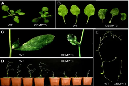
At seedling stage, we found no visible differences between the wild type and the OEMPT3 plants under normal growth conditions. However, OEMPT3 plants at 20 DAP (days after plant-ing) exhibited obvious downward-curled leaves with more anthocyanin, dwarfism and growth re-tardation (Fig 1A, 1B and 1C,S1 Table). The height of OEMPT3 plants was only 1/4 of the wild type plants (Fig 1D and 1E,S1 Table). At reproductive stage, OEMPT3 plants developed pin-like flower with defective sepal, petal, stamen, and consequently reduced fertility (Fig 2A–2E). As
AtMPT3was highly expressed in pollens (Fig 2F and 2G), we speculated that the reduced fertility was probably due to poor pollen viability. By Alexander staining, we found that the OEMPT3 flowers had almost no viable pollens (Fig 2H). L4 and L14 also exhibited similar developmental defects with L5 (S2 Fig). These results demonstrated thatAtMPT3overexpression disturbed the development ofArabidopsisat both vegetative and reproductive growth stages.
Genome-wide expression profile analysis of OEMPT3 plants
Overexpression of
AtMPT3
impairs the mitochondrial function in
Arabidopsis
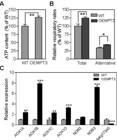
To investigate the possible relationship between the mitochondrial function andAtMPT3, we measured the ATP content and the respiration rate in wild type and OEMPT3 plants. As shown inFig 5A, an increase of approximately 26% ATP content was detected in the OEMPT3 plants under normal conditions. The total respiration rate assay also showed a nearly 1.3-fold increase (Fig 5B), suggesting an enhanced electron transport activity in the mitochondrial inner membrane of OEMPT3 plants, which coincided with the microarray data (Fig 4BandS2 Fig 1. Developmental phenotypes of OEMPT3 plants at seedling stage.(A, B) The rosette leaves of wild type and OEMPT3 plants at 20 DAP. (C) The cauline leaves of wild type and OEMPT3 plants at 40 DAP. (D, E) Phenotypes of OEMPT3 plants compared to wild type at 60 DAP.
doi:10.1371/journal.pone.0129717.g001
Fig 2. Developmental phenotypes of OEMPT3 plants at reproductive stage.Representative flower (A, B), silique (C), stamen (D) and petal (E) of wild type and OEMPT3 plants at 60 DAP. GUS staining of flower (F) and pollen (G) in OEMPT3 plants. (H) Pollen Alexander staining of wild type and OEMPT3 plants.
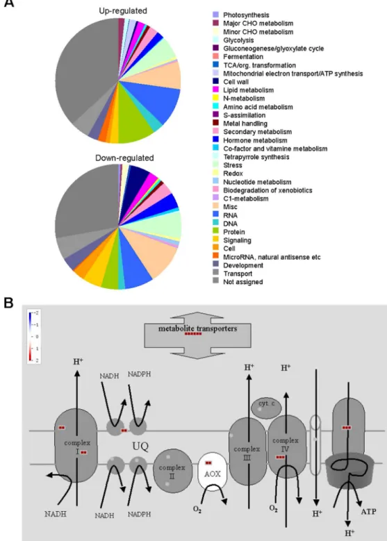
Fig 3. Microarray data analysis of OEMPT3 plants compared to wild type plants.(A) Venn diagram for the proportions of genes affected byAtMPT3at 14 and 40 DAP compared to wild type plants. (B) Biological process classification of the up-regulated and down-regulated genes by GOEAST analysis.
Fig 4.AtMPT3regulates a major proportion of genes involved in mitochondrial electron transport.(A) PageMan classification of genes based on their MapMan ontology term allocation. (B) MapMan scheme of genes for the mitochondrial electron transport chain. Red indicates the transcript levels of the genes are increased.
Table) and a higher ATP level (Fig 5A). The alternative respiratory pathway could also be acti-vated when the electron transport chain dysfunctioned in plants [41]. Then we detected a 1.4-fold increase of alternative respiration rate in OEMPT3 plants (Fig 5B). Consistent with the increased alternative respiration, the expression level ofAOX1A,AOX1B,AOX1C,AOX1D,
NDB2andNDB3were considerably higher in OEMPT3 plants, especiallyAOX1BandNDB3
(Fig 5C). In addition, the tonoplast intrinsicTIP2;2(At4g17340), a marker gene with repressed expression in response to various mitochondrial dysfunctions, was also obviously down-regu-lated (Fig 5C). Collectively, these results showed that overexpression ofAtMPT3indeed led to mitochondrial dysfunction inArabidopsis.
Overexpression of
AtMPT3
induces ROS accumulation
Besides the photosynthesis process in chloroplasts, the respiration in mitochondrion is another major source of ROS production [42]. Given the disturbed mitochondrial respiration process, we presumed that oxidative stress might occur in OEMPT3 plants. To test our hypothesis, we examined the ROS content in wild type and OEMPT3 plants. As shown inFig 6A, DAB stain-ing indicated that both the leaves and flowers of OEMPT3 plants accumulated higher amounts of H2O2than wild type plants. Similarly, qualitative and quantitative analysis all revealed a
Fig 5.AtMPT3impairs mitochondrial function.(A) ATP content and (B) respiration rate of wild type and OEMPT3 plants at 40 DAP. Values represent the average of three replicates. (C) Real time RT-PCR analysis of genes involved in alternative respiration in 40 DAP wild type and OEMPT3 plants. The graphs indicate the induction fold of these genes compared with the control.P-value was determined by Student’sttest (*P<0.05,**P<0.01,***P<0.001).
higher O2-level in OEMPT3 plants (Fig 6A and 6B). In order to exclude the possibility that the increased ROS content was caused by chloroplasts, the photosynthetic parameters of wild type and OEMPT3 plants was detected under normal growth conditions. As shown inS3 Table, the photosynthetic parameters, including Fv/Fm and O2evolution, were indistinguishable between the wild type and OEMPT3 plants under the same growth conditions, suggesting that the chlo-roplasts of OEMPT3 plants were intact and mitochondria would be the major source of excess Fig 6.AtMPT3alters the cellular redox homeostasis and represses the expression of genes involved in leaf and flower development.(A) ROS histological staining analysis of wild type and OEMPT3 plants. (B) NBT-formazan content analysis and (C) MDA content of wild type and OEMPT3 plants at 40 DAP. Real time RT-PCR analysis of ROS responsive genes (D) and leaf and flower development genes (F) in 40 DAP wild type and OEMPT3 plants. Mean values from three independent replicates are normalized to the levels of an internal control,GAPDH. Error bar indicates SD (n = 3).P-value was determined by Student’sttest (*P<0.05, **P<0.01,***P<0.001). (E) Trypan blue staining of the leaves of wild type and OEMPT3 plants at 40 DAP.
ROS in OEMPT3 plants. In addition, the OEMPT3 plants accumulated more MDA (malon-dialdehyde) and transcripts of ROS responsive genes (Fig 6C and 6D). As a result, much sever-er PCD (programmed cell death) occurred in OEMPT3 plants (Fig 6E). Similarly, L4 and L14 plants also showed higher ATP content, faster respiration rate and more ROS compared with wild type plants (S3 Fig). Taken together, these results suggested that overexpression of
AtMPT3disturbed the cellular redox homeostasis and gave rise to ROS accumulation in trans-genicArabidopsis, which was associated with the defects in flower development and male gametogenesis.
The expression of leaf and flower development associated genes are
repressed in OEMPT3 plants
To further explore the mechanisms ofAtMPT3in regulating development, we examined the expression levels of major genes involved in leaf and flower development by real time PCR analysis. The results showed that the expression of these genes,LFY(LEAFY),AP1( APE-TALA1),COI1(CORONATINE INSENSITIVE 1),AS1(ASYMMETRIC LEAVES 1), were com-parable between the wild type and the OEMPT3 plants at 14 DAP (data not shown). However, when reaching the reproductive growth stage (40 DAP), these genes were apparently sup-pressed in OEMPT3 plants (Fig 6F), which was in accordance with the transcript profile analy-sis (S4 Table). These results revealed that in addition to the ROS overaccumulation, the decreased transcription level ofLFY,AP1,COI1andAS1also contributed to the defective de-velopment of leaf and flower in OEMPT3 plants.
Discussion
Mitochondria play vital roles in diverse cellular metabolisms. To date, increasing reports have proved that the altered expression of mitochondrion-associated genes would affect both plant growth and development [4,6]. The concerted function of intramitochondrial and extramito-chondrial metabolism relied on the finely tuned transport of various metabolites, nucleotides, and cofactors across the inner mitochondrial membrane, which was mainly realized by the mi-tochondrial carrier family proteins [9]. Biochemical characterization of plant mitochondrial carrier function over the last 40 years has revealed the operation of carriers for phosphate, ade-nine nucleotides, mono-, di-, and tri-carboxylates, amino acids, and cofactors such as NAD+ and coenzyme A [9,43]. Among them, the MPT, which located in the mitochondrial inner membrane, could catalyze the phosphate (H2PO4-)/proton symport, the phosphate/hydroxyl ion antiport, and the exchange of mitochondrial matrix with cytosolic phosphate [21]. In this study, we provide evidence that overexpression ofAtMPT3exhibited multiple morphological and physiological changes, such as curly leaf, abnormal flower, dwarfism, higher ATP content and respiration rate, which was due to mitochondrial dysfunction (Figs1,2and5). To test whether the development defects were caused by the misregulation of other mitochondrial car-rier genes, we analyzed the expression levels ofAtMPT1,AtMPT2and other three members in OEMPT3 plants, including adenine nucleotide translocator (At3g08580 and At5g13490) and dicarboxylate/tricarboxylate carrier (At5g19760). The results showed that the mRNA levels of these genes were comparable in wild type and OEMPT3 plants, indicating thatAtMPT3did not affect the expression of other mitochondrial carrier (S4 Fig). In addition, we examined the T-DNA insertion mutant forAtMPT3(SALK_010017). However, only heterozygous mutant (no visible developmental defect) could be obtained, suggesting that complete loss ofAtMPT3
(S5 Fig). We suggested thatAtMPT3was more effective and important in regulating plant growth and development compared withAtMPT1andAtMPT2.
Mitochondrial dysfunction usually brings about the excessive generation of ROS, which are well-known signaling molecules in diverse processes [42]. ROS content was closely correlated with plant growth and development, and its accumulation usually resulted in vegetative and re-productive growth defects [25,44,45]. Consistent with this, the OEMPT3 plants accumulated higher H2O2and O2-levels and concomitantly severer PCD than wild type plants under nor-mal conditions (Fig 6A–6E), which accounted for the developmental phenotypes. Moreover, gene chip analysis showed that numerous genes involved in multiple metabolic pathways in-cluding cell development and growth, secondary metabolism, mitochondrion electron trans-port, and stamen and pollen growth were significantly readjusted in OEMPT3 plants (Figs3B and4A). In accordance with the microarray data, real time RT-PCR analysis detected obvious activation of genes involved in cellular redox homeostasis and alternative respiration pathway (Figs5Cand6D), and remarkably suppression of genes participated in plant growth and devel-opment in OEMPT3 plants (Fig 6F), respectively. Besides the experimentally validated aspects, genes involved in other processes, such as response to external stimulus and hormone metabo-lism (Figs3Band4A), may also correlated with the phenotypes of OEMPT3 plants. Taken to-gether, we concluded that the oxidative intracellular status, modified transcription and metabolism in OEMPT3 plants synergistically gave rise to the developmental abnormalities.
This study indicated thatAtMPT3played important roles in regulating plant growth and development inArabidopsis. Based on known data, we proposed that overexpression of
AtMPT3elevated the Pi concentration in mitochondrial matrix, which accelerated the subse-quent processes of electron transport, ATP biosynthesis, ROS accumulation and PCD. In addi-tion, the nucleus received the retrograde signal from mitochondria and reset the entire expression profile accordingly. Then, the final developmental characteristics of OEMPT3 plants were generated. Although this research presented useful clues ofAtMPT3in regulating development, further studies, such as the relationship betweenAtMPT3and oxidative phos-phorylation, the upstream events suppressingLFY,AP1,COI1andAS1, the retrograde signal triggered by mitochondrial dysfunction, are worthy to be conducted to explain the details in the future.
Supporting Information
S1 Fig. Expression levels ofAtMPT3in 2-week-old wild type and transgenic plants. (TIF)
S2 Fig. Phenotypes ofAtMPT3overexpression plants from L4 and L14.(A, B) Phenotypes of L4 and L14 overexpression plants compared to wild type at 30 DAP. (C, D, E) Phenotypes of L4 and L14 overexpression plants compared to wild type at 60 DAP. Representative leaf (F, G) and flower (H, I) of wild type and L4 and L14 overexpression plants at 60 DAP.
(TIF)
S3 Fig. ATP content (A), respiration rate (B) and ROS histological staining analysis (C) of wild type and transgenic plants from L4 and L14.
(TIF)
S5 Fig. Phenotype comparison of wild type andAtMPT1,AtMPT2,AtMPT3overexpression plants.
(TIF)
S1 Table. Comparison of wild type and OEMPT3 plants (P<0.05,P<0.01). (DOC)
S2 Table. List of differentially expressed genes (>2 fold increase andP<0.05) involved in mitochondrial electron transport in OEMPT3 plants.
(DOC)
S3 Table. Photosynthetic parameters of wild type and OEMPT3 plants under normal growth conditions (P<0.05,P<0.01,P<0.001).
(DOC)
S4 Table. Transcription levels ofLFY,AP1,COI1andAS1in genome-wide expression pro-file analysis (OEMPT3 plants compared to wild type plants,P<0.05).
(DOC)
S5 Table. Primers used in this study. (DOC)
Author Contributions
Conceived and designed the experiments: JGH PL CCZ. Performed the experiments: FJJ XMW WZ DS. Analyzed the data: FJJ JGH. Contributed reagents/materials/analysis tools: FJJ XMW WZ DS. Wrote the paper: FJJ JGH. Designed the research: JGH PL CCZ.
References
1. Liu Z, Butow RA. Mitochondrial retrograde signaling. Annu Rev Genet. 2006; 40: 159–85. doi:10.1146/ annurev.genet.40.110405.090613PMID:16771627.
2. Gray MW, Burger G, Lang BF. The origin and early evolution of mitochondria. Genome Biol. 2001; 2 (6): REVIEWS1018. PMID:11423013; PubMed Central PMCID: PMC138944.
3. Gomez-Casati DF, Busi MV, Gonzalez-Schain N, Mouras A, Zabaleta EJ, Araya A. A mitochondrial dysfunction induces the expression of nuclear-encoded complex I genes in engineered male sterile Arabidopsis thaliana. FEBS Lett. 2002; 532(1–2): 70–4. PMID:12459465.
4. Li LG, Sokolov LN, Yang YH, Li DP, Ting J, Pandy GK, et al. A mitochondrial magnesium transporter functions in Arabidopsis pollen development. Mol Plant. 2008; 1(4): 675–85. doi:10.1093/mp/ssn031 PMID:19825572.
5. Marechal A, Parent JS, Sabar M, Veronneau-Lafortune F, Abou-Rached C, Brisson N. Overexpression of mtDNA-associated AtWhy2 compromises mitochondrial function. BMC Plant Biol. 2008; 8: 42. doi: 10.1186/1471-2229-8-42PMID:18423020; PubMed Central PMCID: PMC2377264.
6. Tomaz T, Bagard M, Pracharoenwattana I, Linden P, Lee CP, Carroll AJ, et al. Mitochondrial malate de-hydrogenase lowers leaf respiration and alters photorespiration and plant growth in Arabidopsis. Plant Physiol. 2010; 154(3): 1143–57. doi:10.1104/pp.110.161612PMID:20876337; PubMed Central PMCID: PMC2971595.
7. Walker JE, Runswick MJ. The mitochondrial transport protein superfamily. J Bioenerg Biomembr. 1993; 25(5): 435–46. PMID:8132484.
8. Saraste M, Walker JE. Internal sequence repeats and the path of polypeptide in mitochondrial ADP/ ATP translocase. FEBS Lett. 1982; 144(2): 250–4. PMID:6288471.
9. Picault N, Hodges M, Palmieri L, Palmieri F. The growing family of mitochondrial carriers in Arabidopsis. Trends Plant Sci. 2004; 9(3): 138–46. doi:10.1016/j.tplants.2004.01.007PMID:15003237.
11. Picault N, Palmieri L, Pisano I, Hodges M, Palmieri F. Identification of a novel transporter for dicarboxy-lates and tricarboxydicarboxy-lates in plant mitochondria. Bacterial expression, reconstitution, functional charac-terization, and tissue distribution. J Biol Chem. 2002; 277(27): 24204–11. PMID:11978797. 12. Palmieri F, Rieder B, Ventrella A, Blanco E, Do PT, Nunes-Nesi A, et al. Molecular identification and
functional characterization of Arabidopsis thaliana mitochondrial and chloroplastic NAD+ carrier pro-teins. J Biol Chem. 2009; 284(45): 31249–59. PMID:19745225. doi:10.1074/jbc.M109.041830 13. Palmieri L, Santoro A, Carrari F, Blanco E, Nunes-Nesi A, Arrigoni R, et al. Identification and
characteri-zation of ADNT1, a novel mitochondrial adenine nucleotide transporter from Arabidopsis. Plant Physiol. 2008; 148(4): 1797–808. PMID:18923018. doi:10.1104/pp.108.130310
14. Smith AM, Ratcliffe RG, Sweetlove LJ. Activation and function of mitochondrial uncoupling protein in plants. J Biol Chem. 2004; 279(50): 51944–52. PMID:15456782.
15. Nakamori K, Takabatake R, Umehara Y, Kouchi H, Izui K, Hata S. Cloning, functional expression, and mutational analysis of a cDNA for Lotus japonicus mitochondrial phosphate transporter. Plant Cell Phy-siol. 2002; 43(10): 1250–3. PMID:12407206.
16. Kiiskinen M, Korhonen M, Kangasjarvi J. Isolation and characterization of cDNA for a plant mitochondri-al phosphate translocator (Mpt1): ozone stress induces Mpt1 mRNA accumulation in birch (Betula pen-dula Roth). Plant Mol Biol. 1997; 35(3): 271–9. PMID:9349251.
17. Hamel P, Saint-Georges Y, de Pinto B, Lachacinski N, Altamura N, Dujardin G. Redundancy in the function of mitochondrial phosphate transport in Saccharomyces cerevisiae and Arabidopsis thaliana. Mol Microbiol. 2004; 51(2): 307–17. PMID:14756774.
18. Mayr JA, Merkel O, Kohlwein SD, Gebhardt BR, Bohles H, Fotschl U, et al. Mitochondrial phosphate-carrier deficiency: a novel disorder of oxidative phosphorylation. Am J Hum Genet. 2007; 80(3): 478– 84. PMID:17273968.
19. Huang X, Zhu W, Dai S, Gai S, Zheng G, Zheng C. The involvement of mitochondrial phosphate trans-porter in accelerating bud dormancy release during chilling treatment of tree peony (Paeonia suffruti-cosa). Planta. 2008; 228(4): 545–52. PMID:18566830. doi:10.1007/s00425-008-0757-6
20. Takabatake R, Hata S, Taniguchi M, Kouchi H, Sugiyama T, Izui K. Isolation and characterization of cDNAs encoding mitochondrial phosphate transporters in soybean, maize, rice, and Arabidopis. Plant Mol Biol. 1999; 40(3): 479–86. PMID:10437831.
21. Zhu W, Miao Q, Sun D, Yang G, Wu C, Huang J, et al. The mitochondrial phosphate transporters modu-late plant responses to salt stress via affecting ATP and gibberellin metabolism in Arabidopsis thaliana. PLoS One. 2012; 7(8): e43530. PMID:22937061. doi:10.1371/journal.pone.0043530
22. Kaya H, Nakajima R, Iwano M, Kanaoka MM, Kimura S, Takeda S, et al. Ca2+-activated reactive oxy-gen species production by Arabidopsis RbohH and RbohJ is essential for proper pollen tube tip growth. Plant Cell. 2014; 26(3): 1069–80. PMID:24610725. doi:10.1105/tpc.113.120642
23. Ozgur R, Turkan I, Uzilday B, Sekmen AH. Endoplasmic reticulum stress triggers ROS signalling, changes the redox state, and regulates the antioxidant defence of Arabidopsis thaliana. J Exp Bot. 2014; 65(5): 1377–90. PMID:24558072. doi:10.1093/jxb/eru034
24. Apel K, Hirt H. Reactive oxygen species: metabolism, oxidative stress, and signal transduction. Annu Rev Plant Biol. 2004; 55: 373–99. PMID:15377225.
25. Zhou H, Zhao J, Yang Y, Chen C, Liu Y, Jin X, et al. Ubiquitin-specific protease16 modulates salt toler-ance in Arabidopsis by regulating Na(+)/H(+) antiport activity and serine hydroxymethyltransferase sta-bility. Plant Cell. 2012; 24(12): 5106–22. PMID:23232097. doi:10.1105/tpc.112.106393
26. Busi MV, Maliandi MV, Valdez H, Clemente M, Zabaleta EJ, Araya A, et al. Deficiency of Arabidopsis thaliana frataxin alters activity of mitochondrial Fe-S proteins and induces oxidative stress. Plant J. 2006; 48(6): 873–82. PMID:17092311.
27. Yuan H, Liu D. Functional disruption of the pentatricopeptide protein SLG1 affects mitochondrial RNA editing, plant development, and responses to abiotic stresses in Arabidopsis. Plant J. 2012; 70(3): 432–44. PMID:22248025. doi:10.1111/j.1365-313X.2011.04883.x
28. Murashige T, Skoog F. A revised medium for rapid growth and bio assays with tobacco tissue cultures. Physiologia plantarum. 1962; 15(3): 473–97.
29. Clough SJ, Bent AF. Floral dip: a simplified method for Agrobacterium-mediated transformation of Ara-bidopsis thaliana. Plant J. 1998; 16(6): 735–43. PMID:10069079.
30. Lohse M, Nunes-Nesi A, Kruger P, Nagel A, Hannemann J, Giorgi FM, et al. Robin: an intuitive wizard application for R-based expression microarray quality assessment and analysis. Plant Physiol. 2010; 153(2): 642–51. PMID:20388663. doi:10.1104/pp.109.152553
32. Usadel B, Poree F, Nagel A, Lohse M, Czedik-Eysenberg A, Stitt M. A guide to using MapMan to visual-ize and compare Omics data in plants: a case study in the crop species, Mavisual-ize. Plant Cell Environ. 2009; 32(9): 1211–29. PMID:19389052. doi:10.1111/j.1365-3040.2009.01978.x
33. Orozco-Cardenas M, Ryan CA. Hydrogen peroxide is generated systemically in plant leaves by wound-ing and systemin via the octadecanoid pathway. Proc Natl Acad Sci U S A. 1999; 96(11): 6553–7. PMID:10339626.
34. Kawai-Yamada M, Ohori Y, Uchimiya H. Dissection of Arabidopsis Bax inhibitor-1 suppressing Bax-, hydrogen peroxide-, and salicylic acid-induced cell death. Plant Cell. 2004; 16(1): 21–32. PMID: 14671021.
35. Yaeno T, Matsuda O, Iba K. Role of chloroplast trienoic fatty acids in plant disease defense responses. Plant J. 2004; 40(6): 931–41. PMID:15584958.
36. Peterhansel C, Freialdenhoven A, Kurth J, Kolsch R, Schulze-Lefert P. Interaction Analyses of Genes Required for Resistance Responses to Powdery Mildew in Barley Reveal Distinct Pathways Leading to Leaf Cell Death. Plant Cell. 1997; 9(8): 1397–409. PMID:12237388.
37. Alexander MP. Differential staining of aborted and nonaborted pollen. Stain Technol. 1969; 44(3): 117–22. PMID:4181665.
38. Deikman J, Hammer PE. Induction of Anthocyanin Accumulation by Cytokinins in Arabidopsis thaliana. Plant Physiol. 1995; 108(1): 47–57. PMID:12228453.
39. Meyer EH, Tomaz T, Carroll AJ, Estavillo G, Delannoy E, Tanz SK, et al. Remodeled respiration in ndufs4 with low phosphorylation efficiency suppresses Arabidopsis germination and growth and alters control of metabolism at night. Plant Physiol. 2009; 151(2): 603–19. PMID:19675153. doi:10.1104/pp. 109.141770
40. Carlos G. Bartoli JY, Facundo Go´mez, Laura Ferna´ndez, Lee McIntosh, Christine H. Foyer. Inter-rela-tionships between light and respiration in the control of ascorbic acid synthesis and accumulation in Arabidopsis thaliana leaves. Journal of Experimental Botany. 2006; 57: 1621–31. PMID:16714304 41. Juszczuk IM, Rychter AM. Alternative oxidase in higher plants. Acta Biochim Pol. 2003; 50(4): 1257–
71. PMID:14740012.
42. Mittler R. Oxidative stress, antioxidants and stress tolerance. Trends Plant Sci. 2002; 7(9): 405–10. PMID:12234732.
43. Millar AH, Heazlewood JL. Genomic and proteomic analysis of mitochondrial carrier proteins in Arabi-dopsis. Plant Physiol. 2003; 131(2): 443–53. doi:10.1104/pp.009985PMID:12586869; PubMed Cen-tral PMCID: PMC166821.
44. Potocky M, Jones MA, Bezvoda R, Smirnoff N, Zarsky V. Reactive oxygen species produced by NADPH oxidase are involved in pollen tube growth. New Phytol. 2007; 174(4): 742–51. PMID: 17504458.