Alternative oxidase in the branched
mitochondrial respiratory network:
an overview on structure, function,
regulation, and role
1Laboratory of Bioenergetics, Center of Oxygen Biochemistry,
Institute of Chemistry B6, University of Liege, Liege, Belgium
2Department of Bioenergetics (Prof. Lilla Hryniewiecka),
Adam Mickiewicz University, Poznan, Poland F.E. Sluse1 and
W. Jarmuszkiewicz2
Abstract
Plants and some other organisms including protists possess a complex branched respiratory network in their mitochondria. Some pathways of this network are not energy-conserving and allow sites of energy conservation to be bypassed, leading to a decrease of the energy yield in the cells. It is a challenge to understand the regulation of the partitioning of electrons between the various energy-dissipating and -conserving pathways. This review is focused on the oxidase side of the respiratory chain that presents a cyanide-resistant energy-dissipat-ing alternative oxidase (AOX) besides the cytochrome pathway. The known structural properties of AOX are described including trans-membrane topology, dimerization, and active sites. Regulation of the alternative oxidase activity is presented in detail because of its com-plexity. The alternative oxidase activity is dependent on substrate availability: total ubiquinone concentration and its redox state in the membrane and O2 concentration in the cell. The alternative oxidase activity can be long-term regulated (gene expression) or short-term (post-translational modification, allosteric activation) regulated. Elec-tron distribution (partitioning) between the alternative and cytochrome pathways during steady-state respiration is a crucial measurement to quantitatively analyze the effects of the various levels of regulation of the alternative oxidase. Three approaches are described with their specific domain of application and limitations: kinetic approach, oxygen isotope differential discrimination, and ADP/O method (thermokinetic approach). Lastly, the role of the alternative oxidase in non-thermogenic tissues is discussed in relation to the energy metabo-lism balance of the cell (supply in reducing equivalents/demand in energy and carbon) and with harmful reactive oxygen species forma-tion.
Correspondence F.E. Sluse
Laboratory of Bioenergetics Center of Oxygen Biochemistry Institute of Chemistry B6 University of Liege Sart-Tilman, B-4000 Liege Belgium
Fax: (324) 366-28-78
E-mail: [email protected]
Presented at the XXVI Annual Meeting of the Brazilian Society of Biochemistry and Molecular Biology, Caxambu, MG, Brasil, May 3-7, 1997.
Research supported by the Belgian “Fonds de la Recherche Fondamentale Collective” (No. 2.4568.97), the Polish-Belgian Joint Research Program.
Received January 22, 1998 Accepted February 17, 1998
Key words
•Mitochondria •Alternative oxidase •Structure
•Regulation
Short historical introduction
Cyanide-insensitive respiration was first observed in plants in 1929 by Genevois in sweet pea seedlings (1). Most investigations made in the past were physiological, utiliz-ing whole plants or intact tissues, and indi-cated the occurrence of cyanide resistance linked to heat production (2,3). When iso-lated mitochondria were used, studies were focused on the relative activities of both cyanide-insensitive and cytochrome path-ways by measuring rates of respiration in the presence of an inhibitor of each pathway. In 1978, a cyanide-resistant quinol oxidase was solubilized from Arum maculatum mitochon-dria (4,5), and the alternative cyanide-resist-ant respiration was attributed to an enzyme called the alternative oxidase (AOX). Partial purification of the alternative oxidase was achieved (6,7) but its lability and limited activity have hampered enzymological ap-proaches like kinetic and structural studies for a long time. In 1986, monoclonal anti-bodies to three induced proteins from Sau-romatumguttatum responsible for cyanide-resistant respiration were obtained (8), strongly stimulating the field of the alterna-tive oxidase research. These monoclonal antibodies were used to identify AOX pro-teins in a wide variety of plants (9,10), in
fungi (11,12), in trypanosomes (13), and amoebae (14). Thus, the presence of the cyanide-resistant alternative oxidase is not limited to plant mitochondria and is quite widespread among other types of organisms. Isolation of cDNA and genes encoding the AOX protein has led to the first primary sequences (15), a transmembrane model (16,17) and structural modeling (9,10,18). In the last few years considerable progress has been made in the understanding of the regulation and role of AOX.
The aim of this paper is to provide an overview of the field and to stress the new achievements. For more detailed comprehen-sive reviews of the alternative oxidase field, the reader is referred to reviews by Moore and Siedow (16), McIntosh (19), Siedow and Umbach (10), Day and Wiskich (20), Day et al. (9), Wagner and Krab (21), Krab (22), and Vanlerberghe and McIntosh (23).
Branched respiratory network in mitochondria
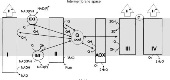
The respiratory chain of plant mitochon-dria, taken as an example, differs in several ways from the mammalian mitochondrial respiratory chain (Figure 1). The dehydro-genase side presents two additional NAD(P)H dehydrogenases, which are not proton pumps
Figure 1 - Organization of the branched respiratory chain of plant mitochondria. Complex I, NADH dehydrogenase; complex II, succinate dehydrogenase;
complex III, cytochrome bc1;
complex IV, cytochrome oxi-dase; AOX, alternative oxioxi-dase; c, cytochrome c; Q, ubiquinone;
QH2, ubiquinol; INT, internal
NAD(P)H dehydrogenase; EXT, external NAD(P)H dehydrogen-ase.
Intermembrane space
(and therefore are not conserving energy), are not inhibited by inhibitors of the proton-pumping energy-conserving complex I (e.g. rotenone) and allow electrons originating from NADH to bypass complex I. Moreover, on the oxidase side a cyanide-resistant alter-native oxidase is present besides the cyto-chrome pathway (complexes III and IV). This oxidase is not linked to proton gradient building and thus dissipates the free energy released during electron flow into heat (16). Ubiquinone (Q) has a central position in the respiratory chain network. This coenzyme links the different branches of the network, receiving electrons from the upstream dehy-drogenases (reducing pathways) and giving electrons to the downstream oxidases (oxidizing pathways). Thus, during the oxidation of NADH formed by the Krebs cycle, one, two or three sites of energy con-servation may be bypassed (i.e., when the rotenone-insensitive dehydrogenase and the cytochrome pathway are involved, when complex I and AOX are involved, or when the rotenone-insensitive dehydrogen-ase and AOX are involved, respectively), leading to a reduction in protonmotive force and finally in the level of ATP synthe-sis. In mitochondria with a branched net-work, the extent to which the dissipative pathways are used affects the energy yield in the cells. It is obviously of crucial interest for these cells to be able to control the relative contribution of each pathway of the network to the total oxygen uptake and it is a chal-lenge for us to understand the mechanisms that regulate the partitioning of electron flow.
Electron flow through AOX can be spe-cifically inhibited by various compounds like hydroxyamic acids (e.g. salicylhydroxamic acid (SHAM) and benzohydroxamic acid (BHAM)) (24), n-propyl gallate (25,26) and disulfiram (27). Then, the level of the cya-nide-resistant oxygen uptake sensitive to one of these inhibitors is the diagnosis of AOX activity.
Characteristics of AOX protein
Primary structure of AOX
The amino acid sequence of a protein can be derived from the cDNA sequences. By analysis of the primary sequence of a protein first insights into its structure can be deduced. AOX primary sequences obtained in such a way contain a mitochondrial transit peptide at the N-terminus. The mature AOX protein from
S. guttatum, as an example, has a calculated molecular mass of 32.2 kDa and contains 283 amino acids (15). Hydropathy plot analysis of several AOX amino acid sequences (15,16) has indicated important conserved features of the protein: a) two hydrophobic regions with a strong α-helical character and highly conserved amino acids have been proposed to be mem-brane-spanning helices, b) these two regions are separated by about 40 amino acids includ-ing an amphipathic helix probably exposed to the intermembrane surface, and c) two hydro-philic regions of about 100 amino acids on both terminal sides with highly conserved short regions at the C-terminus. A topologic model for the AOX protein has been proposed (9,10,16-19), with the amphipathic helix ex-posed on the cytosolic side and the hydrophilic domains extending into the matrix. Both cyto-solic and matricial parts are linked by the two membrane-spanning helices.
Active sites of AOX
recovered by the addition of iron to the culture medium. All of the plant AOX amino acid sequences (in the C-terminal domain) reveal the presence of two copies of the conserved iron-binding motif (E-X-X-H), providing the additional argument that AOX contains iron. By comparison with coupled binuclear iron proteins, an iron-binding four-helical bundle model of the AOX active site has been proposed analogous to that of meth-ane monooxygenase (10,18,31).
The reducing substrate (i.e. ubiquinol (QH2)) binding site has been postulated to be
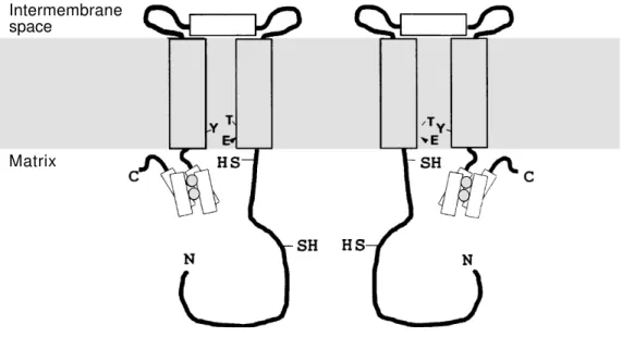
situated on the matrix side of the mitochon-drial membrane in a hydrophobic pocket formed by the two membrane-spanning heli-ces (10,18). The three fully conserved resi-dues (T, E, Y) in the pocket are potential ligands for ubiquinone. Then, in the pro-posed structural model of AOX, the binuclear iron center where oxygen is reduced to water should be close to the postulated binding site of reducing QH2 which itself is in the
vicin-ity of the binding site for the allosteric effec-tor of the enzyme, pyruvate (see below).
Dimeric structure of AOX
The structural property of AOX has been revealed by SDS-PAGE electrophoresis and
immunoblotting using chemical cross-link-ers and oxidizing and reducing agents (32). These experiments have demonstrated that AOX exists as a dimer of 65 kDa in the inner mitochondrial membrane and that two dis-tinct states of the dimer can be identified: an oxidized state in which the dimer is co-valently cross-linked by a disulfide bridge (-S-S-) and a reduced state (-SH HS-) which is maintained through non-covalent interac-tions. The intermolecular disulfide bridge is formed at the level of a conserved cysteine residue (in the plant AOX) situated in the N-terminal domain exposed to the matrix of each monomer (Figure 2). A second con-served cysteine residue (in the plant AOX) implicated in pyruvate binding is situated in the same N-terminal domain close to the membrane surface of each subunit (33).
AOX genetics
The AOX protein is encoded in the nucleus. In many organisms the AOX mono-mer is revealed as multiple bands of approxi-mately 36 kDa on immunoblots using a single monoclonal antibody. In the case of plant AOX protein, depending on the species and tissues, one, two or three bands were ob-served. Therefore, the question was if these
Intermembrane space
Matrix
bands originate from multiple genes (iso-forms) or from a single gene by differential processing or by post-translational modifi-cation of the protein. The existence of three AOX genes has been recently demonstrated in soybean using a polymerase chain reac-tion approach (34,35). Differential expres-sion of these genes has been shown in soy-bean tissues, explaining the presence of mul-tiple bands on immunoblots. Presently, it is suggested that AOX multigene families may be a common feature in plants (34,35).
AOX activity
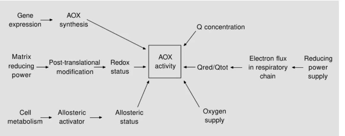
The activity of AOX is controlled by several parameters among which we can distinguish regulatory events and substrate availability (Figure 3).
Regulation of AOX activity can occur at different levels: i) gene expression that af-fects the amount of the protein in the mem-brane and differential gene expression that modifies the ratio between isoforms; ii) post-translational modifications of the protein (i.e. its redox status that affects the nature of the dimer); iii) the action of allosteric effectors like pyruvate.
The substrate availability is linked to the concentration of total quinone in the inner mitochondrial membrane, the redox state of quinone in the membrane, and O2
concentra-tion.
Control of AOX synthesis
Various events can influence the amount of AOX protein present in a cell. The devel-opmental state plays a role in plants, as in thermogenic Araceae, where the amount of AOX increases during flowering (36). In non-thermogenic soybean cotyledons, AOX activity increases immediately after germi-nation and during senescence (20). During the phase of growth in amoeba A. castellanii
cell culture, a parallel exists between AOX activity and protein expression that decreases with the age of the amoeba culture, reaching a stationary phase (14). Under a large variety of stress treatments like chilling in tobacco, wounding in potato tubers, osmotic stress, etc. the expression of the AOX protein is increased (9,19,37). In cell cultures AOX can be induced when the cytochrome path-way activity is decreased, either with specif-ic inhibitors or by disruption of mitochon-drial protein synthesis (9,19). These results indicate that nuclear gene expression is sen-sitive to the activity of the cytochrome path-way and that a communication must exist between mitochondria and the nucleus. In yeast, it has been shown that the ability of an inhibitor of the cytochrome pathway to in-duce AOX synthesis (38) is linked to its ability to cause superoxide formation during mitochondrial respiration. Superoxide anion itself could induce synthesis of AOX protein
Gene expression
AOX synthesis
Matrix reducing
power
Post-translational modification
Redox status
AOX activity
Cell metabolism
Allosteric activator
Allosteric status
Oxygen supply Qred/Qtot Q concentration
Electron flux in respiratory
chain
Reducing power supply
(39) and be the messenger between mito-chondria and the nucleus. In tobacco cell cultures, H2O2 increased levels of AOX
mRNA and AOX activity (40). Salicylic acid, a cellular signal required for the induction of disease resistance in plants, can also be re-sponsible for AOX synthesis (41,42). It has been suggested that salicylic acid-inhibiting catalase activity increases the level of H2O2
which in turn would stimulate gene expres-sion including AOX genes (21).
Differential expression of three AOX genes in soybean has been shown to be tissue-dependent and physiological state-de-pendent (during cotyledon greening) (35). Variation in relative abundance of AOX tran-scripts is generally correlated with the amount of protein isoforms detected by immuno-blotting in roots and cotyledons. The three isoforms (AOX1-3) are found in cotyledons whereas in roots only AOX3 is present. A difference in the proportion of AOX iso-forms leads to variation in the AOX proper-ties between tissues (basal activity, pyruvate stimulation, 18O
2 discrimination; see below).
Moreover, the presence of three types of AOX subunits in the same tissue produces heterodimers (32,35) and could provide a very flexible mechanism of regulation of AOX activity in response to cellular meta-bolic states. AOX isoforms and their hetero-dimerism may be a general feature in plants.
Regulation by redox states
It has been demonstrated that two types of dimeric structure of AOX exist, an oxi-dized form and a reduced form (32). The reduced form can be four- to five-fold more active than the oxidized form (18,32). The ratio of oxidized to reduced protein varies considerably between species and tissues. In cotyledon soybean mitochondria AOX is mainly reduced, while in root soybean mito-chondria it is 50% oxidized (43). In tobacco leaf mitochondria, AOX is largely oxidized and its activity is unmasked by reductants
like dithiothreitol (DTT) (20). Reduction of the AOX protein can also occur during oxi-dation of specific Krebs cycle substrates like isocitrate and malate that are able to reduce NADP+ in plant mitochondria (44). Thus, it
has been proposed that the AOX reduction state depends on NADPH generation and involves NADPH-reduced glutathione or the thioredoxin coupling system (10,20,32,44). It has recently been shown that the redox state of AOX in vivo is different from that determined in isolated mitochondria due to spontaneous oxidation of the reduced spe-cies of the protein during organelle isolation (45). Control of the redox status of AOX could be a powerful mechanism of regula-tion of its activity in vivo that may link AOX activity to the general redox state of the cell (i.e., an increase in the reducing power (NADH, NADPH) induces activation of AOX).
Regulation by allosteric effectors
leaf mitochondria, with a very low AOX activity largely present in the membrane in the oxidized form, no effect of pyruvate is observed until reduction of the protein by DTT occurs. This observation has led to the conclusion that the two systems of AOX regulation (i.e., redox state of the protein and pyruvate binding) interact with each other modulating AOX activity. The AOX dimer must be in its reduced form to be activatable by pyruvate, and the reduced form of the enzyme reveals little activity in the absence of pyruvate.
The effect of pyruvate consists in a low-ering of the range of the ubiquinone redox state over which the AOX is active in iso-lated mitochondria. It has been attributed to lowering the so-called “effective” Km of AOX
for its reduced substrate, ubiquinol (49). This should be understood as an increase in the reactivity of the enzyme towards QH2 which
does not exclude the effect of pyruvate on the catalytic constant of the enzyme. The link between the two regulatory mechanisms (i.e., redox state of the protein and allosteric effect of pyruvate) is such that the active reduced AOX dimer will slowly oxidize ubiquinol until pyruvate is present on its binding site.
In microorganisms such as Acanthamoeba castellanii (50-52), Euglena gracilis (53),
Moniliella tomentosa (54,55), Paramecium tetraurelia (56), Neurospora crassa (57), and Hansenula anomala (58) the alternative oxidase is stimulated by purine nucleotides that probably act from outside the inner mi-tochondrial membrane (56,59). In mitochon-dria of the amoeba A. castellanii, stimulation of AOX activity by GMP resembles the pyru-vate effect on plant mitochondria (affecting AOX activity vs quinone redox state) (60). In amoeba mitochondria, the AOX activity which is not sensitive to pyruvate also seems not to be regulated by the redox status of the protein (14). It may be a general feature of AOX in microorganisms, especially because comparison of known amino acid sequences
of AOX protein from N. crassa (12) and H. anomala (61) indicates lack or displacement of the two regulatory cysteine residues con-served in all plant AOX proteins.
Quinone concentration dependence
As every enzyme, AOX is controlled by its substrate-product concentrations. Thus, the actual concentration of Q and QH2 in the
inner mitochondrial membrane is also im-portant. The Q content in soybean cotyledon mitochondria is 3.5 times higher than the Q content in soybean root mitochondria (35,62). For instance, even if succinate can reduce ubiquinone by 90% in both tissues, the con-centration of ubiquinol present in root mito-chondria will be only 30% of that found in cotyledon mitochondria. This difference in QH2 concentration may be responsible for a
markedly lower AOX activity in root mito-chondria. This may indicate that ubiquinone concentration can be a limiting factor of electron transfer between a dehydrogenase and AOX. However, stimulation by pyru-vate (see above) is more important in root mitochondria whereas it is rather limited in cotyledon mitochondria in the presence of the AOX reductant, DTT (35,43). There-fore, it seems that reduced quinone concen-tration is virtually sufficient to saturate AOX in cotyledon mitochondria while in root mi-tochondria, pyruvate, which lowers the amount of ubiquinol required for AOX ac-tivity (see above), acts in such a way that ubiquinol also saturates AOX. These results illustrate the interplay between substrate (QH2) availability and allosteric effector
(pyruvate) presence indicating some interac-tions between their binding sites.
Quinone redox state dependence
the absence of ADP (state 4) or in the pres-ence of cytochrome chain inhibitors. On the other hand, inhibition of AOX apparently did not modify the activity of the cytochrome pathway. Thus, AOX activity appeared to be controlled by the activity of the cytochrome pathway, giving rise to the overflow para-digm: AOX is only active when the cyto-chrome pathway is saturated or close to being saturated (63-66). Measurements of Q reduc-tion state during succinate oxidareduc-tion by soy-bean cotyledon mitochondria have shown that AOX becomes active only when the Q pool is 40-50% reduced, and then its activity increases sharply and non-linearly with higher Q reduction level (67,68). In contrast, the rate of state 3 respiration is linearly related to the degree of Q reduction (reduced Q (Qred) versus amount of total Q in the membrane (Qtot); Qred/Qtot) and reaches its largest measured values at a low Qred/Qtot ratio at which the AOX activity in the presence of a cytochrome pathway inhibitor is not detect-able (68). In the absence of inhibitors, the rate of state 4 respiration versus Qred/Qtot is linear until AOX becomes active for a Qred/ Qtot ratio higher than 50%, and after this point AOX increases its activity dramati-cally. The relationship between respiratory rate and Qred/Qtot in the cytochrome path-way (in state 4 respiration, with BHAM) is linear (69). To our knowledge, no results concerning the dependence of the cytochrome pathway activity in state 3 respiration (in the presence of BHAM) on ubiquinone reduc-tion level have been published. This general behavior described above is not identical in mitochondria originating from every plant species or tissue. Indeed, different responses of AOX activity to the Q redox state may occur depending on the redox state of the AOX protein and on the presence of allo-steric activators. Hence, the variety of AOX activity widely described with different sub-strates in different mitochondria cannot only be explained by changes in Q redox state. For instance, the difference between external
NADH and succinate in the kinetic relation-ship between AOX activity and Qred/Qtot disappears in the presence of pyruvate (70).
O2 concentration dependence
Because of the O2 concentration in the
incubation medium widely used to study plant mitochondria (about 200-250 µM at air saturation), oxygen reduction by AOX should be independent of the O2 concentrations.
Indeed, the apparent affinity of AOX for O2
is high (apparent oxygen Km around 1-2 µm)
(71-73) and it is lower than the cytochrome oxidase affinity (apparent oxygen Km around
0.1-0.15 µM) (72,74,75). At variance, much higher values of apparent Km of AOX for O2
have been reported (76), ranging from 10 to 20 µM depending on species, tissues and age of the plants investigated. It has also been shown that the apparent Km of AOX for O2
varied with quinone reduction level: the more reduced the quinone pool the lower is the affinity for O2. A kinetic model fitting these
data implies two sequential two-electron re-duction steps of AOX (four-electron-reduced enzyme) and an irreversible activation step of AOX occurs before the reduction of O2 to
water (76).
dur-ing steady-state respiration (i.e. electron par-titioning).
Electron partitioning
The interplay of the various levels of regulation of AOX will influence not only AOX activity but also the way the electrons are distributed between the alternative and cytochrome pathways (i.e. electron parti-tioning). Indeed, it is obvious that an in-crease in AOX activity at a constant quinone-reducing pathway rate due to an increase either in the affinity of AOX for ubiquinol or in the AOX maximal rate will modify the flux of electrons through the cytochrome pathway. Thus, regulation of AOX activity also means regulation of electron partition-ing. The only way to describe quantitatively electron partitioning is to measure the actual contribution of each branching oxidase. This determination is hampered by the fact that both oxidative pathways have the same sub-strates (QH2, O2) and the same products (Q,
H2O). Previous estimations of partitioning
were based on the use of inhibitors of both pathways (63,64). As mentioned before, the usefulness of inhibitor studies was based on the concept that the alternative pathway be-haves as an electron “sink” (overflow) (65,66) and that it is kinetically unfavored, being unable to compete with the cytochrome path-way for QH2. This overflow paradigm used
to be such a strong dogma that it hid the obvious fact that inhibition of one pathway inevitably affects the Qred/Qtot ratio and then modifies electron flux in the other path-way. Fortunately, the recent information on regulation of AOX has led to a reexamina-tion of the overflow assumpreexamina-tions (69,77,78). Numerous studies have demonstrated that several factors enhance AOX activity (see above) and then enable it to compete with an unsaturated cytochrome pathway. Thus, measurement of O2 consumption rates in the
presence of inhibitors to ascertain the elec-tron partitioning between branched
quinol-oxidizing pathways is no longer considered valid. Consequently, most of the electron partitioning determinations made in the past using only rate measurements in the pres-ence or abspres-ence of inhibitors must be con-sidered as misestimated. In order to deter-mine electron partitioning between AOX and the cytochrome pathway it is necessary to measure the true contributions of both path-ways to total respiration when both are ac-tive. These measurements must be performed using well-defined experimental conditions in order to control all the factors that are known to potentially affect both oxidizing pathway activities. Only in this way will an accurate quantitative description of the res-piratory network be obtained. Several strate-gies have been developed to measure the true contributions of branching pathways not only in vitro but also in vivo.
Kinetic approach
de-creases the rate of dehydrogenase activity (product inhibition). The cytochrome path-way curve is obtained in the presence of SHAM by decreasing the overall electron flux with a dehydrogenase inhibitor (e.g. malonate for the succinate dehydrogenase) that decreases the Qred/Qtot ratio and con-sequently the activity of the cytochrome path-way (decrease in substrate availability). The AOX curve is obtained in the presence of KCN by decreasing the overall electron flux with a dehydrogenase inhibitor. The sum of the cytochrome pathway curve and the AOX curve gives the total oxidizing pathway curve when Qred/Qtot decreases. The total non-inhibited steady-state respiration is given by the cross-point of the dehydrogenase curve and the total oxidizing pathway curve. This intersection gives the Qred/Qtot ratio in the absence of inhibitor when both pathways are active. The steady-state when AOX is blocked is given by the intersection of the dehydro-genase curve and the cytochrome pathway curve, while the steady-state when the cyto-chrome pathway is blocked is given by the intersection of the dehydrogenase curve and the AOX curve. The true contribution of the cytochrome pathway to non-inhibited steady-state respiration is the rate obtained on the cytochrome pathway curve for Qred/Qtot without inhibitors, when both oxidizing path-ways are active, while the true contribution of the alternative pathway is the rate ob-tained on the AOX curve for the same Qred/ Qtot ratio. Thus, the kinetic approach de-scribed above allows a prediction of the real contribution of each quinol-oxidizing path-way at any given uninhibited total steady-state rate.
However, although this method is pow-erful, a new set of curves must be rebuilt for every chosen or encountered experimental condition. Apart from this heavy work, the main default of this method is its basic hy-pothesis of a quinone homogenous pool that has never been demonstrated and never disproved. Nevertheless, the kinetic
approach has been useful to emphasize the inadequacy of the classic inhibitor ap-proaches (21,69).
Oxygen isotope differential discrimination
Another method based on the fraction-ation of oxygen isotopes which can occur during plant mitochondrial respiration has been developed (79-83). Fractionation takes place because molecules containing the lighter isotope react a little more readily than molecules that contain the heavier isotope. Respiratory discrimination is determined by observing progressive changes in isotopic composition of oxygen within a closed sys-tem (i.e. the build-up of 18O16O relative to 16O16O after consumption of a substantial
proportion of the available oxygen). A sub-stantial difference found in the oxygen iso-tope discrimination of AOX and cytochrome oxidases, with AOX showing a higher frac-tionation factor (79), has led to development of a mass spectrophotometric technique that is used to estimate directly steady-state par-titioning of electrons between the two oxi-dases. This noninvasive method allows the measurement of the contribution of both oxidizing pathways to total respiration (in the absence of inhibitors) in isolated mito-chondria (43,79,80,83) as well as in intact tissues (81,82).
lnR/Ro D (‰) = _________ x 1000
- lnf
where R is the isotopic ratio of oxygen at the sampling time, Ro is the initial isotopic ratio, and f is the fraction of remaining oxygen in the reaction cell (79). Knowing the values of Dn, Da, and Dc, the partitioning coefficient A can be calculated according to the equa-tion:
(Dn - Dc) A (%) = ____________ x 100
(Da - Dc)
Measurements in material such as Chlamy-domonas reinhardtii which lacks cytochrome oxidase by mutation confirm that the unin-hibited discrimination factor (Dn = 25‰) represents the AOX discrimination factor (Da = 24.2‰), indicating that respiratory flux is solely through AOX and that the obtained values are no artifacts caused by inhibitors (82).
The oxygen isotope fractionation tech-nique showed that AOX can be engaged in respiration under state 3 conditions when the cytochrome pathway is not saturated (83), and it quantified the effect of several regula-tory factors on electron partitioning into AOX. Moreover, this method allows the il-lustration of tissue dependence of this parti-tioning and the comparison of intact tissue and isolated mitochondria partitioning coef-ficients (43,83).
This very elegant method is, at present, the only one that can be used with intact tissues or with whole plants. However, the calibration used in this method requires the use of inhibitors for each respiratory pathway and assumes that change in the total respiration rate does not change the discrimination factor. Moreover, the narrowness between the end points (i.e., Dc and Da) limits the sensi-tivity of the method and the time-consuming measurements will prevent its use for kinetic studies of electron partitioning in various conditions.
ADP/O method
Recently, a method based on the non-phosphorylating property of AOX has been successfully developed and used to deter-mine contributions of the two quinol-oxidiz-ing pathways to total respiration in isolated amoeba mitochondria (84,85). This method involves pair measurements of the ADP/O ratio in the presence or in the absence of an AOX inhibitor (e.g. BHAM) and measure-ments of the overall state 3 respiration in the presence of GMP (an activator of AOX in amoebae) and succinate as oxidizable sub-strate (plus rotenone). This method was first proposed some time ago (63,86) but never used with success although it avoids the use of the rates of electron transport of both pathways in the presence of inhibitors and moreover it is not based on the homogenous quinone pool hypothesis. If
V3 = Vcyt + Valt
where V3 is steady-state 3 respiration, Vcyt is
contribution of the cytochrome pathway, Valt
is contribution of AOX and if
where Ocyt, Oalt = amount of oxygen taken up
related to the activity of the cytochrome and AOX pathways, respectively; then,
Vcyt = V3 x α
Valt = V3 - Vcyt
The ADP/O method is valid only if: i) the ADP/O ratio in the presence of cyanide is equal to zero, ii) BHAM does not induce a proton leak (has no uncoupling effect), and thereby does not affect mitochondrial trans-membrane potential, iii) the ADP/O ratio in the presence of BHAM is independent of the state 3 respiratory rates (within the applied range), and iv) isolated mitochondria are well coupled and stable during the
experi-ADP
---(ADP/O)overall Ocyt + Oalt Ocyt Vcyt
α = --- = --- = --- = ---(ADP/O)cyt (+BHAM) ADP Ocyt + Oalt Vcyt + Valt
mental procedure. All these requirements are positively verified for amoeba mitochon-dria (84,85). The ADP/O method allows the determination of the contributions of both pathways to overall state 3 respiration when it is decreased by increasing the concentra-tion of n-butyl malonate, a non-penetrating inhibitor of succinate uptake by mitochon-dria. When overall state 3 respiration de-clines the AOX contribution decreases sharply and becomes negligible (when state 3 is lower than 100 nmol O min-1 mg
pro-tein-1) while the cytochrome pathway
contri-bution first increases, then passes through a maximum (at state 3 rate around 160 nmol O min-1 mg protein-1) and sharply decreases for
lower state 3 respiration (Figure 4). These results show that AOX is significantly en-gaged before saturation of the cytochrome pathway, and consequently can compete for electrons with the unsaturated cytochrome pathway. This represents the first attempt to examine in detail the steady-state kinetics of the two quinol-oxidizing pathways when both are active and to describe electron partition-ing between them when the steady-state rate of the quinone-reducing pathway is varied.
The ADP/O method, perhaps the best for detailed kinetic and regulation studies of branching oxidases since it is readily appli-cable, cannot, however, be applied to state 4 respiration and could hardly be used with
intact tissues. It has been used with isolated phosphorylating mitochondria and could be applied to permeabilized cells.
In summary, at present, three different methods are available to study electron par-titioning between branching oxidizing path-ways. Each of the described methods has its own domain of application because of its own limitation or restraining basic hypoth-esis. Nevertheless, together they offer the best possibilities to study in depth the mech-anism of regulation of electron partitioning.
Role of the alternative oxidase
The only obvious physiological function of AOX can be recognized in specialized plant thermogenic tissues (spadices of Araceae) as heat generation related to in-crease in temperature taking part in the re-productive processes (87). In non-thermo-genic tissues or cells the role of AOX is now beginning to be better understood thanks to new information concerning its mechanisms of regulation. Indeed, in these cells AOX must play a more fundamental role at the level of metabolism, given the biochemical controls to which the activity of this enzyme is submitted (described above). Metabolic conditions that lead to an increase of the reducing power in the cell will increase the Qred/Qtot ratio, the level of mitochondrial NADPH (therefore the level of reduced AOX diner) and pyruvate (allosteric activator) and thus will increase electron partitioning to AOX. Situations where the phosphate po-tential is high (high ATP/ADP ratio) will lead to the decrease of electron flux into the cytochrome pathway due to proton-electro-chemical potential back pressure and conse-quently increase the partitioning to AOX. Such conditions (high reducing power and ATP/ADP ratio) are consequences of imbal-ances between supply of reducing substrates and energy-carbon demand for biosynthesis, both being coupled by the respiratory chain activity. Operation of AOX may counteract
Contributions
200
150
100
50
0 0
50 100 150 200
V3
Vcyt
+ Valt Vcyt
Valt
Figure 4 - Contributions of the
cytochrome pathway (Vcyt) and
of the alternative pathway (Valt)
these imbalances because it is not directly controlled by the energy status of the cell and may prevent fermentation and favor bio-synthesis. These imbalances could be coun-teracted by fast regulation of AOX activities (allosteric and redox status of the protein) or as a result of slow changes in the environ-ment (various stresses), by modification of the amount of AOX protein (gene expres-sion). The activity of AOX has been shown to prevent the formation of reactive oxygen species by mitochondria at the level of ubiquinol when the cytochrome pathway is slowed down (21,88,89). Thus, AOX may play a protective role in mitochondria by
preventing over-reduction that leads to harm-ful reactive oxygen species production. AOX could also play a role in mitochondrial-chlo-roplastic interactions during photophospho-rylation which increases the reducing power (NADPH) and phosphate potential (ATP) in the cell (90). In conclusion, AOX appears to have a central role in the balance of cell energy metabolism.
Acknowledgments
We thank FAPESP and the Brazilian So-ciety of Biochemistry and Molecular Biol-ogy for their support and invitation.
References
1. Genevois ML (1929). Sur la fermentation et sur la respiration chez les végétaux chlorophylliens. Revue Génétique Botani-que, 41: 252-271.
2. Lance C (1972). La respiration de l’Arum maculatum au cours du développement de l’inflorescence. Annales des Sciences Naturelles Botaniques, 12: 477-495. 3. Meeuse BJD (1975). Thermogenic
respi-ration in Aroids. Annual Review of Plant Physiology, 26: 117-126.
4. Huq S & Palmer JM (1978). Isolation of a cyanide-resistant duroquinol oxidase from Arum maculatum mitochondria. FEBS Letters, 95: 217-220.
5. Rich PR (1978). Quinol oxidation in Arum maculatum mitochondria and its applica-tion to the assay, solubilisaapplica-tion and partial purification of the alternative oxidase. FEBS Letters, 96: 252-256.
6. Bonner Jr WD, Clarke SD & Rich PR (1986). Partial purification and character-ization of the quinol oxidase activity of Arum maculatum mitochondria. Plant Physiology, 80: 838-842.
7. Elthon TE & McIntosh L (1986). Charac-terization and solubilization of the alterna-tive oxidase of Sauromatum guttatum mi-tochondria. Plant Physiology, 82: 1-6. 8. Elthon TE, Nickels RL & McIntosh L
(1989). Monoclonal antibodies to the al-ternative oxidase of higher plant
mito-chondria. Plant Physiology, 89:
1311-1317.
9. Day DA, Whelan J, Millar AH, Siedow JN & Wiskich JT (1995). Regulation of the
alternative oxidase in plants and fungi. Australian Journal of Plant Physiology, 22: 497-509.
10. Siedow JN & Umbach AL (1995). Plant mitochondrial electron transfer and mo-lecular biology. Plant Cell, 7: 821-831. 11. Lambowitz AM, Sabourin JR, Bertrand H,
Nickels R & McIntosh L (1989). Immuno-logical identification of the alternative oxi-dase of Neurospora crassa mitochondria. Molecular Cell Biology, 9: 1362-1364. 12. Sakajo S, Minagawa N, Komiyama T &
Yoshimoto A (1991). Molecular cloning of cDNA for antimycin A-inducible mRNA and its role in cyanide-resistant respira-tion in Hansenula anomala. Biochimica et Biophysica Acta, 1090: 102-108. 13. Clarkson AB, Bienen EJ, Pollakis G &
Grady RJ (1989). Respiration of blood-stream forms of the parasite Trypanoso-ma brucei brucei is dependent on a plant-like alternative oxidase. Journal of Biologi-cal Chemistry, 264: 17770-17776. 14. Jarmuszkiewicz W, Wagner AM, Wagner
JM & Hryniewiecka L (1997). Immuno-logical identification of the alternative
oxi-dase of Acanthamoeba castellanii
mito-chondria. FEBS Letters, 411: 110-114. 15. Rhoads DM & McIntosh L (1991).
Isola-tion and characterizaIsola-tion of a cDNA clone encoding an alternative oxidase protein of Sauromatum guttatum (Schott). Proceed-ings of the National Academy of Sciences, USA, 88: 2122-2126.
16. Moore AL & Siedow JN (1991). The regu-lation and nature of the cyanide-resistant
alternative oxidase of plant mitochondria. Biochimica et Biophysica Acta, 1059: 121-140.
17. Siedow JN, Whelan J, Kearns A, Wiskich JT & Day DA (1992). Topology of the alter-native oxidase in soybean mitochondria. In: Lambers H & van der Plas LHW (Edi-tors), Molecular, Biochemical and Physi-ological Aspects of Plant Respiration. SPB Academic Publishing, The Netherlands, 19-27.
18. Moore AL, Umbach AL & Siedow JN (1995). Structure-function relationships of the alternative oxidase of plant mitochon-dria: a model of the active site. Journal of Bioenergetics and Biomembranes, 27: 367-377.
19. McIntosh L (1994). Molecular biology of the alternative oxidase. Plant Physiology, 105: 781-786.
20. Day DA & Wiskich J (1995). Regulation of alternative oxidase activity in higher
plants. Journal of Bioenergetics and
Biomembranes, 27: 379-385.
21. Wagner AM & Krab K (1995). The alterna-tive respiration pathway in plants: role and
regulation. Physiologia Plantarum, 94:
318-325.
22. Krab K (1995). Kinetic and regulatory as-pects of the function of the alternative oxidase in plant respiration. Journal of Bioenergetics and Biomembranes, 27: 387-396.
and Molecular Biology, 48: 703-734. 24. Schonbaum GR, Bonner WD, Storey BT &
Bahr JT (1971). Specific inhibition of the cyanide-insensitive respiratory pathway in plant mitochondria by hydroxamic acids. Plant Physiology, 47: 124-128.
25. Parrish DJ & Leopold AC (1978). Con-founding of alternate respiration by lip-oxygenase activity. Plant Physiology, 62: 470-472.
26. Siedow JN & Grivin ME (1980). Alterna-tive respiratory pathway. Its role in seed respiration and its inhibition by propyl gal-late. Plant Physiology, 65: 669-674. 27. Grover SD & Laties GG (1978).
Character-ization of the binding properties of disul-firam, an inhibitor of cyanide-resistant res-piration. In: Ducet G & Lance C (Editors), Plant Mitochondria. Elsevier/North Hol-land Biomedical Press, Amsterdam, 259-266.
28. Huq S & Palmer JM (1978). Superoxide and hydrogen peroxide production in the cyanide resistant Arum maculatum mito-chondria. Plant Science Letters, 11: 351-358.
29. Siedow JN (1982). The nature of the cya-nide-resistant pathway in plant
mitochon-dria. Recent Advancements in
Phy-tochemistry, 16: 47-83.
30. Minagawa N, Sakajo S, Komiyama T & Yoshimoto A (1990). A 36-kDa mitochon-drial protein is responsible for
cyanide-resistant respiration in Hansenula
ano-mala. FEBS Letters, 264: 149-152. 31. Siedow JN, Umbach AL & Moore AL
(1995). The active site of the cyanide-re-sistant oxidase from plant mitochondria contains a binuclear iron center. FEBS Let-ters, 362: 10-14.
32. Umbach AL & Siedow JN (1993). Cova-lent and non-covaCova-lent dimers of the cya-nide-resistant alternative oxidase protein in higher plant mitochondria and their re-lationship to enzyme activity. Plant Physi-ology, 103: 845-854.
33. Umbach AL & Siedow JN (1996). The re-action of the soybean cotyledon mito-chondrial cyanide-resistant oxidase with sulfhydryl reagents suggests that α-keto acid activation involves the formation of a
thiohemiacetal. Journal of Biological
Chemistry, 271: 25019-25026.
34. Whelan J, Millar AH & Day DA (1996). The alternative oxidase is encoded in a multi-gene family in soybean. Planta, 198: 197-201.
35. Finnengan PM, Whelan J, Millar AH, Zhang Q, Smith K, Wiskich J & Day DA (1997). Differential expression of the mul-tigene family encoding the soybean
mito-chondrial alternative oxidase. Plant Physi-ology, 114: 455-466.
36. Elthon TE, Nickels RL & McIntosh L (1989). Mitochondrial events during de-velopment of thermogenesis in Sauroma-tum guttaSauroma-tum (Schott). Planta, 180: 82-89.
37. Laties GG (1982). The cyanide-resistant, alternative pathway in plant mitochondria. Annual Review of Plant Physiology, 33: 519-555.
38. Minagawa N, Koga S, Nakano M, Sakajo S & Yoshimoto A (1992). Generation of su-peroxide anion detected by chemilumi-nescence method in the cyanide-insensi-tive mitochondria and the induction of the cyanide-resistant respiration in the yeast Hansenula anomala. In: Yagi H, Kondo M, Niki E & Yoshikawa T (Editors), Oxygen Radicals. Elsevier, New York, 203-206. 39. Minagawa N, Koga S, Nakano M, Sakajo S
& Yoshimoto A (1992). Possible involve-ment of superoxide anion in the induction of cyanide-resistant respiration in Hanse-nula anomala. FEBS Letters, 302: 217-219.
40. Vanlerberghe GC & McIntosh L (1996). Signals regulating the expression of the nuclear gene encoding alternative oxidase of plant mitochondria. Plant Physiology, 111: 589-595.
41. Rhoads DM & McIntosh L (1992). Salicylic acid regulation of respiration in higher plants: alternative oxidase expression. Plant Cell, 4: 1131-1139.
42. Rhoads DM & McIntosh L (1993). Cyto-chrome and alternative pathway respira-tion in tobacco: Effects of salicylic acid. Plant Physiology, 103: 877-883. 43. Ribas-Carbo M, Lennon AM, Robinson
SA, Giles L, Berry JA & Siedow JN (1997). The regulation of electron partitioning be-tween the cytochrome and alternative pathways in soybean cotyledon and root mitochondria. Plant Physiology, 113: 903-911.
44. Vanlerberghe GC, Day DA, Wiskich JT, Vanlerberghe AE & McIntosh L (1995). Alternative oxidase activity in tobacco leaf mitochondria: dependence on tricarboxy-lic acid cycle-mediated redox regulation and pyruvate activation. Plant Physiology, 109: 353-361.
45. Umbach AL & Siedow AL (1997). Changes in the redox state of the alternative oxi-dase regulatory sulfhydryl/disulfide sys-tem during mitochondrial isolation: impli-cations for inferences of activity in vivo. Plant Science, 123: 19-28.
46. Millar AH, Wiskich JT, Whelan J & Day DA (1993). Organic acid activation of the
alternative oxidase of plant mitochondria. FEBS Letters, 329: 259-262.
47. Millar AH, Hoefnagel MHN, Day DA & Wiskich JT (1996). Specificity of the or-ganic acid activation of alternative oxidase in plant mitochondria. Plant Physiology, 111: 613-618.
48. Day DA, Millar AH, Wiskich JT & Whelan J (1994). Regulation of alternative oxidase activity by pyruvate in soybean mitochon-dria. Plant Physiology, 106: 1421-1427. 49. Umbach AL, Wiskich JT & Siedow JN
(1994). Regulation of alternative oxidase kinetics by pyruvate and intermolecular disulfide bond redox status in soybean seedling mitochondria. FEBS Letters, 348: 181-184.
50. Hryniewiecka L, Jenek J & Michejda J (1978). Cyanide resistance in soil amoeba Acanthamoeba castellanii. In: Ducet G &
Lance C (Editors), Plant Mitochondria.
Elsevier/North-Holland, Amsterdam, New York, Oxford, 307-314.
51. Hryniewiecka L & Michejda J (1988). In amoeba mitochondria phosphate mono-nucleotides stimulate specially the alter-native respiratory pathway but not the ro-tenone-sensitive NADH dehydrogenase. 5th European Bioenergetic Conference Reports, Aberystwyth, UK, 20-26 July 1998, 286 (Abstract).
52. Edwards SW & Lloyd D (1978). Properties of mitochondria isolated from cyanide-stimulated and cyanide-sensitive cultures of Acanthamoeba castellanii. Biochemical Journal, 174: 203-211.
53. Sharpless TK & Butow RA (1970). An in-ducible alternative terminal oxidase in Euglena gracilis mitochondria. Journal of Biological Chemistry, 245: 58-70. 54. Hanssens L & Verachtert H (1976).
Aden-osine 5’-monophosphate stimulates cya-nide-insensitive respiration in mitochon-dria of Moniliella tomentosa. Journal of Bacteriology, 125: 829-836.
55. Vanderleyden J, Van den Eynde E & Verachtert H (1980). Nature of the effect of adenosine 5’monophosphate on the cyanide-insensitive respiration in
mito-chondria of Moniliella tomentosa.
Bio-chemical Journal, 186: 309-316. 56. Doussiere J & Vignais PV (1984).
AMP-dependence of the cyanide-insensitive pathway in the respiratory chain of Para-mecium tetraurelia. Biochemical Journal, 220: 787-794.
57. Vanderleyden J, Peeters C, Verachtert H & Bertrandt H (1980). Stimulation of the alternative oxidase of Neurospora crassa
by nucleoside phosphates. Biochemical
58. Sakajo S, Minagawa N & Yoshimoto Y (1997). Effects of nucleotides on cyanide-resistant respiratory activity in mitochon-dria isolated from antimycin A-treated yeast Hansenula anomala. Bioscience of Biotechnology and Biochemistry, 61: 397-399.
59. Vanderleyden J, Kurth J & Verachtert H (1979). Characterization of cyanide-insen-sitive respiration in mitochondria and
sub-mitochondrial particles of Moniliella
tamentosa. Biochemical Journal, 182: 437-443.
60. Jarmuszkiewicz W, Wagner AM & Hryniewiecka L (1996). Regulation of the activity of the alternative oxidase in amoeba A. castellanii mitochondria. Bio-chimica et Biophysica Acta, 9th European Bioenergetic Conference Reports, Louvain la Neuve, Belgium, 17-22 August 1996, 36 (Abstract C. V-5).
61. Li Q, Ritzel RG, McLean LLT, McIntosh L, Ko T, Bertrand H & Nargang FE (1996). Cloning and analysis of the alternative oxi-dase gene of Neurospora crassa. Genet-ics, 142: 129-140.
62. Ribas-Carbo M, Wiskich JT, Berry JA & Siedow JN (1995). Ubiquinone redox be-haviour in plant mitochondria during elec-tron transport. Archives of Biochemistry and Biophysis, 317: 156-160.
63. Bahr JT & Bonner WD (1973). Cyanide-insensitive respiration. I. The steady states of skunk cabbage spadix and bean
hypocotyl mitochondria. Journal of
Bio-logical Chemistry, 248: 3441-3445. 64. Bahr JT & Bonner WD (1973).
Cyanide-insensitive respiration. II. Control of the alternative pathway. Journal of Biological Chemistry, 248: 3446-3450.
65. Lambers H (1980). The physiological sig-nificance of cyanide-resistant respiration in higher plants. Plant Cell and Environ-ment, 3: 293-302.
66. Lambers H (1982). Cyanide-resistant res-piration: a non-phosphorylating electron transport pathway acting as an energy overflow. Physiologia Plantarum, 55: 478-485.
67. Moore AL, Dry IB & Wiskich JT (1988). Measurement of the redox state of the ubiquinone pool in plant mitochondria. FEBS Letters, 235: 76-80.
68. Dry JB, Moore AL, Day DA & Wiskich JT (1989). Regulation of alternative pathway activity in plant mitochondria: nonlinear relationship between electron flux and the redox poise of the quinone pool. Archives of Biochemistry and Biophysics, 273: 148-157.
69. Van den Bergen CWM, Wagner AM, Krab K & Moore AL (1994). The relationship between electron flux and the redox poise of the quinone pool in plant mitochondria. European Journal of Biochemistry, 226: 1071-1078.
70. Hoefnagel MHN & Wiskich JT (1996). Al-ternative oxidase activity and the ubiqui-none redox level in soybean cotyledon
and Arum spadix mitochondria during
NADH and succinate oxidation. Plant
Physiology, 110: 1329-1335.
71. Ikuma H, Schindler FJ & Bonner WD (1964). Kinetic analysis of oxidases in tightly coupled plant mitochondria. Plant Physiology, 39: S-1x.
72. Bendall DS & Bonner WD (1971). Cya-nide-insensitive respiration in plant mito-chondria. Plant Physiology, 47: 236-245. 73. Millar AH, Bergersen FJ & Day DA (1994).
Oxygen affinity of terminal oxidases in
soybean mitochondria. Plant Physiology
and Biochemistry, 32: 847-852. 74. Barzu O & Satre M (1970). Determination
of oxygen affinity of respiratory systems using oxyhemoglobin as oxygen donor. Analytical Biochemistry, 36: 428-433. 75. Rawsthorne S & LaRue TA (1986).
Me-tabolism under microaerobic conditions of mitochondria from cowpea nodules. Plant Physiology, 81: 1097-1102. 76. Ribas-Carbo M, Berry JA, Azcon-Bieto J &
Siedow JN (1994). The reaction of the plant mitochondrial cyanide-resistant al-ternative oxidase with oxygen. Biochimi-ca et BiophysiBiochimi-ca Acta, 1188: 205-212. 77. Day DA, Krab K, Lambers H, Moore AL,
Siedow JN, Wagner AM & Wiskich JT (1996). The cyanide-resistant oxidase: to inhibit or not to inhibit, that is the ques-tion. Plant Physiology, 110: 1-2. 78. Millar AH, Atkin OK, Lambers H, Wiskich
JT & Day DA (1995). A critique of the use of inhibitors to estimate partitioning of electrons between mitochondrial
respira-tory pathways in plants. Physiologia
Plantarum, 95: 523-532.
79. Guy RG, Berry JA, Fogel ML & Hoerinng TC (1989). Differential fractionation of oxy-gen isotopes by cyanide-resistant and cya-nide-insensitive respiration in plants. Planta, 177: 483-491.
80. Guy RG, Berry JA, Fogel ML, Turpin DH & Weger HG (1992). Fractionation of the stable isotopes of oxygen during respira-tion by plants - the basis for a new tech-nique. In: Lambers H & van der Plas LHW
(Editors), Molecular, Biochemical and
Physiological Aspects of Plant Respira-tion. SPB Academic Publishing, Hague,
442-453.
81. Robinson SA, Yakir D, Ribas-Carbo M, Giles L, Osmond CB, Siedow JT & Berry JA (1992). Measurements of the engage-ment of cyanide-resistant respiration in
crassulacean acid metabolism plant
Ka-lanchoe daigremontiana with the use of on-line oxygen isotope discrimination. Planta, 164: 415-422.
82. Robinson SA, Ribas-Carbo M, Yakir D, Giles L, Reuveni Y & Berry JA (1995). Beyond SHAM and cyanide: opportuni-ties for studing the alternative oxidase in plant respiration using oxygen isotope dis-crimination. Australian Journal of Plant Physiology, 22: 487-496.
83. Ribas-Carbo M, Berry JA, Yakir D, Giles L, Robinson SA, Lennon AM & Siedow JN (1995). Electron partitioning between the cytochrome and alternative pathways in plant mitochondria. Plant Physiology, 109: 829-837.
84. Jarmuszkiewicz W, Sluse-Goffart CM, Hryniewiecka L, Michejda J & Sluse FE (1996). Determination of the respective contributions of the cytochrome and
al-ternative oxidase pathways in
Acantha-moeba castellanii mitochondria. In: Westerhoff HV, Snoep JL, Wijker J, Sluse FE & Kholodenko BN (Editors), BioThermoKinetics of the Living Cell. BTK Publishing, Amsterdam, The Netherlands, 31-34.
85. Jarmuszkiewicz W, Sluse-Goffart CM, Hryniewiecka L, Michejda J & Sluse FE (1998). Electron partitioning between the two branching quinol-oxidizing pathways in Acanthamoeba castellanii mitochondria during steady-state 3 respiration. Journal of Biological Chemistry (in press). 86. Lambowitz AM, Smith EW & Slayman CW
(1972). Oxidative phosphorylation in Neu-rospora mitochondria. Studies on wild-type, poky and chloramphenicol induced wild type. Journal of Biological Chemis-try, 247: 4859-4865.
87. Meese BJD (1975). Thermogenic
respira-tion in aroids. Annual Review of Plant
Physiology, 26: 117-126.
88. Purvis AC & Shewfelt RL (1993). Does the alternative pathway ameliorate chill-ing injury in sensitive plant tissues? Physi-ologia Plantarum, 88: 712-718.
89. Purvis AC (1997). Role of the alternative oxidase in limiting superoxide production by plant mitochondria. Physiologia Planta-rum, 100: 165-170.