Overview of Technical Elements of Liver
Segmentation
Nazish Khan*
Institute of Management Sciences, Peshawar, Pakistan
Imran Ahmed
Institute of Management Sciences, Peshawar, Pakistan
Mehreen Kiran
Institute of Management Sciences, Peshawar, Pakistan
Awais Adnan
Institute of Management Sciences, Peshawar, Pakistan
Abstract—Liver diseases are life-threatening, it’s important to detect it tumor in early stages. So, for tumor detection Segmentation of the liver is a first and significant stride. Segmentation of the liver is a yet difficult undertaking in view of its intra patient variability in intensity, shape and size of the liver. The aim of this paper is to assemble a wide assortment of techniques and used CT scan dataset information for liver segmentation that will provide a decent beginning to the new researcher. There are different strategies from basic to advance like thresholding, active contour, region growing to graph cut is briefly abridge to give an outline of existing segmentation strategies. We review the concept of particular strategies and review their original ideas. Our idea is to provide information under which condition a chosen strategy will work or utilize.
Keywords—component; CT Scan; Liver; Dataset; Segmentation technique
I. INTRODUCTION
The liver is key and biggest organ among different organs of the body and provide exceptionally crucial task to our body to keep it free from toxins and harmful substances such as alcohol and medications. Its primary vital functions are: to filter the blood coming from digestive track, it supports all other organs of the body in some way, regulate the supply of body fuel by managing glucose level in our body, cleanse the blood by metabolizing alcohol and obliterating and neutralizing destructive substances, produce bile, which helps in digestion, manufacture many primary and essential proteins which provide resistance to infection and help in blood clotting and thickening. Its direct the supply of vitamins and mineral in our body.
According to Global Cancer Statistics [26] liver cancer fifth the most commonly diagnosed and the second driving reason for death among men and seventh in women. Distinct methods for liver cancer is a blood test, screening and biopsy. A biopsy is intrusive procedure and is exceptionally painful diagnostic technique for patients. That is why researchers are attempting to develop noninvasive techniques. In this way, before the detection of the liver pathology, liver must be segmented accurately.
Liver segmentation is challenging task to develop robust strategies for liver segmentation. Researchers are coping with this challenge to automatically segment the liver; exceedingly unique shapes and volume of liver, similar intensity value among adjacent organs (stomach, spleen, Aorta and abdominal wall), complicated liver structure and contrast media injection cause liver tissue to have different grey level value. Liver segmentation is still an open problem as a result of these challenges. At the time being, researchers are dealing with challenging tasks to increase accuracy in diagnosis and maintain the strategic distance from the need for biopsy (a small tissue of tumor is removed and analyzed) and surgery. These systems do not replace the radiologist, but only provide the second opinion in diagnosis and support radiologist to settle on their choice. Segmentation methods are categorized into 2 main categories; automatic and interactive method. An automatic method has no user intervention and are fully automated and free from user error. It potentially saves the time of operators. The semi-automatic required user intervention like refinement of binary mask and in the selection of seed points. General approach to deal with follow in automated and semi-automatic CAD (Computer Aided System) system is: Pre-processing, Liver segmentation, Lesion Detection, Feature extraction, Classification, Evaluation. Some paper applies the post processing also as indicated by their prerequisite. In pre-processing phase filters are utilized to enhance the image quality and features, furthermore to remove noise and contortion from images. liver segmentation, the liver is sectioned from other encompassing organs. Lesion are detected from segmented liver. Then features are extracted from the lesion area to categorize them either benign or malignant, primary or secondary tumors.
imaging it is ideal to utilize .PNG or .BMP. Many researchers work on Liver segmentation and propose different techniques. Each technique has its own merits and shortcomings.
The rest of the Paper is organized as follows section II Literature Review, section III we organize liver segmentation techniques overview, section IV we include conclusion to summarize our views. we place the dataset comparison table and techniques overview table at the end of paper.
II. LITERATURE REVIEW
Lim et al [2] Develop automatic liver segmentation using previous knowledge of liver position and use a deformable contour method based on morphological filtering operation. On the gradient label map Algorithm [2] perform deformable contouring. To reduce computational complexity and to decide suitable threshold histogram analysis is performed in the ELP (Estimated liver position). Proposed method uses multi scale morphological filter recursively with region labeling and clustering to detect search rang for deformable contouring. They use private dataset of 10 patients. Results are compared with manual segmentation by a radiologist. Graphical representation is shown below in Fig. 1
Fig. 1. Final Segmentation result
Suzuki et al [4] developed automatic liver extraction technique for contrast enhance CT images. The anisotropic diffusion filter uses for image denoising and preserving the shape of the liver. Scale specific gradient magnitude use to enhance liver boundary. These preprocessing results are passed to fast marching level set algorithm that initially refines the liver boundary and use as a rough estimate of liver shape and geodesic active contour combine with level set to extract liver shape. And estimate liver volume. Liver manually trace by the expert radiologist is use as a gold standard to compare the evaluation results of liver volume. Their local dataset consists of 15 patients. Overall accuracy is 98.4 %. Sensitivity, specificity and percent volume error is 91.1%, 99.1%, and 7.2 respectively. Limitation of the paper is small dataset. Graphical representation is shown in Fig. 2
Fig. 2. (a) Original CT image (b)Anisotropic diffusion noise reduction (c) Scale specific gradient magnitude calculation (d) Non-linear grey scale conversion(e) Geodesic active contour segmentation
Massoptier et al [7] Develop innovative statistical model based approach. Active contour and gradient vector flow (GVF)are also used for hepatic segmentation. They analyzed nonlinear anisotropic diffusion and mean shift filter based on the processing time. To save processing time they follow mean shift filter. Clustering technique was used with their powerful initialization method for hepatic lesion segmentation. For lesion detection 82.6% Sensitivity and 87.6% specificity were achieved. Liver volume overlapping was evaluated by DSC (Dice Similarity Coefficient), FNR (false negative ratio), FPR (false positive ratio). They use their own private data set of 21 patient CT data set. From 46 lesions, they diagnosed different type of tumor 6 were HCC (Hepatocellular carcinoma), 2HDG (Hemangioma), 8HM (Hepatic metastases) and 5 have healthy patients.
paper is, they face under segmentation problem when lesions are close to liver boundary. The dataset is very small.
Militzer et al [12] Proposed a novel system for automatically detecting and segmenting focal liver region from CT images. For classification, it utilizes a probabilistic boosting and thus provide fully automated detection and segmentation of the liver lesion simultaneously. They use hierarchical mesh-based shape representation for liver segmentation. Features selected in this paper are gray level statistical feature and Haar like features. Detection rate 77% could be achieved with the specificity of 0.93% and a sensitivity of 0.95% at the same time for lesion segmentation at the same setting.
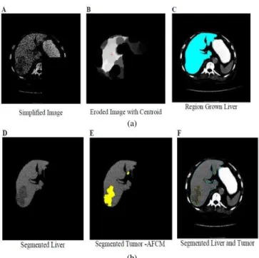
S.S Kumar [15] presented an automatic segmentation of the liver lesion from CT radiographs. This paper utilizes medium filter, erosion, dilation, largest connected component as a pre-processing step. In post-processing, morphological operators are utilized to additionally refine the image and utilize basic region growing techniques for live segmentation and an alternate Fuzzy C-Means Clustering for tumor segmentation. He used 10 cases in his research work. The Technique result was contrasted and evaluated with the manual segmentation based on false positive rate, false negative rate, volume measurement error, spatial overlap and visual overlap. Pictorial results are shown in Figure .1 below.
(a)
(b)
Fig. 3. (a) results of final liver segmentation using AFCM (b) results of final liver segmentation using AFCM
Belgherbi et al [17] A semi-automatic method developed for liver lesion extraction using mathematical morphology from CT images. In pre-processing for refinement of the liver they use dilation, erosion and anisotropic diffusion filter. For liver lesion, they use mathematical morphology, especially on the water shed technique. They use private data set. The proposed scheme achieves 92% Sensitivity & 99% Specificity. Brief graphical representation is shown in Fig.4. Here we
focus only on the graphical representation of the liver. Pictorial representation of liver lesion is not represented here.
Fig. 4. Original image Histogram Thresholding Liver extraction
Marcin Ciecholewski [18] present novel method which automatically segments the liver shape. In CT scan images lumber section of the spine is utilized as seed point. After seed point selection, joint polylines are drawn to approximate the liver contour. These component polylines frames the components of two polygons eliminated from the image, which leaves only the segmented liver shape inside the image.
Results are accessed on 13,30 images using Dice’s similarity.
Fig. 5 shows the results graphically.
(a) (b) (c) (1)
CT images Ground truth proposed method (2)
Fig. 5. (1) (a)Polygon with polyline L(b) Polygon with polyline R(c)segmented liver (2) Segmentation of liver shape using connected polylines
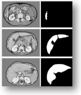
Aldeek et al [21] develop semiautomatic method using a Bayesian classifier for liver segmentation. In post processing, median filter and some filling operation are performed to further refine the classifier results. Dataset consists of 44 cases. Average area overlaps accuracy is over 87% They evaluate their results with the manual segmentation done by the expert radiologist. Graphical representation is shown in Fig.6.
Fig. 6. Bayesian classifier segmentation results
Altarawneh et al. [30]in this paper researcher modify DLSR (Distance regularization level set) [1] method because it does not work well in case of weak or without edges of liver images. [30] overcome this issue by introducing new Balloon force that control weak and without the edges region by slowing down and controlling the evaluation process. Balloon force was created by utilizing probability density energy function to control the speed and energy of the evaluation process. Experiments were performed on 10 cases of 512*512 pixels Slices. Graphical representation of comparison are shown in Fig. 7 below.
CT Slice Ground Truth Original DSLR Proposed DSLR
Fig. 7. Comparison of DLSR with proposed DSLR with balloon force
III. LIVER SEGMENTATION TECHNIQUES
A. Active contour
Active Contour is the energy minimizing, deformable curves that moves due to the effects of internal and external object forces to detect object boundary. Internal forces prevent deformation. “The contours are classified broadly into two categories as: parametric active contours and geometric active
contours. Parametric active contours are again classified into three types: Traditional snake, GVF snake and Ballon forces. Geometric active contours are Level set”.
Basically, snakes use energy minimization to match a deformable model of an image.
General snake or active contour faces two problems: Initial model must be close to the boundary, otherwise
it will show wrong result
This model confronting issue to progress into boundary concavities.
AC [9][6][7] [13] model generally need manual initialization which is close to the image contour, otherwise it will give under or over segmentation problem. In case of liver segmentation, it causes over segmentation problem because liver share same intensity with the adjacent organs. It easily streams to the neighboring organ and cause over segmentation problem. Its performance is degraded if there should be an occurrence of irregular and higher intensities. Some of online available matlab codes are: [34] [35][36].
B. Gradient vector flow
Gradient Vector Flow [5] deal with two issues of the active contour model. It can converge into boundary concavities by calculating both concave and convex features. The traditional snake model must start close to the boundary while GVF [6][7] have the ability to start far from the boundary and till can converge to the image boundary. By and large, GVF demonstrate insensitivity to the initialization. GVF contour can handle broken object edges and subjective contour. Some of available code for GVF are in [37]
C. Level set
LS [13][16][30]semi-automatic techniques since it oblige user to select seed point.LS performance is very rely on the initial position, its performance increase when initial contour is placed close to the hepatic boundary .Its performance is degraded when the object is without edges or have weak edges[1]. The liver has the same intensity as the neighboring organs, hence cause over segmentation issues. Level set is time consuming for large computation. Level set strategy is additionally utilized for refinement of the liver segmentation. Some Matlab code accessible online is [38].
D. Graph cut
using different algorithms. In case of liver tumor segmentation general active contour come up short when tumor is near liver surface, graph cut handle this kind of active contour issues extremely well.
E. Adaptive thresholding
Adaptive Thresholding [3] is additionally called local or dynamic thresholding. The principal idea of adaptive threshold is to apply different threshold on the different region of the image. Adaptive threshold divide the image into small areas and apply different threshold in different areas. On adaptive threshold value at each pixel location is depends on the neighboring pixel intensity. This type of thresholding is functioning admirably in irregular intensities and can handle the lightening condition extremely well. Adaptive threshold work on a pixel level, it sets all the pixel as foreground whose intensity value is greater than a certain threshold and all other pixels as background. Adaptive threshold work on color and grey scale image by converting it into a binary image. Adaptive threshold follow two approaches to find the threshold for every pixel: (i) the Chow and Kaneko approach and (ii) local thresholding. General online link for adaptive matlab code in [39].
F. Region growing
Region Growing [15][20][23][19]technique is semi-automatic strategy, since user interaction is required as the seed point is chosen by the user. RG method divides the image into regions according to predefine basis. This technique is initiates utilizing seed point, and examine the neighboring pixel either utilizing 4 connectivity or 8 connectivity, it iteratively adds the pixel to different region as indicated by predefined criteria. The criteria could be pixel intensity, gray level texture or color. In case of liver segmentation this technique gives great outcomes in contrast enhance images. Its effectiveness is relying on the determination of the seed point. Some of the RG code available online are [31][32][33].
G. Fuzzy clustering mean(fcm)
FCM is developed by dunn [40] is widely utilized in medical image processing. Its originate from the k mean algorithm. In k mean algorithm, each pixel is belonging to only one k cluster which is not feasible in case the of liver segmentation. FCM overcomes this issue by utilizing membership function which shows the belongings of the pixel to the cluster. FCM is fuzzy clustering technique which allows a pixel to belong to one or more clusters. FCM is a semi-automatic method. Some parameters like the selection of centroid, the degree of fuzziness and stopping criteria highly effects the performance of the FCM. In case of liver segmentation. FCM is mostly used for tumor segmentation [15] from the CT data.
H. Statistical shape model(ssm)
SSM [28][29][11]are find extremely effective in liver segmentation. The liver has an exceptionally varying shape. In this approach, probabilistic model is made to adapt to the varying shape of the liver. SSM can deal with the limitation of gray level techniques extremely well. During segmentation of liver, grey level techniques frequently appear under or over segmentation issues when the tumor is close to liver boundary.
This constraint of grey level techniques can handle utilizing SSM. Utilizing the prior knowledge SSM can deal unclear boundary of the liver extremely well. SSM focus on the shape of the liver. Limitation of shape based method and classifier based method [8][23]is the they require great number of training dataset.
IV. CONCLUSION
The different segmentation techniques review has been done in this paper. Here the review of preprocessing, main segmentation technique, and their dataset information is provided in the form of a table. Comparative evaluation of different method is not possible in light of the fact that every author utilizes small private dataset set and different performance measure criteria are utilized. For objective comparison, for the most part acknowledge performance measures are required. From review of the techniques, it is concluded that there is still need to discover robust and efficient method for liver segmentation. As the liver is exceptionally difficult organ to handle and segment. Each existing method has its own advantage and disadvantages they are not full robust. Future work is to cover the classification techniques for liver tumor.
REFERENCES
[1] Li, Chunming, et al. "Distance regularized level set evolution and its application to image segmentation."IEEE transactions on image processing19.12 (2010): 3243-3254.
[2] Lim, Seong-Jae, Yong-Yeon Jeong, and Yo-Sung Ho. "Automatic liver
segmentation for volume measurement in CT Images." Journal of Visual
Communication and Image Representation 17.4 (2006): 860-875.
[3] Massoptier, Laurent, and Sergio Casciaro. "Fully automatic liver
segmentation through graph-cut technique." 2007 29th Annual
International Conference of the IEEE Engineering in Medicine and Biology Society. IEEE, 2007.
[4] Rusko, Laszlo, et al. "Fully automatic liver segmentation for
contrast-enhanced CT images." MICCAI Wshp. 3D Segmentation in the Clinic: A
Grand Challenge 2.7 (2007).
[5] Gui, Tianyi, Lin-Lin Huang, and Akinobu Shimizu. "Liver segmentation
for CT images using an improved GGVF-snake." SICE, 2007 Annual
Conference. IEEE, 2007.
[6] Alomari, Raja S., Suryaprakash Kompalli, and Vipin Chaudhary.
"Segmentation of the liver from abdominal CT using Markov random
field model and GVF snakes." Complex, Intelligent and Software
Intensive Systems, 2008. CISIS 2008. International Conference on. IEEE, 2008.
[7] Massoptier, L., & Casciaro, S. (2008). A new fully automatic and robust
algorithm for fast segmentation of liver tissue and tumors from CT
scans.European radiology, 18(8), 1658-1665.
[8] Mala, K., V. Sadasivam, and S. Alagappan. "Neural network based
texture analysis of liver tumor from computed tomography images." International Journal of Biological and Life Sciences 2.1 (2006): 33-40.
[9] Jiang, Huiyan, and Qingshui Cheng. "Automatic 3D segmentation of CT
images based on active contour models." Computer-Aided Design and Computer Graphics, 2009. CAD/Graphics' 09. 11th IEEE International Conference on. IEEE, 2009.
[10]Yussof, Wan Nural Jawahir Hj Wan, and Hans Burkhardt. "Integration of
Morphology and Graph-based Techniques for Fully Automatic Liver Segmentation." Majlesi Journal of Electrical Engineering 4.3 (2010): 59-66.
[11]Lamecker, Hans, Thomas Lange, and Martin Seebass. Segmentation of
[12]Militzer, Arne, et al. "Automatic detection and segmentation of focal liver lesions in contrast enhanced CT images." Pattern Recognition (ICPR), 2010 20th International Conference on. IEEE, 2010
[13]Suzuki, Kenji, et al. "Computer-aided measurement of liver volumes in CT by means of geodesic active contour segmentation coupled with level-set algorithms." Medical physics 37.5 (2010): 2159-2166.
[14]Wang, Ning, Lin-Lin Huang, and Baochang Zhang. "A fast hybrid method for interactive liver segmentation." Pattern Recognition (CCPR), 2010 Chinese Conference on. IEEE, 2010.
[15]Kumar, S. S., R. S. Moni, and J. Rajeesh. "Automatic segmentation of liver and tumor for CAD of liver." Journal of advances in information technology 2.1 (2011): 63-70.
[16]Li, Bing Nan, et al. "Integrating FCM and level sets for liver tumor segmentation." 13th International Conference on Biomedical Engineering. Springer Berlin Heidelberg, 2009.
[17]Belgherbi, A., I. Hadjidj, and A. Bessaid. "A Semi-automated Method for the Liver Lesion Extraction From a CT Images Based on Mathematical Morphology." Journal of Biomedical Sciences 2.2 (2013).
[18]Ciecholewski, Marcin. "Automatic liver segmentation from 2D CT images using an approximate contour model." Journal of Signal Processing Systems 74.2 (2014): 151-174.
[19]López-Mir, Fernando, et al. "Liver Segmentation on CT Images. A Fast Computational Method Based on 3D Morphology and a Statistical Filter." IWBBIO. 2013.
[20]Anter, Ahmed M., et al. "Automatic computer aided segmentation for liver and hepatic lesions using hybrid segmentations techniques." Computer Science and Information Systems (FedCSIS), 2013 Federated Conference on. IEEE, 2013.
[21]ALDEEK, NIDAA, et al. "LIVER SEGMENTATION FROM ABDOMEN CT IMAGES WITH BAYESIAN MODEL." Journal of Theoretical & Applied Information Technology 60.3 (2014).
[22]Mostafa, Abdalla, et al. "CT liver segmentation using artificial bee colony optimisation." Procedia Computer Science 60 (2015): 1622-1630. [23]Cheng, Ching-Hsue, and Liang-Ying Wei. "Rough Classifier Based on
Region Growth Algorithm for Identifying Liver CT Image." 淡江理工學 刊 19.1 (2016): 65-74.
[24]Sayed, Gehad Ismail, Aboul Ella Hassanien, and Gerald Schaefer. "An Automated Computer-aided Diagnosis System for Abdominal CT Liver Images." Procedia Computer Science 90 (2016): 68-73.
[25]Sayed, Gehad Ismail, and Aboul Ella Hassanien. "Abdominal CT Liver Parenchyma Segmentation Based on Particle Swarm Optimization." The
1st International Conference on Advanced Intelligent System and Informatics (AISI2015), November 28-30, 2015, Beni Suef, Egypt. Springer International Publishing, 2016.
[26]Jemal, Ahmedin, et al. "Global cancer statistics." CA: a cancer journal for clinicians 61.2 (2011): 69-90.
[27]Wu, Kesheng, Ekow Otoo, and Arie Shoshani. "Optimizing connected component labeling algorithms." Medical Imaging. International Society for Optics and Photonics, 2005.
[28]Heimann, Tobias, H. Meinzer, and Ivo Wolf. "A Statistical Deformable Model for the Segmentation of Liver CT Volumes Using Extended Training Data." Proc. MICCAI Work (2007): 161-166.
[29]Li, Guodong, et al. "Automatic liver segmentation based on shape constraints and deformable graph cut in CT images." IEEE Transactions on Image Processing 24.12 (2015): 5315-5329.
[30]Altarawneh, Nuseiba, et al. "Liver Segmentation from CT Images Using a Modified Distance Regularized Level Set Model Based on a Novel Balloon Force| NOVA. The University of Newcastle's Digital Repository." (2014).
[31] https://www.mathworks.com/matlabcentral/fileexchange/50782-region-growing-segmentation
[32] https://www.mathworks.com/matlabcentral/fileexchange/32532-region-growing--2d-3d-grayscale-
[33] https://www.mathworks.com/matlabcentral/fileexchange/19084-region-growing
[34] https://www.mathworks.com/matlabcentral/fileexchange/19567-active-contour-segmentation
[35] https://www.mathworks.com/matlabcentral/fileexchange/23445-chan-vese-active-contours-without-edges
[36] http://www.shawnlankton.com/2008/04/active-contour-matlab-code-demo/
[37] https://www.mathworks.com/matlabcentral/fileexchange/45896-fast-gradient-vector-flow--gvf-
[38] https://www.mathworks.com/matlabcentral/fileexchange/12711-level-set-for-image-segmentation
[39] https://www.mathworks.com/matlabcentral/fileexchange/8647-local-adaptive-thresholding
TABLE I. COMPARISION OF DATASET
Ref Author Year Images used Datatypes Image size
[2] Lim et al 2005 10cases Private dataset 512*512
[3] Massoptier et al 2007 10cases Private dataset -
[4] Rusko et al 2007 10cases - -
[5] Gui et al 2007 4cases ,200images Private dataset 170*170
[6] Alomari et al 2008 13cases Private dataset 512*512
[7] Massoptier et al 2008 21cases Private dataset 512*512
[8] Mala et al 2008 105 images Private dataset 256*256
[9] Jiang et al 2009 5 cases - -
[10] Yussof et al 2010 10cases - 512*512
[11] Akram et al 2010 100images Private dataset 512*512
[12] Militzer et al 2010 15cases Private dataset -
[13] Suzuki et al 2010 15cases Private dataset -
[14] Wang et al 2010 100images Private dataset 512*512
[15] S.S Kumar 2011 10cases - 512*512
[16] Li et al 2009 15cases LTSCdataset
NUHdataset 512*512
[17] Belgerbi et al 2013 - Private dataset -
[18] Ciecholewski 2014 1330 images
120 cases Private dataset -
[19] Lopez-mir et al 2013 30 cases Private dataset 512*512
[20] M.Anther et al 2013 112cases 860 images 2 Private datasets 630*630
[21] ALDEEK et al 2014 44cases Private dataset 512*512
[22] Mostafa, et al 2015 38CT images Private dataset -
[23] Cheng et al 2016 800images Open NBIA
Private dataset 512*512
[24] Sayed et al 2016 62 Images - 256*256
TABLE II. LIVER SEGMENTATION TECHNIQUES OVERVIEW
REF NO PRE-PROCESSING LIVER SEGMENTATION EVALUATION
[3] Mean shift filter
Adaptive threshold Graph cut
Accuracy 96%
[4]
Hough transform Erosion/dilation Largest CCL
Region growing Accuracy 76%
[5]
Canny edge detector Hermit spline curve Anisotropic diffusion filter
GGVF
-
[2] Multilevel thresholding Morphological filter
Gradient label map K mean clustering
Label based search algorithm
Accuracy 96%
[6] Histogram analysis Markov random field
Gradient vector flow
Active contour Similarity matrix
[7] Adaptive threshold Mean shift filter
Gradient vector flow Active contour
A volume overlap of liver 94.2% Sensitivity &specificity for tumor 82.6%& 87.5% respectively
[9] Sobel operator
Erosion/dilation Active contour Accuracy 94%
[10]
Opening
Multiscale filtering Anisotropic diffusion filter
CCL
Graph Cut -
[11]
Median filter Adaptive Histogram Power law transformation
Closing Largest area Global threshold
Accuracy 96%
[13]
Anisotropic diffusion filter Median filter
Scale specific gradient magnitude filter
Fast marching Level set
Geodesic active contour
Manual tracing method
[14] Gradient magnitude Anisotropic diffusion filter Random walk algorithm Watershed TP, TN, FP, FN Manual tracing method
[15] Median filter/ Erosion Largest CCL
Region growing
Alternate FCM Manual tracing method
[16] Morphological operations Fuzzy Clustering Mean/ Level set
Balloon force -
[17]
LIVER: H max transform Dilation/erosion Lesion:
Anisotropic diffusion filter Hmaxima transform filter
Watershed algorithm Sensitivity & specificity 92% & 99 % respectively
[18] - Connected poly lines DICE similarity co efficient 81.3%
[19] Adaptive filter
Dilation/erosion Region Growing -
[20]
Erosion/dilation Adaptive threshold
CCL
Region Growing Watershed
Accuracy 96%
[21] Convert intensity value into Hounsfield units
Median filter Bayesian Model Accuracy 87%
[22]
Median filter Contrast Stretching
Thresholding to separate Ribs
Artificial bee colony
Optimization algorithm Accuracy93.73%
[24] Median filter
FCM
Grey Wolf Optimization SVM