Korean J Pediatr 2012;55(7):249-253 eISSN 1738-1061 • pISSN 2092-7258
249
A fatal case of acute pulmonary embolism caused
by right ventricular masses of acute lymphoblastic
lymphoma-leukemia in a 13 year old girl
We report a case of a 13-year-old girl with acute lymphoblastic lym-phoma-leukemia, who presented with a cardiac metastasis in the right ventricle, resulting in a pulmonary embolism. At the time of her leukemia diagnosis, a cardiac mass was incidentally found. The differ-ential diagnosis for this unusual cardiac mass included cardiac tumor, metastasis, vegetation, and thrombus. Empirical treatment was initiated, including anticoagulation and antibiotics. She underwent plasmapheresis and was administered oral prednisolone for her leukemia. Five days later, she experienced sudden hemodynamic collapse and required extracorporeal membrane oxygenation insertion and emergency surgery. These interventions proved futile, and the patient died. Pathology revealed that the cardiac mass comprised an aggregation of small, round, necrotic cells consistent with leukemia. This is the irst known case of acute lymphoblastic leukemia presenting as a right ventricular mass, with consequent fatal acute pulmonary em bolism. A cardiac mass in a child with acute leukemia merits investigation to rule out every possible etiology, including vegetation, thrombus, and even a mass of leukemic cells, which could result in the fatal complication of pulmonary embolism.
Key words: Leukemia, Metastasis, Pulmonary embolism, Child
Yu Mi Ko, MD1, Soo Hyun Lee, MD1, June Huh, MD, PhD1, Hong Hoe Koo, MD, PhD1, Ji
Hyuk Yang, MD, PhD2
Departments of 1Pediatrics and 2Thoracic and Car
dio vascular Surgery, Samsung Medical Center, Sungkyunkwan University School of Medicine, Seoul, Korea
Received: 1 August 2011, Revised: 10 October 2011 Accepted: 15 December 2011
Corresponding author: June Huh, MD, PhD
Department of Pediatrics, Samsung Medical Center, Sung-kyunkwan University School of Medicine, 50 Irown-dong, Gangnam-gu, Seoul 135-710, Korea
Tel: +82-2-3410-3526, Fax: +82-2-3410-0043 E-mail: [email protected]
Copyright © 2012 by The Korean Pediatric Society
his is an openaccess article distributed under the terms of the Creative Commons Attribution NonCommercial License (http://creativecommons.org/licenses/by nc/3.0/) which permits unrestricted noncommercial use, distribution, and reproduction in any medium, provided the original work is properly cited.
Introduction
Although leukemia is one of the most common neoplasms that metastasize to the heart, the occurrence of an intracardiac mass in acute leukemia is extremely rare1,2). The differential diagnosis of intracardiac mass lesions in leukemia should include intracardiac metastasis, thrombus, and vegetation2,3). An intracardiac mass may
complicate a rare but fatal pulmonary embolism. Single pediatric case
of primary cardiac lymphoma has been reported so far, but there are no reported pediatric cases of patients who presented with a cardiac mass resulting in a pulmonary embolism in South Korea4).
distress. She was awake, alert, and speaking in full sentences.
here was a palpable lymph node at the right supraclavicular area; it was 1×2 cm in size with tenderness upon palpation and movable. It had no redness and was not warm to the touch. here was another small palpable lymph node. Cardiac examination revealed regular tachycardia and a nondisplaced point of maximal impulse. There were no murmurs, rubs, gallops, or jugular venous distension. Chest wall movement was symmetrical, with clear and equal breath sounds. There were no motor or sensory deficits. She exhibited good coordination. The remainder of the physical examination was unremarkable.
Bone marrow analysis showed blasts of 30%, which was compatible with acute leukemia (Figs. 1, 2). he chest radiograph was normal, and the electrocardiogram showed normal sinus rhythm. Twodimensional echocardiography before chemotherapy revealed a 16×18×18 mm sized cardiac mass that was highly mobile and attached to the right ventricular free wall and chordae of the tricuspid valve. Its shape was bizarre, with heterogeneous echogenecity (Fig. 3). The mass looked like an avascular mass on cardiac magnetic
Fig. 1. Bone marrow biopsy showing blast cells, consistent with acute
leukemia (H&E, ×400).
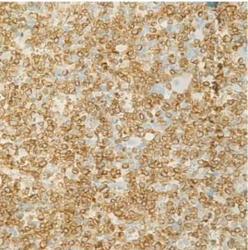
Fig. 2. Bone marrow biopsy with immunohistochemistry special stain
(CD3). CD3 is positive in the leukemic blast cells (×400).
Fig. 3. Echocardiography showing multiple masses. The masses were
resonance imaging (Fig. 4). RV thrombus, vegetation, or metastasis were considered as possible etiologies. To rule out cardiac vegetation and diagnose infective endocarditis, blood cultures were done, but no organism was isolated. A blood test to rule out thromboembolism was conducted: Ddimer was 60 ug/mL, and another coagulation test, International Normalized Ratio, was 1.52 (reference range, 0.9 to 1.1). In addition, ibrinogen was 87 mg/dL (reference range, 182 to 380) and antithrombin III activity was 67% (reference range, 83 to 123). here was no bleeding tendency.
he patient received intravenous antibiotics and antifungal agents in order to treat possible infective endocarditis with vegetation. Heparinization was also started for possible thrombus. Treatment for acute leukemia including intravenous hydration and alkalization, plasmapheresis, and low dose oral prednisolone was started along with other treatments. On the following day, she developed sudden chest pain and dyspnea, which turned out to be caused by pulmonary embolism, which was discovered by chest computed tomography (Fig. 5). Despite heparinization and antibiotics, the RV mass size appeared to grow, as shown by a followup 2dimentional
echocardiogram. She was transferred to the pediatric intensive care unit because of progressive dyspnea. Sudden hemodynamic collapse developed the next morning. Extracorporeal membrane oxygenation (ECMO) support and emergency surgery was performed due to acute pulmonary embolism and for removal of the cardiac mass.
On surgical indings, the mass was fragile and easily crumbled and attached to a dysplastic tricuspid valve (Fig. 6). Also, irregular shaped masses were found in both pulmonary arteries. Pathology revealed that the cardiac mass and pulmonary artery mass consisted of an aggregation of small round necrotic cells that were compatible with leukemic cells (Figs. 7, 8).
In spite of aggressive postoperative ECMO support, the patient died of disseminated intravascular coagulation and heart failure the day after surgery.
Discussion
This is the first case of acute lymphoblastic leukemia presenting as a right ventricular mass and consequent fatal acute pulmonary embolism. his case had diiculties in the diferential diagnosis and choosing appropriate management.
In acute leukemia, immature cells commonly infiltrate the myo cardium, causing conduction abnormalities and rhythm disturbances5). There have been only a few cases of Bcell lineages as masses reported in the literature. Cardiac infiltration is very rare, and in leukemia only a few cases of pericardial efusion, heart muscle infiltration, or intracavitary masses have been described. Cardiac metastases from nonsolid tumors secondary to primary leukemia have accounted for 4.4%6). Although cardiac masses in recently reported cases of leukemia are in the right side of the heart, there has been no evidence of a common site for cardiac masses. A high WBC
Fig. 4. The mass appeared to be avascular on cardiac magnetic
resonance imaging.
Fig. 5. Chest computed tomography showing pulmonary embolism.
Fig. 6. A fragile and easily-crumbled mass (arrow) attached to a
count in leukemia is reported to have a high risk for leukemic cell aggregation and a cardiac mass, and most patients who have had a cardiac mass diagnosis have had acute myeloid leukemia (AML)2,6). Cardiac masses in acute lymphoblastic leukemia are not common. A case of intracardiac thrombi in AML was reported in adults2). here
induction, and the remaining 10% occur during consolidation or therapy intensification protocols. The recently published meta analysis by Caruso et al.8) did not demonstrate a statistically signiicant diference in venous thromboembolism prevalence according to the corticosteroid type and dexamethasone used in induction. herefore, in the patient with acute leukemia with venous thromboembolism, primary prophylaxis is warranted during chemotherapy agent ex posure.
Laboratory indings and imaging studies could not give a deinite diagnosis for this cardiac mass. here was no helpful imaging study to aid in the diferential diagnosis. Lack of deinite diagnostic evidence necessitated treatments covering all possible causes of cardiac mass and underlying disease of acute lymphoblastic leukemia. Rapid deterioration related to pulmonary embolism might have been related to the efect of the chemotherapy agent (prednisolone), which made the cardiac leukemic mass disseminate into pulmonary embolism.
In conclusion, cardiac mass in a child with acute leukemia should be investigated to rule out every possible causes, including vegetation, thrombus, and even cardiac mass of leukemic cells resulting in the fatal complication of pulmonary embolism. Clinicians should be alert to the possibility of the rapid progression to pulmonary embolism after chemotherapy.
References
1. Cheruvu B, Cheruvu P, Boyars M. An unusual case of metastasis to the left side of the heart: a case report. J Med Case Rep 2011;5:23.
2. Nanjappa MC, Shankarappa RK, Kalpana SR, Bhat P, Moorthy N. Intracardiac thrombi in acute myeloid leukemia: an echocardiographic and autopsy correlation. Echocardiography 2010;27:E48.
3. Sakuma M, Fukui S, Nakamura M, Takahashi T, Kitamukai O, Yazu T, et al. Cancer and pulmonary embolism: thrombotic embolism, tumor embolism, and tumor invasion into a large vein. Circ J 2006;70:7449. 4. Bassi D, Lentzner BJ, Mosca RS, Alobeid B. Primary cardiac precursor
B lymphoblastic lymphoma in a child: a case report and review of the literature. Cardiovasc Pathol 2004;13:1169.
5. Bone J, Claver A, Guallar I, Plaza AM. Allergic proctocolitis, food
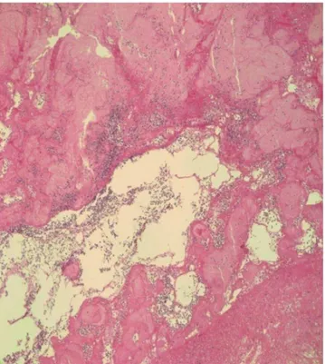
Fig. 7. Atypical hyperchromatic cells observed on H&E stain. The
cardiac mass comprised an aggregation of small, round, necrotic cells consistent with leukemic cells (×400).
Fig. 8. Cardiac mass biopsy with immunohistochemistry special stain
induced enterocolitis: immune mechanisms, diagnosis and treatment. Allergol Immunopathol (Madr) 2009;37:3642.
6. Rigamonti F, Beris P, SanchezPareja A, Meyer P, Ashrafpoor G, Zaza S, et al. Atypical presentation of acute myeloid leukemia: cardiac myeloid sarcoma. Int J Hematol 2009;89:6938.
7. NowakGottl U, Kenet G, Mitchell LG. hrombosis in childhood acute
lymphoblastic leukaemia: epidemiology, aetiology, diagnosis, prevention and treatment. Best Pract Res Clin Haematol 2009;22:10314.