CASE REPORT Pub. 60
ISSN 1679-9216
Received: 6 April 2014 Accepted: 7 August 2014 Published: 15 August 2014 1Serviço de Oncologia Veterinária, Departamento de Clínica e Cirurgia Veterinária - Faculdade de Ciências Agrárias e Veterinárias (FCAV), UNESP de
Jaboticabal, SP, Brazil. 2Serviço de Patologia Veterinária, Departamento de Clínica Veterinária, Faculdade de Medicina Veterinária e Zootecnia (FMVZ),
UNESP de Botucatu, SP. CORRESPONDENCE: R. Laufer-Amorim [[email protected] - Fone: +55 (14) 3811-6293]. Rua Luzia De Masseno Pontes n. 494, Jardim Itamaraty. CEP 18608-032 Botucatu, SP, Brazil.
T-Cell Lymphoma in the Tongue of a Dog with Cutaneous
and Striated Forelimb Muscle Involvement
Linfoma de células T em língua de cão com envolvimento cutâneo e de musculatura estriada de membro torácico
Talita Mariana Morata Raposo-Ferreira1, Paulo Cesar Jark1, Giovanna Rossi Varallo1, Sofi a Borin-Crivellenti1,
Mirela Tinucci-Costa1, Renée Laufer-Amorim2 & Andrigo Barboza De Nardi1
ABSTRACT
Background: Primary tongue tumors rarely affect dogs and correspond to 4% of tumors involving the oropharynx. Until now, primary tongue lymphoma had not been reported. However, lymphoma involvement in the skeletal muscle, although quite unusual, was described in the literature in four cases. Cutaneous lymphoma is another rare extranodal manifestation. The objective of this report is to describe a case of T immunophenotype lymphoma occurrence, whose manifestation is atypical, not only because it is situated in the tongue muscle but also because of the subsequent involvement of the striated musculature of the left forelimb and the skin, which showed unfavorable evolution.
Case: A female seven-year-old mongrel was seen showing a regular lump in the base of the tongue, 3 cm in diameter, not ulcerated and of fi rm consistency, with halitosis as the only clinical sign of the disease. Incisional biopsy of the lump was performed and histopathology verifi ed that it was large cell lymphoma. The material was sent for immunohistochemical evaluation and was characterized as T immunophenotype lymphoma by positive CD3 and negative CD79a marking. The CHOP (cyclophosphamide, doxorubicin, vincristine and prednisone) chemotherapy protocol was established as treatment and after the fi rst chemotherapy session there was partial remission of the mass, measuring 2 cm in diameter. The lump, however, remained stable in the following sessions. Thirty days after the diagnosis of lymphoma, the animal began to show lameness of the left forelimb and swelling near the head of the left humerus. A muscle mass, fi rm in consistency, progress-ing fast, presented a signifi cant increase, just three weeks after its appearance. Two skin lesions, arcuate, erythematous and pruritic also appeared in the dorsocervical and ventral-abdominal region. Incisional biopsy of these lesions was performed and the histopathological diagnosis confi rmed muscle and cutaneous large cell lymphoma and immunophenotype compatible with T cells (positive CD3 and negative CD79a). Due to disease advance, even during chemotherapy, a rescue protocol of L-asparaginase administration followed by lomustine and prednisone was proposed. Even with the rescue protocol there was no remission of the tumors and the case was classifi ed as progressive. The animal of this report died after completing the fi rst cycle of chemotherapy protocol, with a survival of 92 days.
Discussion: Despite the fact that clinical behavior of primary lymphoma in dogs’ skeletal muscle is unknown, it is believed that, as in humans, it can be associated with chronic infl ammation or neoplastic cell invasion by proximity of the tumor or metastasis, which could justify the dissemination of the lymphoma reported here from the tongue to other tissues. However, appearance of concurrent independent lymphomas cannot be ruled out. As observed in the three cases of primary muscu-lar lymphoma, the dog of this report had low response to therapy and short survival. This report presents the fi rst case of lymphoma in tongue with subsequent skin and left forelimb skeletal muscle involvement described in the literature. The clinical outcome corroborates the aggressiveness of muscular lymphoma observed in the other reports and also suggests that both tongue and other skeletal muscle tumors should be included in the differential diagnosis of canine lymphoma.
Keywords: dog, lymphoma, tongue, skeletal muscle.
INTRODUCTION
Primary neoplasms of the tongue are extremely rare, accounting for about 4% of all tumors that in-volve the oropharynx, the most commonly found in the tongue being the squamous cell carcinoma [2], followed by melanoma [4].
Other tumors have been described, although less often, like papilloma, rhabdomyoma, rhabdomyosarcoma, hemangioma, hemangiosarcoma, granular cell tumors, his-tiocytoma, plasmacytoma, mast cell tumor, fi brosarcoma, chondrosarcoma and liposarcoma [2,3,5,6,10,12,13].
Despite the occurrence of lymphoma in the tongue having already been described as a possible oral cavity extranodal manifestation [11], we found no reported case on this primary involvement. In general, the involvement of striated skeletal muscle by lymphoma is quite unusual, both in humans and in dogs, probably because typical skeletal muscle does not contain lymphoid tissue [8,16].
Four cases of lymphoma in skeletal muscle have been reported to this date [1,8,14,15]. The association of cutaneous lymphoma with skeletal muscle tissue has also been described [1]. Cutaneous lymphoma is another, rather uncommon, extranodal manifestation, accounting for ap-proximately 3-8% of lymphoma cases affecting dogs [18]. The aim of this study was to report a case of lymphoma with atypical manifestations, in the skeletal muscle of the tongue and involving the striated muscle of the left forelimb and the skin, and with a very ag-gressive course and little responsive to antineoplastic chemotherapy. From the reviewed literature, this is the fi rst case of primary tongue lymphoma with subsequent involvement of the forelimb skeletal muscle and skin.
CASE
A female, seven years old mongrel, weighing 4 kg was seen. The animal showed a volume increase at the base of the tongue, during a periodontal disease treatment a week earlier. Upon inspection of the patient’s oral cavity, the mass was found to be regular, 3 cm in diameter, not ulcerated and of fi rm consistency (Figure 1). The only clinical sign of the patient was halitosis.
Complete blood count and serum biochemistry (creatinine, urea, alanine aminotransferase and alkaline phosphatase) analyses results were within the normal range for the species, and imaging tests (chest radi-ography and abdominal ultrasonradi-ography) revealed no changes in other organs.
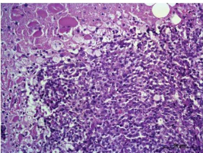
Subsequently, the patient underwent incisional biopsy for histopathological analysis, and large cell lym-phoma was diagnosed (Figure 2). Immunohistochemical analysis of the lingual nodule was performed using anti-human CD3 polyclonal antibody1 and anti-human CD79a
monoclonal antibody1. Antigen retrieval was performed
using citrate buffer, pH 6.0, in pressure cooker (Pascal®)1
for 20 min. The blocking of endogenous peroxidase was performed in methyl alcohol solution of 8% hydrogen per-oxide. The primary antibody was incubated for 18 hours at 4°C, followed by incubation with Advance HRP polymer and application of the chromogen 3,3’-diaminobenzidine1
for 5 min. The sections were counterstained with Harris hematoxylin for 40 s. The marking was positive for CD3 and negative for CD79a, confi rming the diagnosis of T-cell lymphoma (Figure 3).
Once lymphoma was diagnosed, the CHOP pro-tocol began. After the fi rst treatment session, with admin-istration of vincristine2 (0.7 mg/m2/IV), partial remission
was noted and the lump in the tongue decreased to 2 cm in diameter. The animal remained stable during subsequent sessions, and no hematological changes were observed during the evaluation before chemotherapy. Returning for the fourth treatment session, 30 days after
sis, the patient presented lameness of the left forelimb and soreness next to the humeral head, without having trauma history or reports of previous illness. Limb lateral and cranial ventral radiography showed no bone or joint involvement, only a slight increase of adjacent soft tissue.
When the patient returned three weeks later, we found a signifi cant increase in the forelimb muscle mass, of fi rm consistency (Figure 4), and two arcuate, erythematous, and pruritic skin lesions, on the right dorsocervical and ventral abdominal area (Figure 5). Thus, incisional biopsy of the muscles and the skin le-sions was performed. The histopathological diagnosis
of the lesions revealed a large cell lymphoma, and im-munohistochemical analysis was positive for T cells (CD3 positive, CD79a negative).
Therefore, the fi rst chemotherapy protocol was replaced by combination treatment of
L-aspar-aginase3 (400 IU/kg, subcutaneously, weeks 1 and
4), lomustine4 (70 mg/m2 via orally, week 1) and
prednisone5 (2 mg/kg, every 24 h, orally, with weekly
dose reduction up to 1 mg/kg). However, until the patient’s natural death after the fi rst treatment cycle, no remission of cancer was observed. The patient had a survival of 92 days.
Figure 2. Large cell Lymphoma in the base of the dog’s tongue (HE, x200).
Figure 4. Muscle mass of the left forelimb, with rapid progression.
Figure 5. Arcuate, erythematous cutaneous lesions. A. Dorsocervical region. B. Ventral abdominal region.
DISCUSSION
The clinical behavior of skeletal muscle pri-mary lymphoma in dogs is unknown [14]. In humans, it is believed that chronic infl ammation is related to the carcinogenic process [9]. Another suspected mechanism could be through the invasion of cancer cells from a pre-existing site, by proximity or metastasis [15]. This is the fi rst case of lymphoma manifestation in the tongue, possibly primary, which also culminated with cutaneous and left forelimb skeletal muscle involvement.
The other four cases of muscular lymphoma described in the literature involved forelimbs and hindlimbs, cervical muscles and one was a diffuse
manifestation associated to polymyopathy [1,8,14,15]. In one of them a previous appearance of cutaneous lymphoma was also observed [1], whereas in this case it developed after the muscle lymphoma.
Lymphoma muscle involvement may manifest as a single mass or in diffuse form, the latter being rather unusual, and may even be related to a polymy-opathy [1,15].
to justify a possible infl ammation as a predisposing factor for the development of lymphoma, suggesting the involvement by metastatic neoplastic cell invasion, even more so, since all masses have been character-ized as large cell T-lymphoma. However, one cannot rule out the concomitant emergence of independent lymphomas [1].
The muscle lymphoma cases reported were mostly males, aged between 16 months and eight years. Three animals were large and one midsize [1,8,14,15]. The dog of this report, however, was female, seven years old and small breed.
Surgical resection is the best treatment for lingual tumors [5]. However, once lymphoma is considered a systemic disease and responsive to che-motherapy, it is treated conservatively through the combination of multiple anticancer agents for better response and disease control [17]. However, T-cell tends to be more aggressive than B-cell lymphoma with a poorer prognosis [7].
As in cutaneous lymphoma [11], low survival was observed in most muscular lymphoma cases, being related mainly to high-grade and T-cell lymphomas [1,15]. Only the case described by Takeuchi et al. [14]
presented a low grade T lymphoma with a survival of 713 days. The case at hand also had a short survival, since it demonstrated rapid progression and the che-motherapy treatments were ineffective.
This case illustrates an atypical case of lingual lymphoma, which culminated with cutaneous and left forelimb skeletal muscle involvement, and which showed aggressive behavior, corroborating other re-ports of lymphoma cases with muscle involvement. Tongue and skeletal muscle masses should be consid-ered in lymphoma differential diagnosis. New forms of treatment must be explored to improve survival of these patients with an atypical presentation of the disease.
SOURCES AND MANUFACTURERS
1DakoCytomation, Manchester, UK. 2Oncovin®, ABL, Cosmópolis, SP, Brazil. 3Elspar®, Lundbeck, Rio de Janeiro, RJ, Brazil.
4CeeNU®, Bristol-Mayers Squibb, Santo Amaro, SP, Brazil. 5Prednisona®, Legrand, São Paulo, SP, Brazil.
Declaration of interest. The authors report no conflicts of interest. The authors alone are responsible for the content and writing of the paper.
REFERENCES
1 Bennett S.L., Slocombe R.F., Holloway S.A., Charles J.A. & Sandy J.R. 2005. Lymphoma(s) showing epitheli-otropism and diffuse skeletal muscle involvement presenting as a polymyopathy in a young dog. Australian Veterinary Journal. 83(10): 612-615.
2 Brockus C.W. & Myers R.K. 2004. Multifocal rhabdomyosarcomas within the tongue and oral cavity of a dog.
Veterinary Pathology. 41(3): 273-274.
3 Burton J.H., Powers B.E. & Biller B.J. 2012. Clinical outcome in 20 cases of lingual hemangiosarcoma in dogs: 1996-2011. Veterinary Comparative Oncology. doi: 10.1111/j.1476-5829.2012.00351.x.
4 Culp W.T., Ehrhart N., Withrow S.J., Rebhun R.B., Boston S., Buracco P., Reiter A.M., Schallberger S.P., Aldridge C.F., Kent M.S., Mayhew P.D. & Brown D.C. 2013. Results of surgical excision and evaluation of factors associated with survival time in dogs with lingual neoplasia: 97 cases (1995-2008). Journal of the American Veterinary Medical Association. 242(10): 1392-1397.
5 Daleck C.R., De Nardi A.B., Silva V.M.C., Eurides D. & Silva L.A.F. 2007. Neoplasias de língua em cinco cães.
Ciência Rural. 37(2): 578-582.
6 Dennis M.M., Ehrhart N., Duncan C.G., Barnes A.B. & Ehrhart E.J. 2006. Frequency of and risk fators associ-ated with lingual lesions in dogs: 1,196 cases (1995-2004). Journal of the American Veterinary Medical Association. 228(10): 1533-5137.
7 Fan T.M. 2003. Lymphoma updates. Veterinary Clinics of North America-Small Animal. 33(3): 455-471.
8 Harkin K.R., Kennedy G.A., Moore W.E. & Schoning P. 2000. Skeletal muscle lymphoma in a bullmastiff. Journal of the American Animal Hospital Association. 36(1): 63-66.
9 Masaoka S. & Fu T. 2002. Malignant lymphoma in skeletal muscle with rhabdomyolysis: a report of two cases. Journal of Orthopaedic Science. 7(6): 688-693.
www.ufrgs.br/actavet
CR 60
574-576.
11 Moore P.F., Affolter V.K., Graham P.S. & Hirt B. 2009. Canine epitheliotropic cutaneous T-cell lymphoma: an in-vestigation of T-cell receptor immunophenotype, lesion topography and molecular clonality. Veterinary Dermatology. 20(5/6): 569-576.
12 Piseddu E., De Lorenzi D., Freeman K. & Masserdotti C. 2011. Cytologic, histologic, and immunohistochemical features of lingual liposarcoma in a dog. Veterinary Clinical Pathology. 40(3): 393-397.
13 Silva A.E., Moreira E.L.T., Ocarino N.M., Franco A.L., Santos A.P., Souza T.S. & Serakides R. 2007. Condros-sarcoma primário de língua em cão. Arquivo Brasileiro de Medicina Veterinária e Zootecnia. 59(2): 530-532.
14 Takeuchi Y., Fujino Y., Goto-koshino Y., Ohno K., Uchida K., Nakayama H. & Tsujimoto H. 2010. Long term survival of primary skeletal muscle lymphoma in a miniature dachshund. Journal of Veterinary Medical Science. 72(5): 673-677.
15 Thuilliez C., Watrelot-Virieux D., Chanut F., Fournel-Fleury C., Ponce F. & Marchal T. 2008. Presumed primary muscular lymphoma in a dog. Journal of Veterinary Diagnostic Investigation. 20(6): 824-826.
16 Travis W.D., Banks P.M. & Reiman H.M. 1987. Primary extranodal soft tissue lymphoma of the extremities. American Journal of Surgical Pathology. 11(5): 359-366.
17 Vail D.M., Pinkerton M.E. & Young K.M. 2012. Hematopoietic Tumours. In: Withrow S.J., Vail D.M. & Page R.L. (Eds). Small Animal Clinical Oncology. 5th edn. St Louis: Elsevier, pp.622-627.
18 Vonderhaar M.A. & Morrison W.B. 2002. Lymphosarcoma. In: Morrison W.B. (Ed). Cancer in Dogs and Cats: