Intraoral spindle-cell lipoma with chondroid
differentiation: importance in the diagnosis of oral
lesions presenting chondroid tissue
Lipoma de células fusiformes intraoral com diferenciação condroide:
importância no diagnóstico de lesões orais contendo tecido cartilaginoso
Ademar Takahama Junior1; Michele F. Brantes2; Jorge E. Leon3; Adriele F. Gouvêa2; Oslei P. Almeida4
1. Universidade Estadual de Londrina (UEL), Paraná, Brazil. 2. Universidade Federal Fluminense (UFF), Rio de Janeiro, Brazil. 3. Universidade de São Paulo (USP), São Paulo, Brazil. 4. Universidade Estadual de Campinas (Unicamp), São Paulo, Brazil.
First submission on 22/03/16; last submission on 22/03/16; accepted for publication on 08/04/16; published on 20/06/16
ABSTRACT
Lipomas are benign neoplasms of adipose tissue presenting several histologic variants, which can be rarely found in the oral cavity. We present a case of a 62-year-old woman with a submucous nodule located in the tongue. Histopathological examination revealed an encapsulated tumor composed of myxoid tissue, spindle cells and mature adipocytes in transition to cartilaginous tissue. The inal diagnosis was spindle-cell lipoma with myxoid change and chondroid differentiation. No sign of recurrence was found after ive years. The diagnosis of intraoral mesenchymal lesions with chondroid differentiation requires careful histologic examination, mainly to differentiate between benign and malignant lesions.
Key words: lipoma; diagnosis; microscopy; cartilage.
INTRODUCTION
Lipomas are benign neoplasms of mature adipose tissue, most commonly found in areas where adipose tissue is present, above all in the subcutaneous or submucous regions. They are somewhat rare in the oral region, representing about 1% to 4% of all cases(1). Lipomas
often present as a slow-growing mass, almost always asymptomatic(2).
Their most common location in the oral region is the buccal mucosa, followed by loor of the mouth, tongue and lips(1, 2).
Microscopically, classic lipomas are composed of encapsulated,
mature adipose tissue with variably sized adipocytes(1). According
to microscopical features, several variants of oral lipomas have been described, including ibrolipoma, angiolipoma, myolipoma, spindle-cell/pleomorphic lipoma, salivary gland lipoma, osteolipoma and chondroid lipoma. Spindle-cell/pleomorphic lipomas are characterized by circumscribed lesions, composed of a variable admixture of adipocytes and spindle cells, hyperchromatic
rounded cells, and multinucleated giant cells associated with ropey collagen(3, 4).
Mesenchymal differentiation of lipomas into bone and cartilage is a rare event(2, 5). Chondroid lipoma is the main variant
presenting chondroid tissue, with features of both embryonal fat and embryonal cartilage, and its recognition is important because it can be morphologically similar to sarcomas, particularly
myxoid liposarcoma and myxoid chondrosarcoma(6).
In order to avoid misclassiication of different histological types of lipoma, spindle-cell lipoma with cartilaginous differentiation must be distinguished from chondroid lipoma. To the best of our knowledge, only two cases of spindle-cell lipoma with chondroid tissue have been published in the English language literature. Therefore, the purpose of this article is to report a case of spindle-cell lipoma with cartilaginous differentiation affecting the tongue, emphasizing the distinction between these lipomas and chondroid lipoma or other tumors with chondroid tissue.
CASE REPORT
A 62-year-old woman was referred for evaluation of a tongue nodule that had been present for about six months. Her medical history and systemic review were not signiicant. The patient had no history of trauma in the region. Physical intraoral examination revealed a painless well-delimited submucous nodule, irm on palpation, measuring about two centimeters in diameter noted on the
left lateral border of the tongue (Figure 1). The main diagnostic consideration was a benign neoplasm, including salivary gland tumors or mesenchymal neoplasms such as lipoma, granular cell tumor, neuroibroma, schwannoma or ectomesenchymal chondromyxoid tumor. According to these hypotheses, an excisional biopsy was indicated. During surgery, a well-circumscribed yellowish mass was found. On gross examination the lesion did not loat on formaldehyde. On microscopic examination, the lesion consisted of an encapsulated tumor with myxoid areas admixed with spindle and stellate cells and mature adipocytes in transition to cartilaginous tissue, which displayed well-differentiated areas preferentially located in the
central portion (Figure 2). Immunohistochemical stains for S100
protein were positive in the adipocytic cells and cartilaginous tissue,
and CD34 was found in spindle cells of the myxoid areas (Figure 3).
Based on these histopathological and immunohistochemical features, a diagnosis of spindle-cell lipoma with prominent myxoid change and chondroid differentiation was rendered. No recurrence was identiied during a ive-year follow-up period.
DISCUSSION
Lipomas are the most common benign soft tissue mesenchymal tumors, and several microscopical variants have been reported,
FIGURE 1 − Submucous nodule on the left lateral border of the tongue
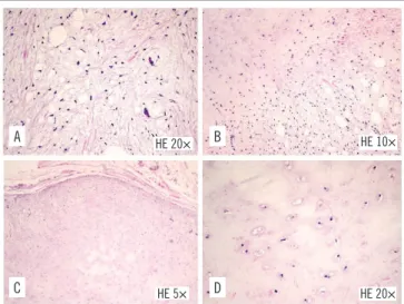
FIGURE 2 − Microscopical features of the spindle-cell lipoma with prominent myxoid change and cartilaginous differentiation
A) areas of lipoblast-like cells in a myxoid stroma; notice the atypical stromal cells; B) transition between the myxoid and cartilaginous areas; C) presence of a fibrous capsule; D) cartilaginous tissue showing mature-appearing cells in a homogeneous stroma. HE: hematoxilin and eosin.
HE 20× B
D A
C
HE 20× HE 10×
HE 5×
FIGURE 3 − Positive immunohistochemical staining for S100 protein (A) and CD34 (B)
B
A CD34 20×
S100 20×
including ibrolipoma, angiolipoma, myolipoma, sialolipoma, osteolipoma, spindle-cell/pleomorphic lipoma and chondroid lipoma. Spindle-cell lipoma is an uncommon variant, irst reported by Enzinger and Harvey in 1975(7). Microscopically, it is
composed of mature fat cells, collagen-forming spindle cells with immunoreactivity for CD34 in a ibrocollagenous and myxoid
background(7, 8). Some cases of spindle-cell lipoma show prominent
myxoid changes(9). Spindle-cell lipoma accounts for approximately
1.5% of all adipocytic neoplasms(10), and typically occurs in elderly
men as a solitary lesion in the posterior neck and back(11). It is less
commonly found in the oral cavity(12, 13). Searching in the literature
for reported cases of oral spindle-cell lipomas, we found 37 cases,
which are summarized in the Table. From these 37 cases, 20 were
in vacuolated cells. Chondroid lipoma should be distinguished from chondrolipoma, which is a lipoma with cartilaginous metaplasia. In chondrolipomas there is absence of lipoblasts and myxoid matrix, and a clear separation between the cartilaginous
tissue and the fatty component(5). Cases of chondrolipomas have
also been reported in the intraoral region(35-39). Our case does not
represent a chondroid lipoma, due to lack of lipoblast-like cells. The presence of chondroid tissue in spindle cell lipoma is very
uncommon. Lau et al. (2015)(30) reported eight cases of
spindle-cell lipoma of the tongue, and they found two cases containing a tissue imparting chondroid appearance.
The presence of chondroid differentiation in an oral lesion requires special attention. A variety of benign and malignant tumors, including pleomorphic adenoma, ectomesenchymal chondromyxoid tumor, myxoid liposarcoma, well-differentiated liposarcoma, pleomorphic liposarcoma and extraskeletal myxoid chondrosarcoma, may present chondroid
differentiation(1, 32, 33). Therefore, some histopathological details
may be important to differentiate them. Pleomorphic adenoma can resemble, clinically and microscopically, lipoma with chondroid differentiation, mainly because of the myxoid and chondroid stroma. However, this salivary gland tumor shows ductal/epithelial elements which are not found in lipomas(40).
Ectomesenchymal chondromyxoid tumor is a rare benign intraoral mesenchymal neoplasm almost exclusively seen on
the dorsum of the tongue(41). Histopathologically, this tumor is
usually unencapsulated but well-demarcated, with lesional cells proliferating in a lobular pattern and arranged in cords, strands, and sheets in a myxoid to chondromyxoid background. The cells are either round, oval, polygonal or spindled in morphology. They may have multilobulated nuclei and may occasionally
show atypia(42). However, mature fat cells are not seen in
ectomesenchymal chondromyxoid tumors. The presence of ropey collagen, seen in spindle-cell/pleomorphic lipomas, is useful for differential diagnosis since it is not seen in well-differentiated, myxoid or pleomorphic liposarcoma; moreover, spindle-cell lipomas lack lipoblasts, which can be seen in liposarcomas(43).
Myxoid liposarcoma is a malignant tumor composed of uniform round- to oval-shaped primitive mesenchymal cells and a variable number of small signet-ring lipoblasts in a prominent
myxoid stroma(6, 32). Well-differentiated liposarcoma, also known
as atypical lipomatous tumor, is a locally aggressive malignant mesenchymal neoplasm composed of mature adipocytic proliferation showing signiicant malignant cytological features; it can sometimes show areas of chondroid metaplasia but without
extensive myxochondroid matrix(44). Extraskeletal myxoid
chondrosarcoma is a malignant soft-tissue tumor characterized
TABLE −Summary of spindle-cell/pleomorphic lipoma of the oral region reported in the English-language literature
Authors publicationYear of Location Gender Age McDaniel et al.(14) 1984 Floor of the mouth
Tongue
F
M
33 52 Christopoulos et al.(15) 1989 Hard palate M 58
Levy and Goding(16) 1989 Floor of the mouth F 74
Lombardi and Odell(17) 1994 Tongue F 68
Khoo et al.(18) 1995 Cheek M 23
Tosius et al.(9) 1995 Cheek M 55
Dutt et al.(19) 1999 Tongue F 42
Piatelli et al.(20) 1999 Cheek M 75
Piatelli et al.(21) 2000 Cheek M 63
Said-Al-Naief et al.(12) 2001 Tongue Tongue
NI NI
NI NI
Agoff et al.(22) 2001 Gingivobuccal sulcus F 61
Darling et al.(23) 2002 Alveolar mucosa M 69
Piatelli et al.(24) 2005 Floor of the mouth M 50
Billings et al.(13) 2006 Tongue
Tongue Tongue Tongue Lip Floor of the mouth
Buccal mucosa M M F F F F M 45 67 31 75 55 84 88
Coimbra et al.(25) 2006 Floor of the mouth F 29
Imai et al.(8) 2008 Tongue M 72
Vecchio et al.(26) 2009 Buccal mucosa M 52 Stokes et al.(27) 2011 Maxilla (intraosseous) M 35
Junior et al.(28) 2013 Tongue F 64
Al-Sheddi et al.(29) 2014 Mucogingival sulcus M 68
Lau et al.(30) 2015 Tongue Tongue Tongue Tongue Tongue Tongue Tongue Tongue M M F M F M F M 62 62 61 80 65 35 47 47
Lin et al.(31) 2015 Tongue F 62
F: female; M: male; NI: no information.
The presence of cartilaginous tissue within a lipoma is a
relatively rare inding(5). Chondroid lipoma and chondrolipoma
are the main variants that exhibit chondroid tissue formation. Chondroid lipoma is a rare variant, mainly in the head and neck region, and was included in the World Health Organization
(WHO) classiication of soft tissue tumors in 2002(3). Microscopic
examination of chondroid lipomas shows mature nests of vacuolated lipoblasts, in a prominent myxoid to hyalinized chondroid matrix, and a variable amount of mature adipose tissue. The tumor is usually circumscribed by a ibrous capsule and there
is no cell atypia or mitotic igures(32-34). Chondroid lipomas may
RESUMO
Lipomas são neoplasias benignas de tecido adiposo que podem apresentar diversas variantes e raramente são encontradas na cavidade oral. Apresentamos o caso de uma mulher de 62 anos de idade com queixa de um nódulo na língua. A análise histopatológica da lesão revelou tumor encapsulado composto de tecido mixoide, células fusiformes e adipócitos maduros em transição para tecido cartilaginoso. Nenhum sinal de recorrência foi observado após acompanhamento de cinco anos. O diagnóstico de lesões mesenquimais intraorais contendo diferenciação condroide requer atenção especial, principalmente para a diferenciação de lesões benignas e malignas.
Unitermos: lipoma; diagnóstico; microscopia; cartilagem.
by a multinodular architecture, abundant myxoid matrix, and
malignant chondroblast-like cells(45). Lipomas with chondroid
differentiation may be confused with this malignant neoplasm
REFERENCES
1. Furlong MA, Fanburg-Smith JC, Childers EL. Lipoma of the oral and maxillofacial region: site and subclassiication of 125 cases. Oral Surg Oral Med Oral Pathol Oral Radiol Endod. 2004; 98(4): 441-50. 2. Fregnani ER, Pires FR, Falzoni R, Lopes MA, Vargas PA. Lipomas of the oral cavity: clinical indings, histological classiication and proliferative activity of 46 cases. Int J Oral Maxillofac Surg. 2003; 32(1): 49-53. 3. Miettinen MM, Mandahl N. Adipocytic tumours. In: Fletcher CDM, Unni KK, Mertens F, editors. Pathology and genetics of tumours of soft tissue and bone. Lyon: IARC Press; 2002.
4. Prado FO, Ito FA, Di Hipólito O Jr, Vargas PA, Almeida OP, Lopes MA. Pleomorphic lipoma of the face: case report. Oral Dis. 2006; 12(1): 73-6. 5. Weiss SW, Goldblum JR. Benign lipomatous tumors. In: Weiss SW, Goldblum JR, editors. Enzinger and Weiss’s soft tissue tumors. 4th ed. St. Louis (MO): Mosby; 2001. p. 575.
6. Thway K, Flora RS, Fisher C. Chondroid lipoma: an update and review. Ann Diagn Pathol. 2012; 16(3): 230-4.
7. Enzinger FM, Harvey DA. Spindle cell lipoma. Cancer. 1975; 36(5): 1852-9.
8. Imai T, Michizawa M, Shimizu H, Imai T, Yamamoto N, Yura Y. Bilateral multiple spindle cell lipomas of the tongue. Oral Surg Oral Med Oral Pathol Oral Radiol Endod. 2008; 106(2): 264-9.
9. Tosios K, Papanicolaou SI, Kapranos N, Papadogeorgakis N. Spindle cell lipoma of the oral cavity. Int J Oral Maxillofac Surg. 1995; 24(5): 363-4.
10. Fletcher CD, Martin-Bates E. Spindle cell lipoma: a clinicopathological study with some original observations. Histopathology. 1987; 11(8): 803-17.
11. Enzinger FM, Weiss SW. Spindle cell lipoma. In: Enzinger FM, editor. Soft tissue tumors. 3rd ed. St Louis (MO): Mosby; 1995. p. 395.
due to the presence of scattered nests of lipoblast-like cells and myxochondroid matrix. However, lipomas are well-circumscribed
tumors and always contain a mature fatty component(1).
12. Said-Al-Naief N, Zahurullah FR, Sciubba JJ. Oral spindle cell lipoma. Ann Diagn Pathol. 2001; 5(4): 207-15.
13. Billings SD, Henley JD, Summerlin DJ, Vakili S, Tomich CE. Spindle cell lipoma of the oral cavity. Am J Dermatopathol. 2006; 28(1): 28-31. 14. McDaniel RK, Newland JR, Chiles DG. Intraoral spindle cell lipoma: case report with correlated light and electron microscopy. Oral Surg Oral Med Oral Pathol. 1984; 57(1): 52-7.
15. Christopoulos P, Nicolatou O, Patrikiou A. Oral spindle cell lipoma: report of a case. Int J Oral Maxillofac Surg. 1989; 18(4): 208-9. 16. Levy FE, Goding GS Jr. Spindle-cell lipoma: an unusual oral presentation. Otolaryngol Head Neck Surg. 1989; 101(5): 601-3. 17. Lombardi T, Odell EW. Spindle cell lipoma of the oral cavity: report of a case. J Oral Pathol Med. 1994; 23(5): 237-9.
18. Khoo SP, Lian CB. Intraoral spindle-cell lipoma: report of a case and literature review. Ann Dent. 1995; 54(1-2): 53-5.
19. Dutt SN, East DM, Saleem Y, Jones EL. Spindle-cell variant of intralingual lipoma – report of a case with literature review. J Laryngol Otol. 1999; 113(6): 587-9.
20. Piattelli A, Fioroni M, Rubini C. Spindle cell lipoma of the oral cavity: report of a case. J Oral Maxillofac Surg. 1999; 57(5): 624-5.
21. Piattelli A, Rubini C, Fioroni M, Iezzi G. Spindle-cell lipoma of the cheek: a case report. Oral Oncol. 2000; 36(5): 495-6.
22. Agoff SN, Folpe AL, Grieco VS, Garcia RL. Spindle cell lipoma of the oral cavity. Report of a rare intramuscular case with ine needle aspiration indings. Acta Cytol. 2001; 45(1): 93-8.
23. Darling M, Thompson I, Schneider J. Spindle cell lipoma of the alveolar mucosa: a case report. Oral Surg Oral Med Oral Pathol Oral Radiol Endod. 2002; 93(2): 171-3.
26. Vecchio G, Amico P, Caltabiano R, Colella G, Lanzafame S, Magro G. Spindle cell/pleomorphic lipoma of the oral cavity. J Craniofac Surg. 2009; 20(6): 1992-4.
27. Stokes SM, Wood JP, Castle JT. Maxillary intraosseous spindle cell lipoma. J Oral Maxillofac Surg. 2011; 69(6): e131-4.
28. Cavezzi Jr O, Aguiar EC, Sartori JH, Lima FO. Spindle cell lipoma of the tongue: a case report of unusual occurrence. J Oral Maxillofac Pathol. 2013; 17(1): 148.
29. Al Sheddi MA, Assari A, Mosadomi H. Spindle cell lipoma of the mandibular mucogingival junction: a case report of unusual oral neoplasm. Int J Oral Sci. 2014; 6(3): 185-7.
30. Lau SK, Bishop JA, Thompson LD. Spindle cell lipoma of the tongue: a clinicopathologic study of 8 cases and review of the literature. Head Neck Pathol. 2015; 9(2): 253-9.
31. Lin HP, Liu CJ, Chiang CP. Spindle cell lipoma of the tongue. J Formos Med Assoc. 2015; 114(5): 477-9.
32. Meis JM, Enzinger FM. Chondroid lipoma. A unique tumor simulating liposarcoma and myxoid chondrosarcoma. Am J Surg Pathol. 1993; 17(11): 1103-12.
33. Kindblom LG, Meis-Kindblom JM. Chondroid lipoma: an ultrastructural and immunohistochemical analysis with further observations regarding its differentiation. Hum Pathol. 1995; 26(7): 706-15.
34. Vreeze RS, van Coevorden F, Boerrigter L, et al. Delineation of chondroid lipoma: an immunohistochemical and molecular biological analysis. Sarcoma. 2011; 2011: 638403.
35. Maes A, Eulderink F. Chondrolipoma of the tongue. Histopathology. 1989; 14(6): 660-2.
36. Fujimura N, Enomoto S. Lipoma of the tongue with cartilaginous change: a case report and review of the literature. J Oral Maxillofac Surg. 1992; 50(9): 1015-7.
37. Hietanen J, Mäkinen J. Chondrolipoma of the tongue. A case report. Int J Oral Maxillofac Surg. 1997; 26(2): 127-8.
38. Bezerra MF, Costa FW, Pereira KM, Cavalcante RB, Pouchain EC. Chondrolipoma of the posterior tongue. J Craniofac Surg. 2010; 21(6): 1982-4.
39. Berg T, Gorsky M. Chondrolipoma of the tongue: a case report of a rare diagnosis. Int J Dermatol. 2010; 49(4): 441-2.
40. Hornick JL, Fletcher CD. Myoepithelial tumors of soft tissue: a clinicopathologic and immunohistochemical study of 101 cases with evaluation of prognostic parameters. Am J Surg Pathol. 2003; 27(9): 1183-96.
41. Aldojain A, Jaradat J, Summersgill K, Bilodeau EA. Ectomesenchymal chondromyxoid tumor: a series of seven cases and review of the literature. Head Neck Pathol. 2015; 9(3): 315-22.
42. Allen CM. The ectomesenchymal chondromyxoid tumor: a review. Oral Dis. 2008; 14(5): 390-5.
43. Azzopardi JG, Iocco J, Salm R. Pleomorphic lipoma: a tumour simulating liposarcoma. Histopathology. 1983; 7(4): 511-23.
44. Laurino L, Furlanetto A, Orvieto E, Dei Tos AP. Well-differentiated liposarcoma (atypical lipomatous tumors). Semin Diagn Pathol. 2001; 18(4): 258-62.
45. Lucas DR, Heim S. Chondro-osseous tumours. In: Fletcher CDM, Unni KK, Mertens F, editors. Pathology and genetics of tumours of soft tissue and bone. Lyon: IARC Press; 2002. p. 213.
CORRESPONDING AUTHOR
Ademar Takahama Junior