Imbalance of
gd
T17/
gd
Treg cells in the pathogenesis
of allergic asthma induced by ovalbumin
Xia Yang
1*, Jing-Hong Zhang
1*, Wang-Sheng Deng
1and Chao-Qian Li
1,21Department of Emergency, the First Af
filiated Hospital of Guangxi Medical University, The Guangxi Talent Highland for Emergency and Rescue Medicine, Nanning, Guangxi, China 2Department of Respiratory Medicine, Guangxi Vocational and Technical College of Health, Nanning, Guangxi, China
Abstract
We aimed to explore the imbalance between the T helper 17gdT cells (gdT17) and the regulatorygdT cells (gdTreg) in asthmatic mice. Male Balb/c mice were randomly divided into the normal control group and the asthmatic model group. The asthmatic model group mice were intraperitoneally injected with the mixture of ovalbumin (OVA)/Al(OH)3and then activated by exposure of the animals to OVA atomization. Airway hyperresponsiveness (AHR) was determined by a non-invasive lung function machine. Hematoxylin and eosin and Alcian blue-periodic acid Schiff staining were done for histopathological analysis. Interleukin (IL)-17 and IL-35 levels in bronchoalveolar lavagefluid were detected by ELISA. The percentage of IL-17+gdT cells
and Foxp3+gdT cells in spleen cells suspension were detected and the transcription levels of RORgt and Foxp3 in the lung
tissue were determined. Compared with the normal control, the severity of airway inflammation and AHR were higher in the asthmatic mice. Furthermore, mice in the asthmatic group displayed significant increases of IL-17+gdT cells, expression of
IL-17A, and RORgt, whereas control mice displayed marked decreases of Foxp3+gdT cells, expression of IL-35, and
transcription factor Foxp3. In addition, the mRNA expression of RORgt was positively correlated with the percentage of IL-17+gdT cells, and the mRNA level of Foxp3 was positively correlated with the percentage of Foxp3+gdT cells. The
imbalance ofgdT17/gdTreg in the asthmatic mice may contribute to the pathogenesis of OVA-induced asthma.
Key words: Bronchial asthma;gdT17 cells;gdTreg cells; Airway inflammation
Introduction
Bronchial asthma is a chronic inflammatory airway disease characterized by airway inflammation, airway hyper-responsiveness (AHR) and airway wall remodel-ing (1). Despite robust clinical studies and experimental animal models, the pathogenesis of asthma remains poorly defined. Some studies have indicated that the imbalance of T helper (Th) 1/Th 2 cells (Th1/Th2) immune pattern and Th17/regulatory T (Th17/Treg) dysregulation (2,3) may play a crucial role in the development and outcome of asthma; however, these imbalances cannot fully explain all the phenomena in asthma.
Emerging evidence has suggested that gdT cells, a T cell subset, have a critical role in the pathogenesis of asthma, such as airway inflammation and remodeling through the release of cytokines (4,5). Our previous studies have confirmed that gdT cells not only have a Th1/Th2 immune pattern (6), but also display a predominantly Th17 phenotype (7). It has previously been reported that gdT17 cells, as important proinflammatory cells that produced the
IL-17 cytokine, were associated with the pathogenesis of various inflammation diseases, such as bronchial asthma, cancer, skin inflammation, and others (8–10). On the other hand, regulatorygdT cells (gdTreg) are the recently reported subset of gdT cells characterized by both expressions of TCRgd and Foxp3, with potential immunosuppressive functions (11). Studies have shown the potential role of gdregulatory T cells in inhibiting antitumor immune responses in patients diagnosed with multiple myeloma (12). However, studies on the relation ofgdT17 cells andgdTreg cells in ovalbumin (OVA)-induced asthma are poorly reported.
Therefore, the relationship between gdT17 cells and gdTreg cells is postulated to be an important component in association with the pathogenesis of asthma. The aim of this study was to explore the imbalance of gdT17/gdTreg cells and related changes in cytokine expression in the pathogenesis of asthmatic mice and to further elucidate whether this imbalance is associated with alternation of inflammation in OVA-induced asthma.
Correspondence: Chao-Qian Li:<[email protected]>
*These authors contributed equally to this work and should be considered as co-first authors.
Material and Methods
Experimental animals and protocols
Male Balb/c mice (4–6 weeks old, 18–22 g body weight) maintained under specific pathogen-free regions were obtained from the Laboratory Animal Center of Guangxi Medical University (Nanning, China). Room temperature and humidity were set at 23±3°C and 55.5±10%,
respectively. All mice received a standard laboratory diet and waterad libitumand were maintained on a 12-h light-dark cycle. Animal experimental protocols were approved by the Ethical Principles in Animal Research adopted by the Guangxi Medical University for Animal Experimentation.
Experimental models
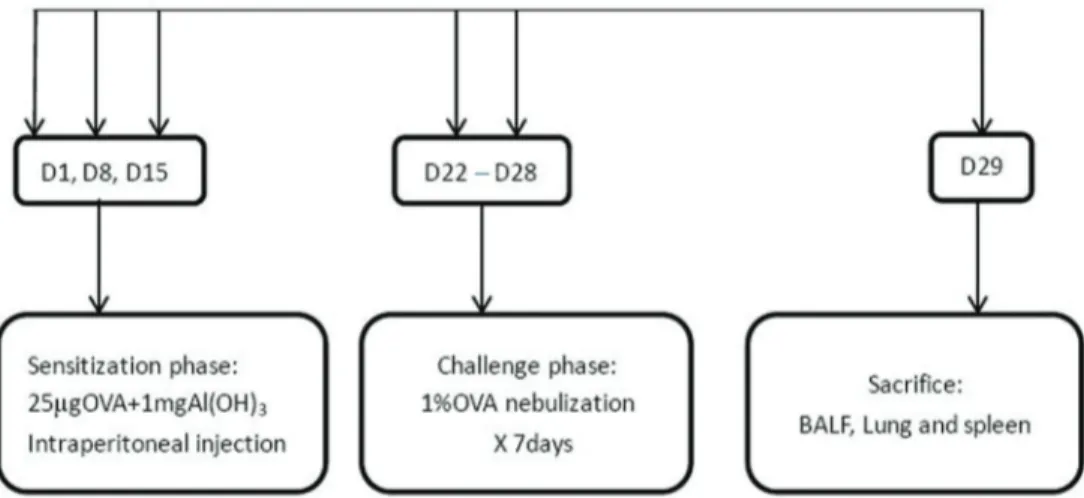
Experimental animals were randomly divided into two groups: normal control and asthmatic model, with 6 mice in each group. The normal control group was treated with saline at the sensitization and challenge stages. The asthmatic model group was sensitized and challenged with OVA to establish the asthmatic model, according to methods proposed by our previous report with minor modifications (13) (Figure 1). Mice were sensitized with 25mg of OVA (grade V, Sigma-Aldrich, USA) emulsified in 1 mg of aluminum hydroxide (Chengdu Kelong Chemical Reagent Factory, China) in a total volume of 200 mL on days 1, 8, and 15 by intraperitoneal administration. Begin-ning on the 22nd day, the mice were challenged with 1% OVA (mg/mL) for 30 min per day by an ultrasonic nebulizer (WH-2000, China) in a closed chamber for 7 days to establish models.
Airway responsiveness
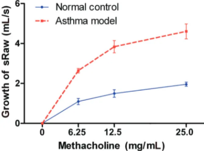
AHR was assessed using a double-chamber plethys-mography device (TBL4500, Buxco, USA) based on the increase in the specific airway resistance (sRaw). In brief,
the mice were exposed to nebulized PBS for 3 min to establish baseline sRaw values, followed by exposure to increasing concentrations of nebulized methacholine (6.25–25 mg/mL; Sigma, USA) using an Aerosonic ultrasonic nebulizer. Following each nebulization cycle, recordings were obtained for 3 min. The sRaw values measured during each 3-min sequence were averaged and reported for each methacholine concentration. The increase in sRaw was calculated as follows: sRaw with each methacholine concentration – sRaw with PBS) / sRaw with PBS.
Sample collection and processing
Twenty-four hours after the last challenge, all animals were anesthetized with pentobarbital. Specimens of bron-choalveolar lavagefluid (BALF), lung, and spleen were harvested. BALF (1200mL) was collected as previously described (14), centrifuged at 1500gfor 10 min at 4°C, and the supernatant was immediately frozen at 80°C for measurement of cytokine levels.
In order to obtain spleen cell suspensions, spleens were removed, cut into small pieces, ground gently into single-cell particles, andfiltered through nylon mesh. The cell suspension was centrifuged at 300 g for 10 min at 4°C. After that, erythrocytes were removed as described previously, and the spleen cell pellets were washed twice with cold PBS.
The right lung tissue was quick-frozen by immersion in liquid nitrogen, and then stored until quantitative real-time PCR was performed.
Histology and morphometry assay
The left lung tissue was fixed with 4% paraformalde-hyde, embedded in paraffin, cut sagittally into 4-mm sections, and stained with hematoxylin and eosin (H&E) and Alcian blue-periodic acid Schiff (AB-PAS) for histological analysis.
The micro-sections were stained for the examination of inflammation and mucus production under a microscopic observation (Olympus, Japan). For each animal, 10fields at a magnification of 200 were measured randomly from HE staining and were measured randomly from AB-PAS staining (400). Two investigators independently measured the origin of stained tissues in a blinded manner.
Cytokine measurement
The concentrations of IL-17 and IL-35 in BALF of OVA-induced asthmatic mice were measured by commercial ELISA kits according to the manufacturer’s instructions (Cusabio Biotech, China). ELISA kit of IL-17 detects from 47 pg/mL to 3000 pg/mL, and ELISA kit of IL-35 detects
from 15.6 pg/mL to 1000 pg/mL. The absorbance was meas-ured at 450 nm by a micro-plate ELISA reader (BioTek, USA). All samples were assayed in duplicate.
Flow cytometry
The expression markers on T cells from spleen were determined by flow cytometry using the following anti-bodies: Percp-CD3, APC-gdT, PE-IL-17, and PE-Foxp3 purchased from BD Pharmingen (USA) or eBioscience (USA). Cell surface staining was performed according to the standard procedures. For intracellular detection of cytokines, cells were stimulated with phorbol-myristate-acetate (25 ng/mL; Sigma-Aldrich) and ionomycin (1 ng/mL; Sigma-Aldrich) in the presence of GolgiPlugt (BD Phar-mingen) for 4 h at 37°C in 5% CO2. The cells were then
washed and stained with fluorescent antibodies against CD3 at room temperature in the dark. After surface staining, cells were fixed/permeabilized in fixation/permeabilization solution (Cytofix/Cytopermt; BD Pharmingen) according to the manufacturer’s protocol, and stained with anti-IL-17 mAbs/anti-Foxp3-mAbs for 30 min at 4°C. Cells were then washed with Perm/Wash Buffer (BD Pharmingen) and resuspended in PBS+2% FBS forflow cytometry analysis. Flow cytometry was performed on a BD FACS Canto II (BD Biosciences, USA) and analyzed using FCS Express 4 software (De Novo Software, USA).
Real-time quantitative PCR
For quantifying the transcription levels of RORgt and Foxp3, total RNA (1mg) was extracted from the right lung tissue with TRIzol (Invitrogen, Life Technologies, USA) according to the manufacturer’s instructions. Complemen-tary DNA (cDNA) was prepared using oligo(dT) primers (PrimeScripttRT, reagent Kit, Takala, Japan). Quantitative RT-PCR was performed by duplicate with SYBR Green I (SYBRsPremix Ex Taqt, Takala) using an Applied Figure 2.Change in specific airway resistance (sRaw) of mice
after being sensitized and challenged with ovalbumin and inhalation of different concentrations of methacholine compared with the control group (Po0.05). Data are reported as means ±SD (Student’st-test).
Biosystems 7500 (ThermoFisher Scientific, USA) according to the manufacturer’s instructions. DNA was amplified under the following conditions: denaturation at 95°C for 30 s, extension at 95°C for 5 s, 60°C for 34 s, and the samples were amplified for 40 cycles. The following primers were used: 50-AATTCCATCATGAAGTGTGA-30, 50-ACTCC
TGCTTGCTGATCCAC-30 forb-actin; 50- ACggCCCTggT
TCTCATCA -30and 50- CCAAATTgTATTgCAgATgTTCCA
C -30for RORgt; 50-AGTTCCTTCCCAGAGTTCTTCCA-30
and 50-GCTCAGGTTGTGGCGGATG-30for Foxp3. Mouse
b-actin was used as an internal control, and levels of each gene were normalized tob-actin expression using the delta-delta Ct method. The identity of the amplified products was examined using agarose gel electrophoresis and melt curve analysis.
Statistical analysis
Data are reported as means±SD. Differences between
groups were compared with unpaired Student’st-test. Correlations between variables were determined by Spear-man’s rank correlation test. Analysis was completed using SPSS version 19.0 statistical software (USA), and Po0.05 was considered to indicate statistical significance.
Results
Symptoms of the asthmatic mice
No mice died before sacrifice. During the challenge phase, OVA-induced asthmatic mice were found with different degrees of shortness of breath, cyanosis of lips, reduction of spontaneous activities, loss of weight, and
Figure 4.Alcian blue-periodic acid Schiff staining (400; bar, 25mm) showing the normal control group with rare goblet cell and no mucus secretion (A) and the asthmatic model group (B) with increased goblet cell hyperplasia in airway epithelia and mucus secretion.
incontinence, whereas mice in the control group displayed no manifestations.
Airway resistance
Compared with the normal control group, sRaw showed a significant increase at each concentration of methacholine in the asthmatic model group (Po0.05, Figure 2).
Histology and morphometry assay
and no mucus secretion, and the asthmatic model group had more goblet cell hyperplasia in airway epithelia and more mucus secretion (Figure 4A and B). The above results demonstrated that OVA administration successfully induced asthma.
Cytokines analysis
As shown in Figure 5, IL-17 expression was signifi -cantly higher in the asthmatic model group than in the normal control group (control group: 515.5±254.3 pg/mL,
asthmatic group: 1171.2±315.1 pg/mL, P=0.003). However,
IL-35 expression was significantly decreased in the
asthmatic model group, compared with the normal control group (control group: 45.1±23.4 pg/mL, asthmatic group:
17.7±9.2 pg/mL, P=0.024). These data indicated that
changes in the expression of IL-17 and IL-35 may con-tribute to thegdT17 andgdTreg imbalance.
Percentage ofcdT17 andcdTreg cells
As shown in Figures 6 and 7, the asthmatic model group exhibited an increase ingdT17 cells (25.0±5.5%)
compared with the normal control group (11.7±0.8%).
On the other hand, the normal control group exhibited a population ofgdTreg cells of 6.0±0.8%. Compared with
the normal control group, OVA-induced mice showed a significantly lower proportion ofgdTreg cells (3.2±0.4%).
These findings indicated that OVA could affect the percentage of gdT17 and gdTreg cells in the spleen of animals.
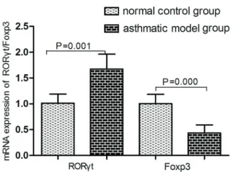
mRNA expression of RORct and Foxp3
As shown in Figure 8, compared with the normal control group, the mRNA expression of RORgt was significantly increased in the asthmatic model groups. Conversely, the mRNA expression Foxp3 decreased in the model group after OVA administration compared with the normal control group.
Correlation between mRNA expressions andcdT17 andcdTreg
As shown in Figure 9, there were positive correlations between the mRNA of RORgt and the percentage of gdT17 cells (r=0.907, Po0.05), as well as between Foxp3 and the percentage of gdTreg cells (r=0.86, Po0.05) in the asthmatic model group (Figure 9A and B). It appears that RORgt and Foxp3 expression were closely Figure 7. Comparative analysis ofgdT17 cells (A) andgdTreg
cells (B). Data are reported as means±SD (n=6). *Po0.05vs normal control group (t-test).
related to the proportion ofgdT17 andgdTreg cells in the lungs of mice with OVA-associated bronchial asthma.
Discussion
Asthma is a chronic lung disease of which the mechan-ism is little known. As human experiments are impossible, animal experiments play an important role in the under-standing of the mechanism of asthma. The ideal evaluation index of the asthmatic mouse model is AHR and airway inflammation (15). To determine the effects of OVA on the balance betweengdT17 andgdTreg cells, we used classical methods to prepare murine models of asthma. Thefindings of our study were in accordance with the study of Da-zhen Wei and colleagues (16). Taken together, our results con-firm that we established a successfully asthmatic model.
Evidence has emerged indicating that the pathogen-esis of asthma is mainly related with dysregulated immune responses by Th1/Th2 cells and Th17/Treg cells (17). How-ever, with the discovery of T cells, its mechanism is con-stantly changing. Recent studies have indicated gdT cells play a crucial role in the occurrence and development of autoimmune diseases and pathogen-induced immune res-ponses, but the specific mechanisms need further investi-gation (18).
Emerging evidence suggests that Th17/Treg imbal-ance contributes to the development of asthma (19), showing an increase of Th17 cells and suppression of Treg development. Th17 cells have a role in chronic infl am-mation. As a subset ofgdT cells,gdT17 cells are regarded
of RORgt and the percentage of gdT17 cells, as well as between Foxp3 and the percentage ofgdTreg cells in the asthmatic model group. It appears that RORgt and Foxp3 expressions were closely related to the proportion of gdT17 and gdTreg cells in the lungs of mice with OVA-associated asthma.
Consequently, the above results demonstrated that the relationship between gdT17 and gdTreg cells may be involved in the pathogenesis of asthma induced by OVA. There is a possibility that thegdT17-related immune
reaction became unbalanced although moregdT17 cells were involved. In contrast, gdTreg cells frequency was significantly negatively associated with the process. Fur-ther studies may lead to the effects of immunoFur-therapy on asthma.
Acknowledgments
The research received funding from the National Natural Science Foundation of China (No. 81470230).
References
1. Galli SJ, Tsai M, Piliponsky AM. The development of allergic inflammation. Nature 2008; 454: 445–454, doi: 10.1038/ nature07204.
2. Ming M, Luo Z, Lv S, Li C. Inhalation of inactivated-Mycobacterium phlei prevents asthma mediated airway hyperresponsiveness and airway eosinophilia in mice by reducing IL-5 and IL-13 levels. Mol Med Rep 2016; 14: 5343–5349, doi: 10.3892/mmr.2016.5865.
3. Li C, Sheng A, Jia X, Zeng Z, Zhang X, Zhao W, et al. Th17/ Treg dysregulation in allergic asthmatic children is asso-ciated with elevated notch expression.J Asthma2018; 55: 1–7, doi: 10.1080/02770903.2016.1266494.
4. Glanville N, Message SD, Walton RP, Pearson RM, Parker HL, Laza-Stanca V, et al. gdT cells suppress inflammation and disease during rhinovirus-induced asthma exacerbations.
Muco-sal Immunol2013; 6: 1091–10100, doi: 10.1038/mi.2013.3.
5. Hahn YS, Taube C, Jin N, Sharp L, Wands JM, Aydintug MK, et al. Different potentials of gamma delta T cell subsets in regulating airway responsiveness: V gamma 1+cells, but not V gamma 4+cells, promote airway hyperreactivity, Th2 cytokines, and airway inflammation.J Immunol2004; 172: 2894–2902, doi: 10.4049/jimmunol.172.5.2894.
6. Zhang J, Li C, Guo S. Effects of inhaled inactivated Myco-bacterium phlei on airway inflammation in mouse asthmatic models.J Aerosol Med Pulm Drug Deliv2012; 25: 96–103, doi: 10.1089/jamp.2011.0904.
7. Ming M, Li C, Luo Z, Lv S, Sun Q. The effect of inhaled inactived Mycobacterium phlei as a treatment for asthma.Mol
Med Rep2017; 15: 777–783, doi: 10.3892/mmr.2016.6087.
8. Nakada EM, Shan J, Kinyanjui MW, Fixman ED. Adjuvant-dependent regulation of interleukin-17 expressinggdT cells and inhibition of Th2 responses in allergic airways disease.
Respir Res2014; 15: 90, doi: 10.1186/s12931-014-0090-5.
9. Zhong F, Cui D, Tao H, Du H, Xing C. IL-17A-producing T cells and associated cytokines are involved in the progres-sion of gastric cancer.Oncol Rep2015; 34: 2365–2374, doi: 10.3892/or.2015.4246.
10. Cai Y, Shen X, Ding C, Qi C, Li K, Li X, et al. Pivotal role of dermal IL-17-producinggdT cells in skin inflammation.Immunity 2011; 35: 596–610, doi: 10.1016/j.immuni.2011.08.001. 11. Kang N, Tang L, Li X, Wu D, Li W, Chen X, et al.
Identi-fication and characterization of Foxp3(+) gammadelta T cells in mouse and human.Immunol Lett2009; 125: 105– 113, doi: 10.1016/j.imlet.2009.06.005.
12. Ma Y, Lei H, Tan J, Xuan L, Wu X, Liu Q. Characterization of
gdregulatory T cells from peripheral blood in patients with
multiple myeloma.Biochem Biophys Res Commun 2016; 480: 594–601, doi: 10.1016/j.bbrc.2016.10.098.
13. Jinghong Z, Chaoqian L, Sujuan G, Yi L. Inhaled inactivated-Mycobacterium phlei modulates gdT cell function and alleviates airway inflammation in a mouse model of asthma.
Asian Pac J Allergy Immunol 2013; 31: 286–291, doi:
10.12932/AP0323.31.4.2013.
14. Li C, Jiang X, Luo M, Sun Q, Feng G, Chen Y. Myco-bacterium vaccae nebulization can protect against asthma in Balb/c mice by regulating Th9 expression. PLoS One 2016; 11: e0161164, doi: 10.1371/journal.pone.0161164. 15. Kim KS, Cho DH, Yang HJ, Choi EK, Shin MH, Kim KH,
et al. Effects of the inhaled treatment of liriope radix on an asthmatic mouse model.Am J Chin Med2015; 43: 425–441, doi: 10.1142/S0192415X15500275.
16. Wei DZ, Guo XY, Lin LN, Lin MX, Gong YQ, Ying BY, et al. Effects of angelicin on ovalbumin (OVA)-induced airway inflammation in a mouse model of asthma. Inflammation 2016; 39: 1876–1882, doi: 10.1007/s10753-016-0423-2. 17. Shi YH, Shi GC, Wan HY, Jiang LH, Ai XY, Zhu HX, et al.
Coexistence of Th1/Th2 and Th17/Treg imbalances in patients with allergic asthma.Chin Med J (Engl)2011; 124: 1951–1956. 18. Lu H, Li DJ, Jin LP.gdT Cells and Related Diseases.Am J
Reprod Immunol2016; 75: 609–618, doi: 10.1111/aji.12495.
19. Zhao J, Lloyd CM, Noble A. Th17 responses in chronic allergic airway inflammation abrogate regulatory T-cell-mediated toler-ance and contribute to airway remodeling.Mucosal Immunol 2013; 6: 335–346, doi: 10.1038/mi.2012.76.
20. Gupta PK, Wagner SR, Wu Q, Shilling RA. Th17 cells are not required for maintenance of IL-17A producinggdT cells in vivo.Immunol Cell Biol2017; 95: 280–286, doi: 10.1038/ icb.2016.94.
21. Wu Q, Gupta PK, Suzuki H, Wagner SR, Zhang C, W Cummings O, et al. CD4 T Cells but not Th17 cells are required for mouse lung transplant obliterative bronchiolitis.
Am J Transplant2015; 15: 1793–1804, doi: 10.1111/ajt.13215.
22. Li X, Kang N, Zhang X, Dong X, Wei W, Cui L, et al. Generation of human regulatory gammadelta T cells by TCR gammadelta stimulation in the presence of TGF-beta and their involvement in the pathogenesis of systemic lupus erythematosus. J Immunol 2011; 186: 6693–6700, doi: 10.4049/jimmunol.1002776.
23. Liu J, Qu H, Li Q, Ye L, Ma G, Wan H. The responses ofgd