Short Report
0103 - 5053 $6.00+0.00
*e-mail: [email protected]
A Simple Method for the Quantitative Analysis of Tyrosol by HPLC in
Liquid Czapek Cultures from Endophytic Fungi
Denise O. Guimarães,a Keyller B. Borges,b Pierina S. Bonatob and Mônica T. Pupo*,a
aDepartamento de Ciências Farmacêuticas and bDepartamento de Física e Química, Faculdade de Ciências
Farmacêuticas de Ribeirão Preto, Universidade de São Paulo, 14040-903 Ribeirão Preto-SP, Brazil
O tirosol é provavelmente uma molécula sinalizadora em fungos endofíticos. A análise do tirosol em cultura líquida Czapek de fungo endofítico foi realizada através de cromatografia líquida de alta eficiência acoplada a detector por arranjo de diodos. As análises foram obtidas em sistema de fase móvel utilizando gradiente, modo linear, iniciando em acetonitrila/água (1:9 v/v) e terminando em acetonitrila 100% em 30 minutos com vazão de 1 mL min-1. Coluna
analítica ZORBAX® ODS (250 × 4,6 mm, 5 µm) à 25 °C foi utilizada. Extração líquido-líquido de
0,5 mL do meio (pH 7,0) com acetato de etila e injeção de 20 µL após concentração do solvente sob ar comprimido originou bons resultados. Os parâmetros validados foram: linearidade 0,0125-5,0 µg mL-1 (r = 0,9967), limite de quantificação 0,0125 µg mL-1 obtidos pela média
das análises; %CV (precisão) e %E (exatidão) com valores abaixo de 15% e recuperação de cerca de 80%. Além disso, o método desenvolvido apresentou valores de validação satisfatórios demonstrando eficiência na análise do tirosol em meio líquido Czapek.
Tyrosol is a possible quorum sensing molecule in endophytic fungi. High-performance liquid chromatography (HPLC) coupled with diode array detector (DAD) was used for the analysis of tyrosol in liquid Czapek fungal cultures. The optimized conditions were gradient mobile phase, in linear mode, consisting initially of acetonitrile/water (1:9 v/v) and increasing up to acetonitrile (100%) in 30 minutes at a flow rate of 1 mL min-1. The column used was a ZORBAX ODS
(250 × 4.6 mm, 5 µm) at 25 oC. Liquid-liquid extraction of 0.5 mL medium (pH 7.0) with
ethyl acetate and injection of 20 µL after solvent evaporation under air flow gave good results. Some validation parameters obtained were: linearity 0.0125-5.0 µg mL-1 medium (r = 0.9967),
quantification limit of 0.0125 µg mL-1 medium, %CV (precision) and %E (accuracy) bellow 15%
and recovery around 80%. Therefore, the developed method presented satisfactory validation parameters and it was efficient for the analysis of tyrosol in Czapek medium.
Keywords: tyrosol, endophytic fungi, HPLC-DAD, validation method
Introduction
Tyrosol (2-(4-Hydroxyphenyl)ethanol) is a well-known phenolic compound with antioxidant properties
that is present in wine and olive oil,1 and it is reported to
have scavenging effects on reactive oxygen and nitrogen species that are implicated in human pathologies such
as cardiovascular and thrombotic diseases.2,3 Tyrosol is
produced by terrestrial fungi and showed antifungal activity
against Lagenidium callinectes4 and Gibberella pulicaris.5
Recently, tyrosol has been reported as a candidate to be used in stroke therapy due to its neuroprotective effect in
rats.1 Moreover, tyrosol was identified as an autoregulatory
molecule with important implication on the dynamics
of growth and morphogenesis in Candida albicans6 in a
process known as quorum-sensing, which is characterized by a cellular density-dependent phenomenon. Quorum-sensing effect is accomplished by the extracellular accumulation of small, self-generated chemical signaling molecules that induce bacterial population to produce the
desired phenotypic effect.7 The first described
quorum-sensing system involved the bioluminescent marine
bacterium Vibrio fischeri.8
Several chemical classes of microbial derived signaling molecules have been identified, and they might be classified
in two main categories: (i) amino acids and short peptides,
commonly utilized by Gram-positive bacteria,9,10 and
Gram-negative bacteria.11,12 Particular emphasis has been placed on the wide range of quorum-sensing systems that
employ N-acyl homoserine lactones (acyl HSLs) as the
signaling molecules that control the expression of diverse
physiological functions.12 Several examples of signaling
molecules illustrate that quorum-sensing molecules have been used by microorganisms, especially bacteria, in order
to control a great variety of functional systems.13 Studies
of bacterial quorum-sensing phenomenon have shown information on how bacterial chemical communication works; how chemical information is integrated, processed and transduced to control gene expression; how intra- and inter species cell-cell communication is accomplished and the intriguing possibility of prokaryote-eukaryote cross-communication. In fungi this phenomenon has been mostly
studied in Candida albicans.
Due to its scavenging effects, tyrosol has been previously
quantified in different matrices such as beverages14 and
biological fluids (low-density-lipoprotein)15 using different
analytical quantitative methods such as HPLC-DAD and HPLC-ESI-MS-MS.
In our prospection study of endophytic fungi from Asteraceae species we have isolated tyrosol from several bioactive endophytic cultures. So, we hypothesized it might have some quorum-sensing role in those microorganisms. In order to check this possibility, we initially set about developing a high-performance liquid chromatography (HPLC) coupled with diode array detector (DAD) method for the analysis of tyrosol in endophytic fungi cultures.
Experimental
Standard solutions and chemicals
Tyrosol utilized for the preparation of standard solutions was obtained from the cultivation of the
endophytic fungus Glomerella cingulata (Stoneman)
Spauld. & H. Schrenk (code VA1) found in association
with the host plant Viguiera arenaria (Asteraceae). The
mycelium of G. cingulata grown on PDA (potato dextrose
agar) Petri dishes was inoculated on 12 Erlenmeyer flasks
containing 200 mL of pre-fermentative medium16 and
incubated at 30 ºC with shaking (120 rpm min-1) for 24 h.
Afterward, the obtained mycelia were transferred to 12 Erlenmeyer flasks containing 400 mL of fermentative
medium Czapek,17 and the fungus was allowed to grow at
30 ºC and 120 rpm min-1 for an additional 144 hour period.
The culture broth was separated from the mycelium through vacuum filtration (400 mL) and submitted to extraction with ethyl acetate (EtOAc) three consecutive
times (150 mL each). The EtOAc crude extract (88.0 mg) was fractionated in a silica gel column (0.063-0.200 mm) with hexane/EtOAc (9:1 v/v); hexane/EtOAc (1:1 v/v); EtOAc and methanol. The sub fraction 25 (6.4 mg), obtained with hexane/EtOAc (1:1 v/v), was submitted to preparative thin layer chromatography in silica gel
PF254 eluted with dichloromethane/methanol (9:1 v/v)
yielding tyrosol (rf: 0.40, 5.0 mg). Tyrosol was extracted
with acetone/methanol (4:1 v/v).18 NMR spectra were
acquired in Bruker spectrometers (400 and
DRX-500), working at 400 and 500 MHz for 1H and at 100 and
125 MHz for 13C. The spectra were recorded in CDCl
3,
and the solvent signals at d 7.26 for proton, and d 77.0 for
carbon, were used as reference. Mass spectra analysis was conducted in a mass spectrometer ESI-MS (Micromass Quattro LC-electrospray ionization). Chromatograms area analyses were compared in order to check the purity of isolated tyrosol and available commercial tyrosol 98% (Sigma-Aldrich Chemie, Steinheim, Germany). The purity index was 95.4% for isolated tyrosol used as standard for the quantitative validation procedures. Tyrosol stock standard solutions were prepared in methanol at concentrations of 0.250, 0.500, 1.0, 5.0, 20.0, 60.0 and
100.0 µg mL-1 (resulting in concentrations of 0.0125,
0.0250, 0.050, 0.250, 1.0, 3.0 and 5.0 µg mL-1 when 25 µL
is used to spike 0.5 mL culture medium). The solutions
were stored at −20 oC, in the absence of light.
HPLC-grade acetonitrile, methanol, ethyl acetate were purchased from Mallinckrodt Baker Inc. (Paris, USA) and ethyl acetate from Merck (Darmstadt, Germany). All other chemicals were of analytical-grade in the highest purity available. Water was distilled and purified using a Millipore Milli Q Plus system (Bedford, USA).
Instrumentation and analytical conditions
The analyses were carried out using a Shimadzu (Kyoto, Japan) HPLC system, consisting of a LC-6AD model
solvent pump, system controller SCL 10AVP, a column
oven CTO-10ASVP, a Rheodyne model 7725 injector with
a 20 µL loop, a SPD-M10AVP diode array detector operating
at 225 nm and a software Class VP for data acquisition. The
determination of tyrosol was performed on a ZORBAX
ODS column (250 × 4.6 mm, I.D., 5 µm particle size, Agilent Technologies, Exton, USA), protected with a 4.6 mm I.D. × 12.5 mm × 4.6 mm, 4-Pack endcapped guard column (Agilent Technologies), using a gradient mobile phase, in linear mode, consisting initially of acetonitrile/ water (1:9 v/v) and increasing up to acetonitrile (100%) in
30 minutes at a flow rate of 1 mL min-1. The column was
Extraction procedure
Preliminary recovery studies were carried out in order to verify a satisfactory pH condition and solvent to be used in the liquid-liquid extraction procedure. Tyrosol standard
solution was prepared at the concentration of 40.0 µg mL-1
(resulting in concentrations of 1.0 and 2.0 µg mL-1 when
25 µL of this solution was used to spike 1 and 0.5 mL culture medium, respectively). Aliquots of 1 mL of Czapek medium at three different pHs: 5.0; 7.0 and 9.0 were spiked with 25 µL of standard tyrosol solution at the concentration of
40.0 µg mL-1 and extracted with 3 mL ethyl acetate (100%)
or toluene/isopropanol (4:1 v/v). The tubes were capped and submitted to vortex mixing for 1 min and then centrifuged at 1800 × g for 4 min. The upper organic phases (2 mL) were transferred to conical tubes and evaporated under compressed air. The dried residues were dissolved in 50 µL of the mobile phase (10% acetonitrile aqueous solution) and 20 µL were chromatographed under the previously described conditions.
In order to verify whether the phosphate buffer 1 mol L-1,
pH 7.0 solution was satisfactory to maintain a neutral pH at 7.0 for liquid-liquid extraction another procedure was tested: aliquots of 0.5 mL of the Czapek matrix spiked with
25 µL of the standard tyrosol solution (40.0 µg mL-1) were
neutralized with 1 mL 1 mol L-1 phosphate buffer solution
pH 7.0 and extracted with 4 mL ethyl acetate. The upper organic phases (3 mL) were transferred to conical tubes and evaporated under compressed air. The dried residues were dissolved in 100 µL of the mobile phase (10% acetonitrile aqueous solution) and 20 µL were chromatographed under the previously described conditions.
Good results were obtained with this last described extraction procedure, which was used for the method validation and method application by samples analyses. In
addition, the room temperature was set at 22 ± 2 oC in order
to prevent solvent evaporation during sample preparation.
Method validation
Calibrations curves were obtained by spiking aliquots of 0.5 mL medium with standard solutions of tyrosol, prepared in methanol, in the range of 0.250-100.0 µg
mL-1, resulting in concentrations of 0.0125-5.0 µg mL-1
in the culture medium. No internal standard was used in this method.
To determine the extraction recovery, medium samples (0.5 mL) were spiked with tyrosol in the concentrations
of 0.05, 1.0 and 5.0 µg mL-1 (n = 3) and submitted to the
extraction procedure. Another set of samples were prepared extracting 0.5 mL aliquots of medium and then spiking
the extract with the same amount of tyrosol. The recovery was determined by comparing the areas obtained before and after extraction and was expressed as percentage of the amount extracted.
The detectability of the method was evaluated by determining the quantification limit (LOQ). The LOQ was defined as the lowest concentration that could be
determined with accuracy and precision below 20%19
over five analytical run and it was obtained using medium samples (0.5 mL, n = 5) spiked with concentrations of
0.0125 µg mL-1 of tyrosol.
The precision and accuracy of the method were evaluated by analyzing spiked matrix with known standard tyrosol solution. The experiments were carried out by intra-day (n = 5) and inter-day (n = 3) assays using medium samples spiked with tyrosol at the concentrations of 0.0250, 0.250 and
3.0 µg mL-1. The results obtained were expressed as coefficient
of variation (CV, %) and relative error (E, %).
The selectivity of the method was evaluated by analyzing sterile medium (blank) and endophytic fungal culture broth under the conditions previously established.
The stability of tyrosol was evaluated by testing the influence of freeze (−20 ºC) and thaw (22 ± 2 ºC) cycles, short-term room temperature and 48- h freeze temperature. Spiked Czapek media were prepared in sextuplicate (n = 6)
at the low (0.05 µg mL-1) and high concentration (3 µg mL
-1). To perform the freeze-thaw cycle stability test, these
samples were stored at −20 ºC for 24 h and thawed at room temperature. When completely thawed, the samples were refrozen for 12 h under the same conditions. The freeze-thaw cycle was repeated once more, and then the samples were analyzed on the second cycle. The 48 h freeze temperature stability test was performed analyzing frozen (−20 ºC) Czapek medium samples over 48 h. This period corresponds to the maximum time the samples were kept frozen. The short-term room temperature stability test was assessed analyzing samples that were kept on the bench-top for 4 h at room temperature (22 ± 2 ºC). After this period, the samples were analyzed. The peak area obtained from the stability tests was compared to the peak area obtained with freshly prepared samples. Student’s t test was applied, with the level of
significance set at p ≤ 0.05.
Endophytic fungus isolation and tyrosol formation
The fungus G. cingulata used in these experiments
was previously isolated as endophyte from V. arenaria
(Asteraceae) and identified by its rDNA sequence.18 The
strain has been maintained on potato dextrose agar slants
strain was deposited in the “Laboratório de Enzimologia Industrial-FCFRP/USP”.
G. cingulata was cultured using a two-step fermentative process. First, suspension of the mycelium was aseptically inoculated into 1000 mL Erlenmeyer flasks containing
200 mL of seed medium.16 The flasks were incubated
for 48 h at 30 oC on a rotary shaker (New Brunswick
Scientific Co., Inc., model INNOVA TM 4300, New
Jersey, USA) operating at 120 rpm. The resulting mycelia obtained after seed medium filtration were transferred into 2000 mL Erlenmeyer flasks containing 400 mL of
Czapek medium.17 Three different pHs were adopted for
the cultivation on Czapek medium: pH 5.0, 7.0 and 9.0.
Experiments were carried out at 30 oC, with shaking at
120 rpm for additional 20 days. Aliquots of the Czapek filtrates (0.5 mL) after mycelium inoculation were submitted to the previously described extraction procedure and analyzed by HPLC.
Results and Discussion
Method development and liquid-liquid extraction
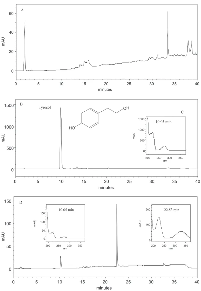
Analyses were carried out using a ZORBAX-ODS column (250 mm × 4.6 mm I.D.) and a linear gradient elution consisting initially of acetonitrile/water (1:9 v/v) and increasing up to acetonitrile (100%) in 30 minutes. Analyses of the blank of the Czapek medium (Figure 1A) showed no interference at the retention time for tyrosol
(tR = 10.05 min) (Figure 1B). In addition, the UV profile
of tyrosol is represented at Figure 1C.
The utilization of the gradient linear mode and the absence of an internal standard were related to the complexity of the endophytic fungi extracts that might be obtained from Czapek culture medium. In this case, an internal standard might interfere in the extracts’ analyses. In addition, the gradient mode contributes for a better separation of the compounds in a complex extract.
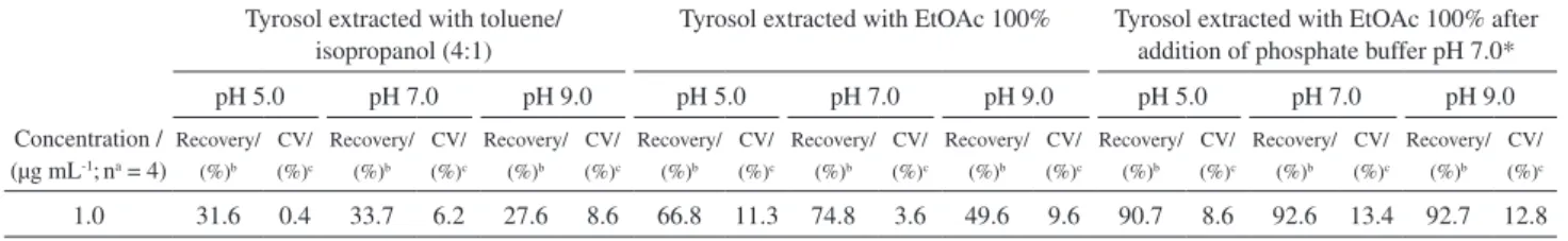
Preliminary recovery studies (Table 1) show that ethyl acetate at pH 7.0 was the best condition for liquid-liquid extraction of tyrosol. In order to establish the pH of the
culture broth after fungal cultivation, we performed the
addition of the phosphate buffer 1 mol L-1, pH 7.0 to the
Czapek medium (pH 5.0, 7.0 and 9.0) before the extraction with ethyl acetate. Recovery values for extraction of tyrosol after addition of the phosphate buffer using three different pHs were similar (Table 1) illustrating that addition of this buffer solution was efficient to maintain the pH of the medium at 7.0 in order to guarantee a satisfactory pH for the tyrosol extraction.
Method validation
The calibration curves were linear over the concentration
range of 0.0125-5.0 µg mL-1 for tyrosol with linear
equation: y = 122534x + 3240.4; correlation coefficient (r) of 0.9967 and coefficient of variation of 13.9%. The lowest concentration quantified by the validated method
was 0.0125 µg mL-1 with coefficient of variation of 13.7%
and error 13.9%. Recoveries were around 80% for tyrosol with coefficients of variation lower than 15% (Table 2). Intra-day (n = 5) and inter-day (n = 3) precision and accuracy presented coefficients of variation and relative errors lower than 15% (Table 3). In spite of not using an internal standard, the precision results were acceptable.
Stability of tyrosol in Czapek medium was evaluated by
Student’s t test with the level of significance set at p ≤0.05.
The p-values obtained were above 0.05 illustrating the stability of tyrosol. Coefficients of variation obtained for low and high concentration used in stability test were lower than 15% (Table 4).
Table 2. Recovery of tyrosol
Concentration / (µg mL-1; na = 3)
Recovery / (%) CVb / (%)
0.05 73.5 11.0
1.0 89.6 2.6
5.0 79.0 3.1
Mean 80.7 11.8
anumber of determinations; bCV, coefficient of variation.
Table 1. Tyrosol optimization of extraction procedure
Tyrosol extracted with toluene/ isopropanol (4:1)
Tyrosol extracted with EtOAc 100% Tyrosol extracted with EtOAc 100% after addition of phosphate buffer pH 7.0*
Concentration / (µg mL-1;na = 4)
pH 5.0 pH 7.0 pH 9.0 pH 5.0 pH 7.0 pH 9.0 pH 5.0 pH 7.0 pH 9.0
Recovery/ (%)b CV/ (%)c Recovery/ (%)b CV/ (%)c Recovery/ (%)b CV/ (%)c Recovery/ (%)b CV/ (%)c Recovery/ (%)b CV/ (%)c Recovery/ (%)b CV/ (%)c Recovery/ (%)b CV/ (%)c Recovery/ (%)b CV/ (%)c Recovery/ (%)b CV/ (%)c
1.0 31.6 0.4 33.7 6.2 27.6 8.6 66.8 11.3 74.8 3.6 49.6 9.6 90.7 8.6 92.6 13.4 92.7 12.8
Figure 1. (A) Chromatogram referring to the analysis of medium culture blank. (B) Chromatogram referring to the analysis of tyrosol tR = 10.05 min; purity: 0.99990 at 1 mg mL-1. (C) UV-Profile of standard solution of tyrosol in 190-370 nm. (D) Chromatogram referring to the analysis of Glomerella
Table 3. Precision and accuracy of the method for analysis of tyrosol in Czapek medium
Intra-day (na = 5) Inter-day (n a = 3)
Nominal concentration / (µg mL-1) 0.0250 0.250 3.00 0.0250 0.250 3.00
Analyzed concentration / (µg mL-1) 0.0256 0.250 2.85 0.0250 0.231 2.66
Precision / (CV, %)b 9.8 8.5 5.9 14.5 11.3 8.9
Accuracy / (E, %)c 2.3 0.1 -4.8 -0.0 -7.6 -11.2
aNumber of determinations: 5 for intra-day assay and 3 for inter-day assay; bexpressed as coefficient of variation, CV; cexpressed as relative error, E.
Table 4. Stability test of tyrosol
Nominal concentration / (µg mL-1, na =6)
tyrosol
Tyrosol concentration (mean) / (µg mL-1) CVb / (%) Ec / (%) p-value
Fresh
0.05 0.0509 2.78 1.92
-3.00 3.1454 7.88 4.84
-Freeze-thaw cycles
0.05 0.0497 1.64 -0.46 0.10
3.00 2.9625 1.71 1.25 0.11
48 h freeze
0.05 0.0497 1.80 -0.48 0.11
3.00 3.0004 10.46 0.01 0.39
Short-term room temperature
0.05 0.0504 0.74 0.86 0.39
3.00 2.9253 0.80 2.49 0.06
anumber of determinations; bCV, coefficient of variation. cExpressed as relative error, E.
Method application
The developed method showed to be efficient for the quantitative analyses of tyrosol in Czapek fermentative medium. The method was applied for a Czapek sample derived from the cultivation of the endophytic fungus
Glomerella cingulata and no interference for tyrosol peak was observed, since the peak purity indexes were above 0.99000 for all analyses. These analyses were carried out after inoculation of the mycelia obtained from seed medium into Czapek medium at three different pHs (5.0, 7.0 and 9.0) without leaving any additional time of incubation (incubation time = 0 h) and with 144 h of incubation (chromatogram of 144 h of incubation time is shown in Figure 1D). Concentration of tyrosol at 0 h of incubation was 0.04 (CV 9.9%); 0.03 (CV 7.4%) and
0.04 (CV 11.1%) µg mL-1 for cultures at pHs at 5.0, 7.0
and 9.0, respectively. For the 144 h incubation time the concentration of tyrosol found was 2.27 (CV 4.1%); 1.18
(CV 3.4%) and 1.12 (CV 7.3%) µg mL-1 for cultures at pH
at 5.0, 7.0 and 9.0, respectively. The presence of tyrosol
at similar concentrations for the three different culturing conditions at 0 hour of incubation means that tyrosol production probably starts during the seed medium culture. The chromatogram in Figure 1D refers to the analysis of
tyrosol at 144 h of incubation of Glomerella cingulata
in Czapek medium. Analysis of the peak purity index
(1.00000) by DAD at tR=10.05 min shows that no influence
of other secondary metabolites were observed. Moreover,
the presence of the peak at tR = 22.53 min illustrates that
the validated method can be used to verify the correlation between tyrosol and other secondary metabolites produced by fungi in Czapek medium. Is is noteworthy that as the production of tyrosol increases in the culture medium, the
production of compound at tR = 22.53 min also increases,
as illustrated by the chromatogram in Figure 1D. It suggests that tyrosol might be acting as a signaling compound for the production of this additional metabolite.
matrices such as human plasma, human low density
lipoproteins15 and ciders14 using basically HPLC or
HPLC-ESI-MS-MS as main techniques have been employed for the quantification of these antioxidant compounds. Both isocratic and gradient linear methods can be found for quantitative analyses of tyrosol and the time of analysis
varies from a short20 through long periods14 depending on
the main objective of the study. To our concern there are no published data for the analysis of tyrosol in Czapek medium.
Conclusions
A suitable high-performance liquid chromatography method was developed and validated for the determination of tyrosol in fermentative Czapek medium. This method was successfully used to quantify the formation of tyrosol
by the endophytic fungus G. cingulata cultured at different
pHs. Therefore, this analytical quantitative method might be used for the detection and quantification of tyrosol in Czapek medium of endophytic fungi under different culturing conditions. In addition, the development of sample preparation and appropriate concentration procedure might extend the applicability of this method to other matrices. Finally, this method will allow further studies in order to verify the role of tyrosol as a quorum-sensing signaling molecule for the secondary metabolites production by endophytic fungi from Asteraceae species.
Acknowledgments
The authors are grateful to Fundação de Amparo à Pesquisa do Estado de São Paulo (FAPESP), sub-program BIOTA/FAPESP (Rede BIOprospecTA), Conselho Nacional de Desenvolvimento Científico e Tecnológico (CNPq) and to Coordenação de Aperfeiçoamento de Pessoal de Nível Superior (CAPES) for financial support and for granting research fellowships (FAPESP grants 03/07535-5, 04/07935-6, 05/03791-2).
References
1. Bu, Y.; Rho, S.; Kim, J.; Kim, M. K.; Lee, D. H.; Kim, S. Y.; Choi, H.; Kim, H.; Neurosci. Lett. 2007, 414, 218.
2. de la Puerta, R.; Martinez Domínguez, M. E.; Ruiz-Gutierrez, V.; Flavill, J. A.; Hoult, J. R.; Life Sci. 2001, 69, 1213. 3. Bertelli, A. A.; Migliori, M.; Panichi, V.; Longoni, B.; Origlia,
N.; Ferretti, A.; Cuttano, M. G.; Giovannini, L.; Ann. N. Y. Acad. Sci. 2002, 957, 295.
4. Sofia, M.; Turnes, G.; Fenical, W.; Biol. Bull. 1992, 182, 105. 5. Slininger, P. J.; Burkhead, K. D.; Schisler, D. A.; J. Ind.
Microbiol. Biotechnol. 2004, 31, 517.
6. Chen, H.; Fujita, M.; Feng, Q.; Clardy, J.; Fink, G. R.; Proc.
Natl. Acad. Sci. U. S. A. 2004, 101, 5048.
7. De Kievit, T. R.; Iglweski, B. H.; Infect. Immun.2000, 68, 4839.
8. Nealson, K. H.; Hastings, J. W.; Microbiol. Rev.1979, 43, 496.
9. Lazazzera, B. A.; Grossman, A. D.; Trends Microbiol. 1998, 6, 288.
10. Shapiro, J. A.; Annu. Rev. Microbiol. 1998, 52, 81.
11. Dunny, G. M.; Winans, S. C.; Cell-cell Signaling in Bacteria; ASM Press: Washington, D. C., 1999.
12. Whitehead, N. A.; Barnard, M. L.; Slater, H.; Simpson, N. J. L.; Salmond, G. P. C.; FEMS Microbiol. Rev. 2001, 25, 365. 13. Gera, C.; Srisvastava, S.; Current Sci. 2006, 90, 666. 14. Suaréz, B.; Palacios, N.; Fraga, N.; Rodríguez, R.;
J. Chromatogr. A 2005, 1066, 105.
15. de la Torre-Carbot, K.; Jauregui, O.; Castellote, A. I.; Lamuela-Raventós, R. M.; Covas, M. -I.; Casals, I.; Lópes-Sabater, M.
C.; J. Chromatogr. A 2006, 1116, 69.
16. Jackson, M.; Karwoswski, J. P.; Humphrey, P. E.; Kohl, W. L.; Barlow, G. J.; Tanaka, S. K.; J. Antibiot. 1993, 46, 34. 17. Alviano, C. S.; Farbiarz, S. R.; Travassos, L. R.; Angluster, J.;
Souza, W.; Mycopathologia 1992, 119,17.
18. Guimarães, D. O.; Borges, W. S.; Kawano, C. Y.; Ribeiro, P. H.; Goldman, G. H.; Nomizo, A.; Thiemann, O. H.; Oliva, G.; Lopes, N. P.; Pupo, M. T.; FEMS Immunol. Med. Microbiol.
2008, 52, 134.
19. http://www.fda.gov/cder/guidance/4252fnl.pdf, accessed in May 2008.
20. Grizis, C.; Atta-Politou, J.; Koupparis, M. A.; J. Liq. Chromatogr.
Relat. Technol. 2003, 26, 599.
Received: May 12, 2008 Web Release Date: November 18, 2008