Sensitive Detection of
Using a High-Throughput, Colourimetric Loop
Mediated Isothermal Amplification (HtLAMP)
Platform: A Potential Novel Tool for Malaria
Elimination
Sumudu Britton1*, Qin Cheng2, Matthew J. Grigg3, Catherine B. Poole4, Cielo Pasay1, Timothy William5, Kimberley Fornace6, Nicholas M. Anstey3, Colin J. Sutherland6, Chris Drakeley6, James S. McCarthy1
1University of Queensland, Brisbane, Australia and QIMR Berghofer Medical Research Institute, Brisbane, Australia,2Australian Army Malaria Institute, Brisbane, Australia,3Menzies School of Health Research and Charles Darwin University, Darwin, Australia,4New England Biolabs, Ipswich, Massachusetts, United States of America,5Jesselton Medical Centre, Kota Kinabalu, Sabah, Malaysia,6London School of Hygiene and Tropical Medicine, London, United Kingdom
Abstract
Introduction
Plasmodium vivaxmalaria has a wide geographic distribution and poses challenges to malaria elimination that are likely to be greater than those ofP.falciparum. Diagnostic tools forP.vivaxinfection in non-reference laboratory settings are limited to microscopy and rapid diagnostic tests but these are unreliable at low parasitemia. The development and valida-tion of a high-throughput and sensitive assay forP.vivaxis a priority.
Methods
A high-throughput LAMP assay targeting aP.vivaxmitochondrial gene and deploying color-imetric detection in a 96-well plate format was developed and evaluated in the laboratory. Diagnostic accuracy was compared against microscopy, antigen detection tests and PCR and validated in samples from malaria patients and community controls in a district hospital setting in Sabah, Malaysia.
Results
The high throughput LAMP-P.vivaxassay (HtLAMP-Pv) performed with an estimated limit of detection of 1.4 parasites/μL. Assay primers demonstrated cross-reactivity withP. know-lesibut not with otherPlasmodiumspp. Field testing of HtLAMP-Pv was conducted using 149 samples from symptomatic malaria patients (64P.vivax, 17P.falciparum, 56P. know-lesi, 7P.malariae, 1 mixedP.knowlesi/P.vivax, with 4 excluded). When compared against
OPEN ACCESS
Citation:Britton S, Cheng Q, Grigg MJ, Poole CB, Pasay C, William T, et al. (2016) Sensitive Detection ofPlasmodium vivaxUsing a High-Throughput, Colourimetric Loop Mediated Isothermal Amplification (HtLAMP) Platform: A Potential Novel Tool for Malaria Elimination. PLoS Negl Trop Dis 10(2): e0004443. doi:10.1371/journal.pntd.0004443
Editor:Alyssa E. Barry, Walter and Eliza Hall Institute, AUSTRALIA
Received:October 5, 2015
Accepted:January 18, 2016
Published:February 12, 2016
Copyright:© 2016 Britton et al. This is an open access article distributed under the terms of the
Creative Commons Attribution License, which permits unrestricted use, distribution, and reproduction in any medium, provided the original author and source are credited.
Data Availability Statement:All relevant data are within the paper and its Supporting Information files.
multiplex PCR, HtLAMP-Pv demonstrated a sensitivity forP.vivaxof 95% (95% CI 87–
99%); 61/64), and specificity of 100% (95% CI 86–100%); 25/25) whenP.knowlesisamples were excluded. HtLAMP-Pv testing of 112 samples from asymptomatic community controls, 7 of which had submicroscopicP.vivaxinfections by PCR, showed a sensitivity of 71% (95% CI 29–96%; 5/7) and specificity of 93% (95% CI87-97%; 98/105).
Conclusion
This novel HtLAMP-P.vivaxassay has the potential to be a useful field applicable molecular diagnostic test forP.vivaxinfection in elimination settings.
Author Summary
Plasmodium vivaxhas a worldwide distribution and is the second most common causative agent of human malaria. The dormant liver stage ofP.vivaxallows the infection to recur unless diagnosed and treated appropriately, which poses a significant challenge to the goals of malaria elimination and eradication as outlined by the WHO. Although highly sensitive molecular diagnostic tools are available in reference laboratory settings, the cur-rently available diagnostic tools outside referral settings for the detection ofP.vivaxare limited to microscopy and rapid diagnostic tests, which are insufficiently sensitive for the detection of low level parasitemia particularly in asymptomatic individuals. Based on a DNA amplification technology called loop-mediated isothermal amplification (LAMP), this study describes the development and validation of a colourimetric, high throughput assay (HtLAMP) suitable for the detection ofP.vivaxinfection in non-referral settings. The sensitivity of the assay combined with its field applicability offers the potential for it to play an important role as a diagnostic tool for the purpose of malaria elimination.
Introduction
Plasmodium vivaxis the most geographically widespread of the Plasmodium species that infect humans [1] and can cause severe and fatal disease [2]. In the 2014 World Malaria Report it was estimated that there were 15.8 million cases ofP.vivaxin 2013, accounting for 47% of malaria cases outside the African region [3]. Asymptomatic sub-microscopicP.vivaxinfection is com-monly reported in endemic countries [4–6], accounting for on average 69.5% ofP.vivax infec-tion relative to those with patent parasitaemias from community surveys [7], compared with 50.8% forP.falciparum[8]. The parasite reservoir ofP.vivaxis also aided by the dormant liver stage which can cause relapsing infection, with fast gametocyte production allowing transmis-sion earlier in the course of the disease, and the development of multidrug resistance [9] posing difficulties for both clinical management and malaria elimination goals.
In non-referral settings in elimination areas, the diagnostic tools currently available for detection ofP.vivaxinfections for case management and surveillance are microscopy and immunochromatographic lateral-flow antigen detection in the form of“rapid diagnostic tests”
(RDTs). Reference laboratories may also offer expert microscopy and PCR. The reliability of RDTs for diagnosingP.vivaxinfections, particularly at low level parasitemia, remains less than that forP.falciparum[4,10]. While the most recent WHO RDT testing report found the high-est performing parasite lactate-dehyrogenase (pLDH) basedP.vivaxRDTs were equivalent to HRP2-basedP.falciparumRDTs at parasitemias of 200/μL [11], they remain inadequately
funders had no role in study design, data collection and analysis, decision to publish or preparation of the manuscript.
sensitive for the detection of lower levelP.vivaxandP.falciparumparasitemia in sub-patent asymptomatic individuals. While microscopy and RDT provide adequate diagnostic accuracy for case management of symptomatic patients in clinical settings, they have been shown to be inadequate in detecting a large proportion of low densityP.falciparuminfections in active com-munity surveillance [12], and for mass screening and treatment programs [13]. Studies using RDTs specifically for detection ofP.vivaxin this context are yet to be performed, although similar results could be expected given the inherently lower parasitemias associated withP.
vivaxinfection. Although molecular based assays such as conventional PCR are capable of detecting very low density infections, they are not suitable for large scale community surveil-lance due to complex procedures that do not allow provision of test results on the day of sam-ple, expensive reagents and the requirement for specialised equipment.
Loop mediated isothermal amplification (LAMP) is a molecular diagnostic technology that has the potential to be a readily-applicable tool in settings such as malaria elimination. LAMP is an isothermal process that relies on theBacillus stearothermophilus(Bst) enzyme and does not require cyclical temperature changes [14]. As such, unlike PCR, it offers an opportunity for field adaptation because of its low technology requirement. The output of a LAMP reaction can be visualised as a magnesium pyrophosphate precipitate detectable by turbidimetry [14], metal ion detectors such as calcein [15], hydroxynaphthol blue [16] and pico-green [17]. In addition to melting curve analysis [18], LAMP end products have also been visualised using a bioluminescent output in real time(BART) [19], a lateral flow dipstick [20], or a portable device with a fluorescence detecting unit (realAmp) [21]. LAMP has also recently been performed on non-instrumented nucleic acid amplification (NINA) platforms [22] improving its potential for field application.
LAMP has been shown to detect allPlasmodiumspecies [23] includingP.knowlesi[24,25], to be amenable to use with crudely extracted DNA from whole blood [26], and to have a limit of detection of 5 parasites/μL for identifyingPlasmodiumgenus andP.falciparum[27].
Com-mercially available Loopamp MALARIA detection kits (Eiken chemical co) usingPlasmodium
genus andP.falciparumhave been found to perform well in regional health facilities, but were not capable of specifically detectingP.vivax[28,29]. Furthermore, for specific detection ofP.
vivax, the three LAMP assays published to date have had low analytic sensitivity, with reported detection limits of 100 plasmid copies/μL for 18s rRNA target [23] (which was equivalent to
500 parasites/μL when tested by Patel et.al [30]), 125 parasites/μL for Pvr64 target (highly
con-served repeat region inP.vivaxgenome) [30] and 100 copies/μL for alpha-tubulin target [31].
Here we describe the development and validation of a novelP.vivaxspecific LAMP assay targeting mitochondrial DNA in a high-throughput, colourimetric platform. Its performance was compared to PCR and microscopy in a district hospital, non reference laboratory setting.
Methods
The clinical samples used retrospectively in this study to validate theP.vivaxspecific primers were obtained with ethics approval granted by the Malaysian Medical Research Ethics Com-mittee, Menzies School of Health Research, Australia, and London School of Hygiene and Tropical Medicine, UK.
Parasite samples used for validating primers and limit of detection (LOD)
In order to determine the analytical sensitivity of the HtLAMP-Pv, a two-fold dilution series of aP.vivaxDNA sample beginning at a starting parasitemia of 90,000 parasites/μL (as
deter-mined by quantitative PCR) was evaluated in duplicate. HtLAMP-Pv performance was com-pared with that of previously publishedP.vivaxprimers [23,30] in the HtLAMP platform. In addition, a sample of starting parasitemia of 2000 parasites/μL (as determined by expert
microscopy) was serially diluted in 50% haematocrit uninfected blood. DNA from each dilu-tion was extracted using Qiagen blood kit and tested in duplicate using the HtLAMP-Pv assay.
Comparison between HtLAMP-Pv and RDT
The limit of detection of the HtLAMP-Pv assay was compared with the SD Bioline Pf/Pan RDT (Alere Standard Diagnostics). This RDT detectsP.falciparumhistidine-rich protein II (HRP-II) andPlasmodiumlactate dehydrogenase (pLDH) with a reported sensitivity of 100% at 200 parasites/μL [11], microscopy and quantitative PCR on a blood sample obtained from a
P.vivaxblood stage clinical trial (ACTRN12614000930684) participant. Informed consent was obtained as per the approval of the QIMR Berghofer HREC. Briefly, a wild typeP.vivaxbank was produced using blood collected from a patient, who had returned to Australia from a malaria endemic country with PCR-provenP.vivaxmalaria infection, prior to treatment with artemether-lumefantrine. The clinical trial was performed as described [32] and a 2 ml EDTA-blood sample was collected from the clinical trial participant at peak parasitemia prior to com-mencement of antimalarial treatment. This sample was serially diluted in 50% haematocrit blood and each dilution was subjected to an LDH ELISA assay, thick film blood smear for expert microscopy, an SD Bioline Pf/Pan RDT (Alere Standard Diagnostics) and 4 x 5μL filter
paper (whatman) blood spots. The filter paper blood spots and the remaining whole blood, which had been stored at -20°C, was extracted in 10μL and 50μL volumes using modified
che-lex-saponin based DNA extraction protocols as described below. The extracted DNA was stored at -20°C until performance of the HtLAMP-Pv assay.
Diagnostic accuracy and field testing of HtLAMP-Pv
The sensitivity and specificity of theP.vivaxHtLAMP was tested retrospectively on clinical samples from patients enrolled in a randomised controlled trial and case-control study per-formed in Sabah, Malaysia commencing in December 2012 as outlined by Grigget al. [33]. Briefly, samples were collected from microscopy positive, symptomatic patients presenting as outpatients to Kota Marudu District Hospital and asymptomatic, microscopy negative, com-munity controls as a result of reactive active case detection from within the village of a case patient from Kota Marudu district, Sabah. These were stored as 20μL filter paper (Whatman)
blood spots. A subset of 149 microscopy positive samples and 112 microscopy negative samples were used to compare the performance of theP.vivaxHtLAMP (HtLAMP-Pv), with micros-copy and PCR. DNA extraction of the filter paper samples from symptomatic patients and HtLAMP-Pv were performed in the Kota Marudu district hospital laboratory, with no standing molecular diagnostic capability, using a plastic bucket adapted into a water bath, a centrifuge and a portable spectrophotometer. Two local staff members were trained in the process of per-forming and interpreting the assay as part of its evaluation.
DNA extraction
TheP.vivaxDNA used for the analysis of sensitivity was extracted from whole blood samples as per Qiagen manufacturing protocol (QIAamp DNA mini kit) with some modifications. Briefly, 500μL of packed red cell blood sample was mixed with 500μL of PBS. To an aliquot of
added. After incubation at 56°C for 10 minutes, 400μL of 100% ethanol was added, mixed then
loaded into a spin column for centrifugation at 8,000 rpm for 1 min. The spin column was then washed once with 650μL of AW1 and then AW2 spinning each wash at 8,000 rpm for 1 min
followed by a dry spin after the AW2 wash at 13,000 rpm for 1 minute. Nucleic acid extract were eluted in 100μL of elution buffer and stored at -20°C until LAMP reactions were
performed.
P.vivax DNAfrom the Kota Marudu clinical filter spot samples from symptomatic patients was extracted using an established chelex protocol [34] with incubations shortened to improve turnaround time. Briefly, 6 mm filter paper punch samples were incubated in PBS containing 0.5% saponin for 2 hours at 37°C, before being centrifuged, washed in PBS, heated at 98°C for 30 minutes in 150μL 6% chelex and centrifuged at 4,000 rpm for 3 minutes. The resultant
100μL DNA supernatant was then stored at -20°C until analysis by PCR and LAMP.
Red cell pellet samples from asymptomatic individuals were extracted using a different che-lex protocol. Briefly, 1 ml of non-ionised water was incubated with 10μL of whole blood for
15–30 minutes at room temperature, followed by centrifugation at 10,000–15,000 x g for 3 minutes. After discarding supernatant, 200μL of 5% chelex was added, vortexed for 5–10
sec-onds, incubated at 55°C for 30–90 minutes and vortexed again for 5–10 seconds. The sample was then heated for 10 minutes at 100°C, vortexed for 5–10 seconds and centrifuged for 3 min-utes at 10,000–15,000 x g. The DNA supernatant was placed in a sterile microfuge tube for stor-age at -20°C.
Whole blood samples, for the comparative study with RDTs, were extracted using a chelex-based DNA extraction methodology [35] modified by the addition of saponin. Briefly, 10μL of
whole blood was mixed with either 200μL of 0.5% saponin and incubated at 37°C for 30
min-utes. Samples were then centrifuged, supernatant discarded and pellet heated at 98°C in 150μL
of 6% chelex for 30 minutes. The resultant supernatant was stored at -20°C. The process was also performed on 50μL of whole blood.
Positive control plasmid
In order to establish specificity of theP.vivaxLAMP primers, a plasmid containingP.vivax cox1gene was constructed. The target region of the gene was amplified using COX1 specific PCR primers. Reactions were performed in 20μL total volume containing 1X NH4 buffer,
2 mM MgCl2, 200μM dNTPs, 200μM primer mix and 0.5 U Taq polymerase (Bioline). The
~500 bp PCR product was visualised following agarose gel electrophoresis. The PCR product was purified using a commercial kit (Roche) and TA cloned using pGEM-T easy as per manu-facturer instructions. RecombinantE.coliwere identified by blue-white colour selection. Pres-ence of theP.vivax cox1gene within the plasmid was confirmed by PCR and Sanger
sequencing.
Estimation of the number of mitochondrial
cox1
genes in the
P
.
vivax
genome
Copy number of thepvcox1 genein theP.vivaxgenome was estimated by quantitative real time PCR SYBR green PCR assay using the Light Cycler 96 (Roche). Two single copy genes coding forP.vivax mdr1(GenBank Acc No. AY618622.1) andP.vivax aldolase(GenBank Acc No. AF247063) were used as reference genes to estimate thepvcox1copy number. PCR reac-tions were set up in triplicates using the Roche Fast Start Essential DNA Green Master Mix (Cat. No. 06 402 712 001), 10μM of each primer (Table 1) and 1μl of each DNA (cox1
C for 3 mins; 45 cycles of 950C 30 secs and 600C for 1 min. This was followed by melt curve analysis to confirm correct products were synthesised.
To assess PCR amplification efficiencies, standard curves containing five ten-fold dilutions of two plasmids containing eitherpvcox1, orpvmdr1andpvaldolase(1:1) Suwanarusk, 2007 [36] were prepared, starting from the same initial concentration of 7.2 x10⁻⁴ng/μl. Differences
in CT value betweenpvcox1andpvmdr1/pvaldolaseat each plasmid concentration was calcu-lated and averaged to deriveΔCTcal. After confirming similar amplification efficiencies, geno-mic DNA extracted from 5 well characterisedP.vivaxisolates with varying DNA
concentrations were used in the copy number assay. These samples were from patients enrolled in a clinical trial and surveillance study, with ethics approval granted by the Malaysian Medical Research Ethics Committee and Menzies School of Health Research, Australia, conducted in Kota Marudu district, Sabah, Malaysia[37,38]. Mean Ct values were calculated from triplicate and analysed using Graph Pad Prism (version 6). Thepvcox1copy number in each sample was calculated as N = 2ΔΔCt +/- SDi.e. N = 2(CTpvmdr1-CTpvcox1)-(CTpvmdr1cal-CTpvcox1cal)as reported in Suwanarusk, 2007 [36].
PCR
Nested PCR forP.vivaxwas performed as previously published [39]. Reactions were per-formed in 20μL total volume containing 1X buffer, 2 mM MgCl2, 200μM dNTPs, 200μM
primer mix (rPLU5new/rPLU6 for nest 1 and rVIV1/rVIV2 for nest 2) and 0.5 U Taq polymer-ase (Bioline). PCR products were visualised on a 2% agarose gel.
Multiplex PCR [40] for the detection ofP.falciparum,P.vivax,P.malariaeandP.ovalewas performed on clinical samples from the 149 symptomatic case samples, withP.knowlesi con-firmed using PCR as described by Imwonget al. [41]. Nested PCR, as described by Singhet al. [42], was performed on the 112 community control samples. Quantitative PCR on theP.vivax
blood stage clinical trial sample was performed as previously described [32].
High-throughput LAMP
High throughput (HtLAMP) was performed on a 96-well standard u-bottom microtitre plate (Sterihealth) as previously described [43]. Briefly, reactions were performed in 25μL total
vol-ume containing 5μL DNA, 1X buffer (20 mM Tris HCL pH 8.8, 10 mM KCl, 8 mM MgSO4,
10 mM(NH4)SO4), 1.25 mM each dNTP, 1.78μM each FIP/ BIP, 0.8μM each LF/ LB, 0.2μM
each F3/ B3), 120μM hydroxynaphthol-blue (Fluka, CAS number 63451-35-4)and 8 unitsBst
polymerase(New England Biolabs, Ipswich, MA). The microtitre plate was incubated in a waterbath at 65°C for 30 minutes before the colour change and precipitate in each well was recorded. A blue colour change with a visible precipitate was scored as a positive result, and purple colour without a precipitate was a negative result (Fig 1). The microtitre plate was then read in an ELISA plate reader at 600 nm wavelength to obtain an optical density (OD) reading
Table 1. Primers used to amplify Pv mdr, aldolase and cox 1 (*Source [36]).
Gene Primer name Sequence
pv mdr* Pvmdr F 5’CTGATACAAGTGAGG AAG AACTAC G 3’
Pvmdr R 5’GTCCACCTGACAACTTAGATGC 3’
pv aldolase* Pvaldo F 5’GACAGTGCCACCATCCTTACC 3’
PvaldoR 5’CCTTCTCAACATTCTCCTTCTTTCC 3’
pv cox 1 V1V2F3 5’GGTACTGGATGGACTTTATAT 3’
V1V2B3 5’GGTAATGTTAATAATAGCATTACAG 3’
of each well. The threshold value for a positive reaction was calculated using the mean plus two standard deviations of the no template control (NTC) wells. A positive or negative OD reading for each sample was then calculated using the threshold value and correlated with the visually detected colour change. Samples that were discordant in terms of colour change and OD threshold were deemed negative.
Statistical analysis
PCR, nested or multiplex, for the detection ofP.vivaxwas used as the gold standard by which the sensitivity and specificity of HtLAMP-Pv was calculated. PCR is the best established molec-ular diagnostic tool available for the detection ofPlasmodiumparasites and therefore an appro-priate choice for comparison of a new molecular diagnostic modality.
Sensitivity was estimated as the number of LAMP positives that were also PCR positive, divided by the number of PCR positives. Specificity was estimated as the number of LAMP negatives that were also PCR negative divided by the total number of PCR negatives.
Results
P
.
vivax
LAMP primers
Two sets of LAMP primers (VIV1 and VIV2) targetingP.vivaxmitochondrial sequences were designed manually. Each set of primers was tested for its ability to amplifyP.vivax-specific DNA.
Fig 1. HtLAMP colour change associated with hydroxynaphtholblue (HNB).Leftclear, purple colour = negative andrightcloudy, blue colour = positive.
doi:10.1371/journal.pntd.0004443.g001
Table 2. P.vivaxVIV2 HtLAMP primer sequences (5’!3’).
Primer Sequence
F3 GGTACTGGATGGACTTTATAT
B3 GGTAATGTTAATAATAGCATTACAG
LF GATAACATCTACTGCAACAGG
LB CTACTGTAATGCATCTAAGATC
FIP CCAGATACTAAAAGACCAACCCACCATTAAGTACATCACT
BIP GCTAGTATTATGTCTTCTTTCACTTAATATACCAAGTGTTAAACC
However, only one set of primers (VIV2) targeting theP.vivaxmitochondrialcox1gene (Table 2
andFig 2), was subject to further validation as the other set failed to amplifyP.vivaxDNA. The specificity of VIV2 primers was investigated by searching for nucleotide similarity using the BLAST algorithm at the NCBI nucleotide database (www.ncbi.nlm.nih.gov/Blast.cgi) and found to have limited sequence identity only to otherPlasmodiumspecies. Given the conserved nature of the mitochondrial genome and the sequence similarity across differentP.vivaxstrains from around the world, no evaluation of VIV2 primers on differentP.vivaxstrains was performed.
Species-specificity of VIV2 LAMP primers
The VIV2 primer set was tested in duplicate on DNA extracts from one PCR-confirmed sample of each of the following species:P.vivax,P.falciparum,P.knowlesi,P.malariae,P.ovale wallikeri and P.ovale curtisi. There was amplification ofP.vivaxandP.knowlesiDNA but no amplifica-tion product was detected forP.falciparum,P.malariae,P.ovale wallikeriorP.ovale curtisi.
Quantification of mitochondrial
cox1
gene copy numbers in the
P
.
vivax
genome
Using the single-copyP.vivax aldolasegene as reference, the estimated copy number ofpvcox1
in five selectedP.vivaxsamples ranged from 9.2–16.47 with a mean of 12.43 (± 1.233). Using
Fig 2.P.vivaxVIV2 HtLAMP primer set superimposed on alignments ofP.vivax(AY598035),P.falciparum(AJ276844) andP.knowlesi (NC_007232)cox1genes.
thepvmdr1gene as reference, the estimated copy number ofpvcox1ranged from 7.32–14.05 with a mean of 10.28 (± 1.182) (Fig 3).
Limit of detection of HtLAMP-Pv
Using a two-fold DNA dilution series of a clinicalP.vivaxsample, with a starting parasitemia of 90,000 parasites/μL as determined by quantitative PCR, the LOD was 1.4 parasites/μL
(Table 3). Using the same dilutions, the LOD ofP.vivaxLAMP primers published by Han [23] and Patel [30], in the HtLAMP platform was determined to be706 parasites/μL and 176
para-sites/μL respectively. HtLAMP-Pv performed on the microscopy-determinedP.vivaxdilution
series in whole blood had an LOD of 2 parasites/μL (Table 4).
HtLAMP-Pv vs RDT
Quantitative PCR analysis of theP.vivaxblood stage clinical trial sample confirmed a peak parasitemia of 12 parasites/μL prior to commencement of antimalarial therapy. HtLAMP-Pv
was positive and the RDT was negative at this level of parasitemia. The limit of detection (LOD) of the HtLAMP-Pv assay varied depending on the type and volume of sample from which DNA was extracted. The LOD for filter paper extracted using saponin and chelex was more than 12 parasites/μL whereas the LOD for 10μL and 50μL of whole blood extracted
using saponin and chelex was 3 parasites/μL and 1.5 parasites/μL respectively (Table 5).
Sensitivity and specificity of HtLAMP-Pv in symptomatic patients
Of the 149 patients with microscopy-confirmed malaria from the district of Kota Marudu, Sabah, Malaysia, 145 were confirmed by PCR: 4 samples were excluded due to lack of micros-copy and PCR data, 56 were identified asP.knowlesi(median parasitemia 2005 parasites/μL,
range 26–143,790), 64 asP.vivax(median parasitemia 4676 parasites/μL, range 53–89,640), 7
asP.malariae, 17 asP.falciparum(median parasitemia 18,725 parasites/μL, range 837–
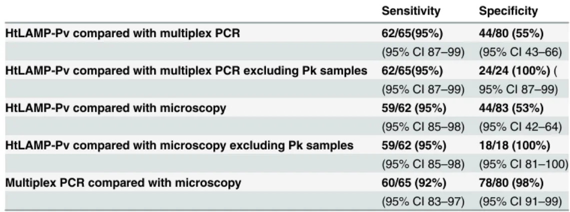
693,922) and 1 as a mixedP knowlesi/P.vivaxinfection. HtLAMP-Pv was compared with mul-tiplex PCR in these clinical samples. The sensitivity of HtLAMP-Pv forP.vivaxwas 95% (62/ 65, 95% CI 87–99) and specificity was 55% (44/80, 95% CI 43–66) respectively compared with multiplex PCR and 94% (59/62, 95% CI 85–98) and 53% (44/83, 95% CI 42–64) respectively compared with expert microscopy (Table 2). The low specificity of the assay can be attributed
Fig 3. EstimatedPlasmodium vivaxcox1 copy number based on comparison with two single copy genes,pv aldolase1andpvmdr1.
to cross-reactivity of the VIV2 primers withP.knowlesi, with 97% sequence homology at the
cox1gene betweenP.vivaxandP.knowlesi. WhenP.knowlesisamples were excluded from the analysis, the specificity was 100% compared with both multiplex PCR and microscopy (Table 6).
Sensitivity and specificity of HtLAMP-Pv in asymptomatic, reactive
active case detection community control patients
HtLAMP-Pv was compared with nested PCR for red cell pellet samples from asymptomatic, microscopy negative community controls from the malaria endemic district of Kota Marudu, Sabah, Malaysia. The sensitivity of HtLAMP-Pv was 71% (95% CI 29–96; 5/7) and specificity was 93% (95% CI 87–97; 98/105) compared with PCR.
Table 3. Analytical sensitivity of HtLAMP-Pv, compared with other publishedP.vivaxLAMP primers, using a DNA dilution series of clinical sample with qPCR confirmed parasitemia.Each sample was tested in duplicate in the HtLAMP platform using each of the threeP.vivaxLAMP primer sets. The dilution at which both duplicates were positive was the limit of detection for each of the primer sets in the HtLAMP platform (Pos*indicates dilutions at which only one of the duplicates was positive). HtLAMP-Pv was able to detect 1.4 parasites/μL compared with previously publishedP.vivaxLAMP primers.
Sample Quantitative PCR (parasites/μL calculated) [23] 18s rRNA target [30] Pvr64 target HtLAMP-Pvcox1target
1 Original 90,000 Pos Pos Pos
2 45,000 Pos Pos Pos
3 22,500 Pos Pos Pos
4 11,250 Pos Pos Pos
5 5625 Pos Pos Pos
6 2813 Pos Pos Pos
7 1406 Pos Pos Pos
8 703 Pos Pos Pos
9 352 Pos* Pos Pos
10 176 Pos Pos
11 88 Pos
12 44 Pos
13 22 Pos
14 11 Pos
15 5.5 Pos
16 2.7 Pos* Pos
17 1.4 Pos
18 0.7 Pos*
19 0.3
doi:10.1371/journal.pntd.0004443.t003
Table 4. Analytical sensitivity of HtLAMP-Pv on microscopy-determined whole bloodP.vivaxdilution series.The limit of detection HtLAMP-Pv is 2 parasites/μL, performed in duplicate and where both
dupli-cates were positive.
P.vivaxParasites /μL HtLAMP-Pv
2000 Pos
200 Pos
20 Pos
2 Pos
0.2 Neg
0 Neg
Turnaround time of assay
The HtLAMP assay turnaround time was 1 hour after DNA extraction, 4 hours when com-bined with whole blood chelex saponin protocol and 6 hours when comcom-bined with filter paper rapid DNA extraction protocol.
Applicability of the assay in a non-reference laboratory in a resource
limited setting
HtLAMP-Pv testing of a total of 149 filter paper samples was performed successfully in a regional hospital laboratory in Kota Marudu district, Sabah, Malaysia. Good workflow set up ensured that there was no contamination despite the lack of formal molecular diagnostic infra-structure. Locally trained staff was able to perform and interpret results of the HtLAMP-Pv assay using only a centrifuge, pipettes, water bath and a portable spectrophotometer.
Discussion
Field-applicable diagnostic tools for the detection ofPlasmodium vivaxare essential compo-nents for the malaria eradication agenda [44]. Given the widespread distribution and unique challengesP.vivaxposes compared withP.falciparum, there is a pressing need for the
Table 5. Comparing microscopy and RDT with the limit of detection of HtLAMP-Pv using filter paper and whole blood at 10μL and 50μL volumes.
0.75 parasites/μL 1.5 parasites/μL 3.0 parasites/μL 6.0 parasites/μL 12 parasites/μL
Microscopy Neg Neg Pos Pos Pos
RDT Neg Neg Neg Neg Neg
Filter paper (5μL) Neg Neg Neg Neg Pos
Whole blood 10μL Neg Neg Pos Pos Pos
Whole blood 50μL Neg Pos Pos Pos Pos
The limit of detection (LOD) of the HtLAMP-Pv assay depends on the starting sample material and the volume of blood extracted. LOD threshold was determined by the presence of 2 positive duplicate tests (ie: 4 positive results) at a particular parasitemia. The LOD for a 5μLfilter paper blood spot
extracted using a chelex-saponin protocol was more than 12 parasites/μL. The LOD for 10μL of whole blood extracted using a chelex-saponin protocol
was 3 parasites/μL compared with 50μL of whole blood which was 1.5 parasites/μL.
doi:10.1371/journal.pntd.0004443.t005
Table 6. Sensitivity and specificity of HtLAMP-Pv forP.vivaxin symptomatic patients in Sabah withP.
falciparum,P.vivaxandP.knowlesi.149 filter paper samples were tested by HtLAMP-Pv; 4 samples were
excluded due to a lack of PCR and microscopy data. Among the 145 samples, PCR confirmation of species was as follows: n = 64P.vivaxn = 56P.knowlesi, n = 17P.falciparum, n = 7P.malariaeand n = 1 mixedP.
vivax/P.knowlesiinfection.
Sensitivity Specificity
HtLAMP-Pv compared with multiplex PCR 62/65(95%) 44/80 (55%)
(95% CI 87–99) (95% CI 43–66)
HtLAMP-Pv compared with multiplex PCR excluding Pk samples 62/65(95%) 24/24 (100%)( (95% CI 87–99) 95% CI 87–99)
HtLAMP-Pv compared with microscopy 59/62 (95%) 44/83 (53%)
(95% CI 85–98) (95% CI 42–64)
HtLAMP-Pv compared with microscopy excluding Pk samples 59/62 (95%) 18/18 (100%)
(95% CI 85–98) (95% CI 81–100)
Multiplex PCR compared with microscopy 60/65 (92%) 78/80 (98%)
(95% CI 83–97) (95% CI 91–99)
development of species- specific molecular diagnostic tools. LAMP is a molecular diagnostic tool which holds much promise in terms of its ability to be deployed in non-referral laboratory settings, given its simplicity and rapid assay turnaround time, ability to be performed on crudely extracted DNA from both whole blood and filter paper and lack of expensive equip-ment. The colourimetric, 96 well microtitre plate-based platform for performing LAMP (HtLAMP) for the detection ofPlasmodiumparasites, as previously described [43], increases the throughput of the LAMP using minimal equipment. The objective of this paper was to develop and validate aP.vivaxspecific HtLAMP assay on this platform with good diagnostic accuracy.
The 6-kb mitochondrial genome of the genusPlasmodiumencodes three mitochondrial proteins- cytochrome B (cytb) and subunit 1 and 3 of cytochrome c oxidase (cox1andcox3), and is estimated to be present in relatively high copy number. The complete mitochondrial genome ofP.vivax(Genbank AY598035) has been shown to be closely related toP.knowlesi
[45]. Previously published standard PCR primers forP.vivaxtargetingcox1have shown 100% sensitivity and specificity [46], but were not evaluated againstP.knowlesi. LAMP primers tar-geting mitochondrial sequences for the detection ofP.genusandP.falciparumdemonstrated an analytical sensitivity of 5 parasites/μL [27] suggesting that mitochondrial DNA offers an
attractive target, presumably due to increased copy number of mitochondrial targets within cells. Recent estimates of genomic sequence coverage have shown that theP.falciparum
genome contains ~20 copies/cell of the mitochondrial genome [47].
This HtLAMP-Pv assay targeting the conservedcox1gene demonstrated excellent analytic sensitivity, being able to detect 1.4 parasites/μL. This is the lowest LOD so far achieved for a
publishedP.vivax–specific LAMP assay. The estimated copy number forcox1inP.vivaxis approximately 11 copies/ cell. Therefore, it is likely that the sensitivity of the HtLAMP-Pv assay is a reflection of the increased number of mitochondrial targets per cell. Previously pub-lishedP.vivaxLAMP primers, which targeted non-mitochondrial genes, when used in the HtLAMP platform had limits of detection of 706 parasites/μL and 176 parasites/μL which
cor-related well with published limits of detection of 125–500 parasites/μL for theseP.vivax
prim-ers sets [30].
Thepkcox1gene ofP.knowlesiexhibits 97% sequence identity withpvcox1at the nucleotide level, and thus the cross-reactivity of the VIV2 primers between these two species was expected. However, there was no cross-reactivity withP.falciparum(87% sequence identity),P.ovale wallikeri(92%),P.ovale curtisi(92%) orP.malariae(93%).
Validation of the HtLAMP-Pv in clinical samples of symptomatic patients with vivax, fal-ciparum and knowlesi malaria demonstrated sensitivity forP.vivaxof 94–95% and a specificity of 53–55% compared with microscopy and multiplex PCR respectively. The poor specificity however was a reflection of the cross-reactivity withP.knowlesi. WhenP.knowlesisamples were excluded from the analysis, the specificity of the HtLAMP-Pv assay improved to 100%, compared with both multiplex PCR and microscopy.
While this cross-reactivity appears to be a limitation of this HtLAMP-Pv assay,P.knowlesi
malaria is uncommon or absent in most areas ofP.vivaxendemicity, so this would be an important consideration only in Malaysia, whereP.knowlesipredominates [48] and in the other countries in south-east Asia whereP.knowlesihuman infection has been documented [49]. In terms of treatment, bothP.vivaxandP.knowlesirespond to artemisinin-based combi-nation therapy (ACT) [50]. However, in elimination programmes utilising primaquine for rad-ical cure ofP.vivaxmalaria, there is a potential risk of inappropriate use of this potentially haemolytic drug in people withP.knowlesiinfections. This is a problem localised to Southeast Asia, and would not pose a problem for LAMP-based detection and radical treatment ofP.
The HtLAMP-Pv assay was also evaluated in a limited sample set of asymptomatic, micros-copy negative, community control patients enrolled from the same village as a case patient, as a result of reactive active case detection. Although the LOD of HtLAMP-Pv appears to be 1.4 parasites/μL, in this sample set its sensitivity was only 71%. This may be due to the very low
parasitemias in these 7 PCR positive samples or variability due to stochastic effects at such low parasitemias. Therefore further validation in a larger sample set is required to confirm the sen-sitivity of HtLAMP-Pv in this population in order to evaluate the potential role for
HtLAMP-Pv as a diagnostic tool in malaria elimination settings.
HtLAMP-Pv showed significantly better sensitivity than the SD Bioline Pf/Pan RDT at low parasitemia. RDTs were negative in the serially diluted samples at 12 parasites/μL.
HtLAMP-Pv was positive at this level. The analytical sensitivity of the assay varied depending on whether filter paper samples or whole blood was used irrespective of the chelex-saponin DNA extraction protocol used. This may have important implications for choosing the type of sample collected in addition to choosing the appropriate diagnostic tool for surveillance or screening policy and protocols for malaria elimination programs.
In this study we also demonstrated that HtLAMP-Pv performed well in the 96-well microti-tre plate platform for increasing the throughput of the assay in a non-referral laboratory in a district hospital in Sabah, Malaysia. DNA extraction was performed in the non-referral labora-tory using a chelex protocol on filter paper blood spots and the HtLAMP-Pv assay was able to process these samples in a simple water-bath within one hour from time of DNA extraction. Positive and negative results were readily identified by two locally trained staff by visual inspec-tion of the colour change. Optical densitometry readings at 600 nm in portable photospectrom-eter were used to provide objective confirmation of the visually detected results. As such the validation of this HtLAMP-Pv assay in a rural district laboratory setting confirms the potential it has as a field-applicable molecular diagnostic tool. Furthermore, the process of assay valida-tion using the combinavalida-tion of visual and optical densitometry values has previously shown that the visually detectable colour change was reliable for determining both positive and negative results [43]. Therefore while the photospectrometer offers objective confirmation, it is not an essential component of the assay.
Some of the limitations to this platform pertain to DNA extraction. Firstly, in order to maintain cost effectiveness of the assay, modified chelex-based protocols were used for whole blood and filter paper extractions. Although these multi-step DNA extraction processes, which relied on a centrifuge, were performed adequately in a resource limited setting, further simplifi-cation of DNA extraction would enhance the feasibility of the HtLAMP-Pv assay. It would also allow a greater number of samples to be processed, as might be required for mass surveillance for malaria elimination, thereby making full use of the high throughput aspect of the HtLAMP platform. Secondly, while equivalent small volumes of blood on filter paper and whole blood have shown whole blood to produce better analytical sensitivity in the HtLAMP platform [43], the limit of detection of HtLAMP-Pv using larger volumes of blood on filter paper is yet to be established.
Acknowledgments
We thank Kim Piera (Menzies School of Health Research) for providing theP.vivaxdilution series and sample shipments. We thank Marina Chavchich (Australian Army Malaria Insti-tute) for providing the plasmid containingpvmdr1andpvaldolase1genes and for assay assistance.
Author Contributions
Conceived and designed the experiments: SB JSM QC TW MJG NMA CP KF CD. Performed the experiments: SB CP KF MJG. Analyzed the data: SB QC JSM CP CJS. Contributed reagents/materials/analysis tools: SB QC JSM NMA MJG TW KF CBP CP CD. Wrote the paper: SB QC JSM NMA MJG KF CBP CP CJS.
References
1. Gething PW, Elyazar IR, Moyes CL, Smith DL, Battle KE, et al. (2012) A long neglected world malaria map: Plasmodium vivax endemicity in 2010. PLoS Negl Trop Dis 6: e1814. doi:10.1371/journal.pntd. 0001814PMID:22970336
2. Anstey NM, Douglas NM, Poespoprodjo JR, Price RN (2012) Plasmodium vivax: clinical spectrum, risk factors and pathogenesis. Adv Parasitol 80: 151–201. doi:10.1016/B978-0-12-397900-1.00003-7 PMID:23199488
3. WHO (2014) WHO Global Malaria Program—World Malaria Report 2014. Switzerland.
4. Harris I, Sharrock WW, Bain LM, Gray KA, Bobogare A, et al. (2010) A large proportion of asymptomatic Plasmodium infections with low and sub-microscopic parasite densities in the low transmission setting of Temotu Province, Solomon Islands: challenges for malaria diagnostics in an elimination setting. Malar J 9: 254. doi:10.1186/1475-2875-9-254PMID:20822506
5. Baum E, Sattabongkot J, Sirichaisinthop J, Kiattibutr K, Davies DH, et al. (2015) Submicroscopic and asymptomatic Plasmodium falciparum and Plasmodium vivax infections are common in western Thai-land—molecular and serological evidence. Malar J 14: 611.
6. Thanh PV, Van Hong N, Van Van N, Van Malderen C, Obsomer V, et al. (2015) Epidemiology of forest malaria in Central Vietnam: the hidden parasite reservoir. Malar J 14: 601.
7. Cheng Q, Cunningham J, Gatton ML (2015) Systematic review of sub-microscopic P. vivax infections: prevalence and determining factors. PLoS Negl Trop Dis 9: e3413. doi:10.1371/journal.pntd.0003413 PMID:25569135
8. Okell LC, Ghani AC, Lyons E, Drakeley CJ (2009) Submicroscopic infection in Plasmodium falciparum-endemic populations: a systematic review and meta-analysis. J Infect Dis 200: 1509–1517. doi:10. 1086/644781PMID:19848588
9. Price R, Tjira E, Guerra C, Yeung S, White N, et al. (2007) Vivax malaria: Neglected but not benign. American Journal of Tropical Medicine and Hygiene 77: 79–87. PMID:18165478
10. Mikhail AF, Leslie TJ, Mayan MI, Zekria R, Mohammad N, et al. (2011) Field trial of three different Plas-modium vivax-detecting rapid diagnostic tests with and without evaporative cool box storage in Afghan-istan. Malar J 10: 169. doi:10.1186/1475-2875-10-169PMID:21696587
11. WHO (2014) Malaria Rapid Diagnostic Test Performance- Summary results of WHO product testing of malaria RDTs: Round 1–5 (2008–2013). Italy: WHO.
12. Tiono AB, Ouedraogo A, Diarra A, Coulibaly S, Soulama I, et al. (2014) Lessons learned from the use of HRP-2 based rapid diagnostic test in community-wide screening and treatment of asymptomatic car-riers of Plasmodium falciparum in Burkina Faso. Malar J 13: 30. doi:10.1186/1475-2875-13-30PMID: 24467946
13. Cook J, Xu W, Msellem M, Vonk M, Bergstrom B, et al. (2015) Mass screening and treatment on the basis of results of a Plasmodium falciparum-specific rapid diagnostic test did not reduce malaria inci-dence in Zanzibar. J Infect Dis 211: 1476–1483. doi:10.1093/infdis/jiu655PMID:25429102
14. Mori Y, Nagamine K, Tomita N, Notomi T (2001) Detection of loop-mediated isothermal amplification reaction by turbidity derived from magnesium pyrophosphate formation. Biochem Biophys Res Com-mun 289: 150–154. PMID:11708792
16. Goto M, Honda E, Ogura A, Nomoto A, Hanaki K (2009) Colorimetric detection of loop-mediated iso-thermal amplification reaction by using hydroxy naphthol blue. Bio Techniques 46: 1637–1172.
17. Wastling SL, Picozzi K, Kakembo ASL, Welburn SC (2010) LAMP for Human African Trypanosomiasis: A Comparative study of detection formats. PLoS Negl Trop Dis 4: e865. doi:10.1371/journal.pntd. 0000865PMID:21072228
18. Yamamura M, Makimura K, Ota Y (2009) Evaluation of a new rapid molecular diagnostic system for Plasmodium falciparum combined with DNA filter paper, loop-mediated isothermal amplification, and melting curve analysis. Jpn J Infect Dis 62: 20–25. PMID:19168954
19. Kiddle G, Hardinge P, Buttigieg N, Gandelman O, Pereira C, et al. (2012) GMO detection using a biolu-minescent real time reporter (BART) of loop mediated isothermal amplification (LAMP) suitable for field use. BMC Biotechnol 12: 15. doi:10.1186/1472-6750-12-15PMID:22546148
20. Yongkiettrakul S, Jaroenram W, Arunrut N, Chareanchim W, Pannengpetch S, et al. (2014) Application of loop-mediated isothermal amplification assay combined with lateral flow dipstick for detection of Plasmodium falciparum and Plasmodium vivax. Parasitol Int 63: 777–784. doi:10.1016/j.parint.2014. 06.004PMID:25038579
21. Lucchi NW, Demas A, Narayanan J, Sumari D, Kabanywanyi A, et al. (2010) Real-time fluorescence loop mediated isothermal amplification for the diagnosis of malaria. PLoS One 5: e13733. doi:10. 1371/journal.pone.0013733PMID:21060829
22. Sema M, Alemu A, Bayih A, Getie S, Getnet G, et al. (2015) Evaluation of non-instrumented nucleic acid amplification by loop-mediated isothermal amplification (NINA-LAMP) for the diagnosis of malaria in Northwest Ethiopia. Malar J 14: 44. doi:10.1186/s12936-015-0559-9PMID:25626339
23. Han E, Watanabe R, Sattabongkot J, Khuntirat B, Sirichaisinthop J, et al. (2007) Detection of four Plas-modium species by genus and species- specific loop mediated isothermal amplication for clinical diag-nosis. Journal of Clinical Microbiology 45: 2521–2528. PMID:17567794
24. Lau YL, Fong MY, Mahmud R, Chang PY, Palaeya V, et al. (2011) Specific, sensitive and rapid detec-tion of human Plasmodium knowlesi infecdetec-tion by loop-mediated isothermal amplificadetec-tion (LAMP) in blood samples. Malar J 10: 197. doi:10.1186/1475-2875-10-197PMID:21774805
25. Iseki H, Kawai S, Takahashi N, Hirai M, Tanabe K, et al. (2010) Evaluation of a loop-mediated isother-mal amplification method as a tool for diagnosis of infection by the zoonotic simian isother-malaria parasite Plasmodium knowlesi. J Clin Microbiol 48: 2509–2514. doi:10.1128/JCM.00331-10PMID:20444968
26. Poon LL, Wong BW, Ma EH, Chan KH, Chow LM, et al. (2006) Sensitive and inexpensive molecular test for falciparum malaria: detecting Plasmodium falciparum DNA directly from heat-treated blood by loop-mediated isothermal amplification. Clin Chem 52: 303–306. PMID:16339303
27. Polley SD, Mori Y, Watson J, Perkins MD, Gonzalez IJ, et al. (2010) Mitochondrial DNA targets increase sensitivity of malaria detection using loop-mediated isothermal amplification. J Clin Microbiol 48: 2866–2871. doi:10.1128/JCM.00355-10PMID:20554824
28. Cook J, Aydin-Schmidt B, Gonzalez IJ, Bell D, Edlund E, et al. (2015) Loop-mediated isothermal ampli-fication (LAMP) for point-of-care detection of asymptomatic low-density malaria parasite carriers in Zan-zibar. Malar J 14: 43. doi:10.1186/s12936-015-0573-yPMID:25627037
29. Vallejo AF, Martinez NL, Gonzalez IJ, Arevalo-Herrera M, Herrera S (2015) Evaluation of the loop medi-ated isothermal DNA amplification (LAMP) kit for malaria diagnosis in P. vivax endemic settings of Colombia. PLoS Negl Trop Dis 9: e3453. doi:10.1371/journal.pntd.0003453PMID:25569550
30. Patel JC, Oberstaller J, Xayavong M, Narayanan J, Debarry JD, et al. (2013) Real-Time Loop-Mediated Isothermal Amplification (RealAmp) for the Species-Specific Identification of Plasmodium vivax. PLoS One 8: e54986. doi:10.1371/journal.pone.0054986PMID:23349994
31. Dinzouna-Boutamba SD, Yang HW, Joo SY, Jeong S, Na BK, et al. (2014) The development of loop-mediated isothermal amplification targeting alpha-tubulin DNA for the rapid detection of Plasmodium vivax. Malar J 13: 248. doi:10.1186/1475-2875-13-248PMID:24981710
32. McCarthy JS, Griffin PM, Sekuloski S, Bright AT, Rockett R, et al. (2013) Experimentally induced blood-stage Plasmodium vivax infection in healthy volunteers. J Infect Dis 208: 1688–1694. doi:10.1093/ infdis/jit394PMID:23908484
33. Grigg MJ, William T, Drakeley CJ, Jelip J, von Seidlein L, et al. (2014) Factors that are associated with the risk of acquiring Plasmodium knowlesi malaria in Sabah, Malaysia: a case-control study protocol. BMJ Open 4: e006004. doi:10.1136/bmjopen-2014-006004PMID:25149186
34. Plowe CV, Djimde A, Bouare M, Doumbo O, Wellems TE (1995) Pyrimethamine and proguanil resis-tance-conferring mutations in Plasmodium falciparum dihydrofolate reductase: polymerase chain reac-tion methods for surveillance in Africa. Am J Trop Med Hyg 52: 565–568. PMID:7611566
36. Suwanarusk R, Russell B, Chavchich M, Chalfein F, Kenangalem E, et al. (2007) Chloroquine resistant Plasmodium vivax: in vitro characterisation and association with molecular polymorphisms. PLoS One 2: e1089. PMID:17971853
37. Grigg MJ, William T, Dhanaraj P, Menon J, Barber BE, et al. (2014) A study protocol for a randomised open-label clinical trial of artesunate-mefloquine versus chloroquine in patients with non-severe Plas-modium knowlesi malaria in Sabah, Malaysia (ACT KNOW trial). BMJ Open 4: e006005. doi:10.1136/ bmjopen-2014-006005PMID:25138814
38. Barber BE, William T, Grigg MJ, Menon J, Auburn S, et al. (2013) A prospective comparative study of knowlesi, falciparum, and vivax malaria in Sabah, Malaysia: high proportion with severe disease from Plasmodium knowlesi and Plasmodium vivax but no mortality with early referral and artesunate ther-apy. Clin Infect Dis 56: 383–397. doi:10.1093/cid/cis902PMID:23087389
39. Snounou G, Viriyakosol S, Zhu XP, Jarra W, Pinheiro L, et al. (1993) High sensitivity of detection of human malaria parasites by the use of nested polymerase chain reaction. Mol Biochem Parasitol 61: 315–320. PMID:8264734
40. Padley D, Moody AH, Chiodini PL, Saldanha J (2003) Use of a rapid, single-round, multiplex PCR to detect malarial parasites and identify the species present. Ann Trop Med Parasitol 97: 131–137. PMID: 12803868
41. Imwong M, Tanomsing N, Pukrittayakamee S, Day NP, White NJ, et al. (2009) Spurious amplification of a Plasmodium vivax small-subunit RNA gene by use of primers currently used to detect P. knowlesi. J Clin Microbiol 47: 4173–4175. doi:10.1128/JCM.00811-09PMID:19812279
42. Singh B, Bobogare A, Cox-Singh J, Snounou G, Abdullah MS, et al. (1999) A genus- and species-spe-cific nested polymerase chain reaction malaria detection assay for epidemiologic studies. Am J Trop Med Hyg 60: 687–692. PMID:10348249
43. Britton S, Cheng Q, Sutherland CJ, McCarthy JS (2015) A simple, high-throughput, colourimetric, field applicable loop-mediated isothermal amplification (HtLAMP) assay for malaria elimination. Malar J 14: 335. doi:10.1186/s12936-015-0848-3PMID:26315027
44. MalERA (2011) A research agenda for malaria eradication: diagnoses and diagnostics. PLoS Med 8: e1000396. doi:10.1371/journal.pmed.1000396PMID:21311583
45. Jongwutiwes S, Putaporntip C, Iwasaki T, Ferreira MU, Kanbara H, et al. (2005) Mitochondrial genome sequences support ancient population expansion in Plasmodium vivax. Mol Biol Evol 22: 1733–1739. PMID:15901839
46. Cunha MG, Medina TS, Oliveira SG, Marinho AN, Povoa MM, et al. (2009) Development of a Polymer-ase Chain Reaction (PCR) method bPolymer-ased on amplification of mitochondrial DNA to detect Plasmodium falciparum and Plasmodium vivax. Acta Trop 111: 35–38. doi:10.1016/j.actatropica.2009.02.003 PMID:19426660
47. Preston MD, Campino S, Assefa SA, Echeverry DF, Ocholla H, et al. (2014) A barcode of organellar genome polymorphisms identifies the geographic origin of Plasmodium falciparum strains. Nat Com-mun 5: 4052. doi:10.1038/ncomms5052PMID:24923250
48. William T, Jelip J, Menon J, Anderios F, Mohammad R, et al. (2014) Changing epidemiology of malaria in Sabah, Malaysia: increasing incidence of Plasmodium knowlesi. Malar J 13: 390. doi:10.1186/ 1475-2875-13-390PMID:25272973
49. Moyes CL, Henry AJ, Golding N, Huang Z, Singh B, et al. (2014) Defining the geographical range of the Plasmodium knowlesi reservoir. PLoS Negl Trop Dis 8: e2780. doi:10.1371/journal.pntd.0002780 PMID:24676231
![Table 1. Primers used to amplify Pv mdr, aldolase and cox 1 (*Source [ 36]).](https://thumb-eu.123doks.com/thumbv2/123dok_br/16323597.187692/6.918.47.877.134.273/table-primers-used-amplify-pv-mdr-aldolase-source.webp)