Accumulation of Pathological Prion Protein
PrP
Sc
in the Skin of Animals
with Experimental and Natural Scrapie
Achim Thomzig1[*, Walter Schulz-Schaeffer2[, Arne Wrede2, Wilhelm Wemheuer3, Bertram Brenig3, Christine Kratzel1, Karin Lemmer1, Michael Beekes1*
1P24 Transmissible Spongiform Encephalopathies, Robert Koch-Institut, Berlin, Germany, 2 Prion and Dementia Research Unit, Department of Neuropathology, Universita¨tsklinikum Go¨ttingen, Go¨ttingen, Germany,3Institute of Veterinary Medicine, Georg-August-Universita¨t Go¨ttingen, Go¨ttingen, Germany
Prion infectivity and its molecular marker, the pathological prion protein PrPSc, accumulate in the central nervous system and often also in lymphoid tissue of animals or humans affected by transmissible spongiform encephalopathies. Recently, PrPSc was found in tissues previously considered not to be invaded by prions (e.g., skeletal muscles). Here, we address the question of whether prions target the skin and show widespread PrPSc deposition in this organ in hamsters perorally or parenterally challenged with scrapie. In hamsters fed with scrapie, PrPScwas detected before the onset of symptoms, but the bulk of skin-associated PrPScaccumulated in the clinical phase. PrPScwas localized in nerve fibres within the skin but not in keratinocytes, and the deposition of PrPScin skin showed no dependence from the route of infection and lymphotropic dissemination. The data indicated a neurally mediated centrifugal spread of prions to the skin. Furthermore, in a follow-up study, we examined sheep naturally infected with scrapie and detected PrPScby Western blotting in skin samples from two out of five animals. Our findings point to the skin as a potential reservoir of prions, which should be further investigated in relation to disease transmission.
Citation: Thomzig A, Schulz-Schaeffer W, Wrede A, Wemheuer W, Brenig B, et al. (2007) Accumulation of pathological prion protein PrPScin the skin of animals with experimental and natural scrapie. PLoS Pathog 3(5): e66. doi:10.1371/journal.ppat.0030066
Introduction
Transmissible spongiform encephalopathies (TSEs), or prion diseases, are fatal neurodegenerative diseases affecting both animals and humans. According to the prion hypothesis, TSEs are caused by infectious prions that consist essentially— if not entirely— of a misfolded form of the prion protein (PrP), which is known as PrPSc [1]. Although the precise molecular composition and structure of prions remains elusive, PrPSchas been shown in many studies to accumulate together with infectivity in target tissues of infection and is therefore considered a reliable biochemical marker for TSE agents [2] as reported for experimentally challenged hamsters [3], other animal species [4], and humans [5,6].
Scrapie of sheep and goats, chronic wasting disease (CWD) of deer, bovine spongiform encephalopathy (BSE) of cattle, and variant Creutzfeldt-Jakob disease (vCJD) of humans represent acquired prion diseases that are caused by exposure to TSE agents in the living environment of the respective host. Different lines of evidence suggest that many, if not the majority, of cases of ovine scrapie, BSE, and purportedly CWD are caused by ingestion of prions and subsequent invasion of the organism via the alimentary tract [7]. This also holds true for vCJD, which is now generally acknowledged to be acquired through consumption of BSE-contaminated foodstuffs [8].
Although the exact mechanism of infection following passage of prions through the alimentary tract has not yet been completely elucidated, findings from different mamma-lian species suggested that the infection ascended retrograd-ually via peripheral nerves to the spinal cord and to the brain
(for reviews see [2,7]). From these sites of initial central nervous system invasion at the level of the thoracic spinal cord and the medulla oblongata, the infection propagated in both ascending and descending directions [2,3,7,9–11]. Centrifugal spread from the central nervous system appeared to be responsible for subsequent infection of further parts of the peripheral nervous system [9,11]. In particular, PrPScwas found associated with nerve fibres or nerve endings innervat-ing peripheral organs and tissues such as muscles [11–14]. This prompted us to look for further tissues which could serve as reservoirs of prions in the mammalian body, and from which these pathogens could be potentially dissemi-nated into the environment and transmitted to other individuals via peroral or alternative routes. In this context, the skin appears to be of utmost importance. The skin
Editor:Neil Mabbott, Institute for Animal Health, United Kingdom
ReceivedFebruary 5, 2007;AcceptedMarch 20, 2007;PublishedMay 25, 2007
Copyright:Ó2007 Thomzig et al. This is an open-access article distributed under the terms of the Creative Commons Attribution License, which permits unrestricted use, distribution, and reproduction in any medium, provided the original author and source are credited.
Abbreviations:BSE, bovine spongiform encephalopathy; BSE-H, hamster-adapted BSE; CWD, chronic wasting disease; dpi, days post infection; f.p., foot pad; H&E, haematoxylin and eosin; i.c., intracerebral; PET, paraffin-embedded tissue; p.o., oral; PrP, prion protein; PrP27–30, protease-resistant core of the pathological prion protein PrPSc; PrPC, cellular isoform of the prion protein; PrPSc, pathological isoform of the prion protein; s.w., steel wires; TSE, transmissible spongiform encephalop-athy; vCJD, variant Creutzfeldt-Jakob disease
* To whom correspondence should be addressed. E-mail: [email protected] (AT); [email protected] (MB)
consists of different strata and appendages which are highly innervated and interspersed with lymphatics and blood vessels [15]. It constitutes the largest organ of humans and many animal species and provides an interface with their environment. However, although PrPSc detection has been reported for mucosal tissue [16,17], the skin has not been extensively studied for the presence of prions and PrPScso far. In 2004, Cunningham et al. reported on the presence of BSE agent in a wide range of tissues from a BSE-infected greater kudu [18]. In one animal of this study, the salivary gland and skin were found to contain infectivity, and the authors suggested that these findings possibly indicate routes by which direct animal-to-animal transmission of the disease may occur.
Here, we examined the skin of prion-infected hamsters for the presence of PrPSc. Our hamster experiments focussed on orally infected animals, which have been previously estab-lished as a relevant rodent model to study the spread of prions in the peripheral nervous system [2,7]. These studies were performed in order to (i) investigate whether anatom-ical structures within the skin may provide a target for PrPSc accumulation, (ii) elucidate the identity of such skin components, and (iii) find out whether prions can be present in the skin prior to the onset of visible TSE symptoms. In a proof-of-concept approach, we extended PrPSctesting of the skin to specimens from sheep naturally infected with scrapie. This follow-up study intended to obtain further insights into the pathophysiology of scrapie and the putative pathways of its natural transmission in the field.
Results
PrPScAccumulation in the Skin following Peroral Infection with Scrapie Becomes Detectable Shortly before the Onset of Clinical Symptoms
To investigate whether and at which stages of scrapie infection PrPScaccumulates in the skin, we performed a time-course study in hamsters orally exposed to 263K scrapie
agent. As established previously for the examination of muscle tissue [11], PrPScwas visualized by sensitive Western blotting after extraction of the protein in the form of its protease-resistant core, PrP27–30, from skin specimens by a high-yield purification method.
We examined samples of skin tissue from hamsters taken at five different time points, i.e., at 70, 100, and 130 days post infection (dpi) in the pre-clinical phase of incubation, at the onset of clinical symptoms, and at the end stage of disease, which occurred after 164 6 6 d (expressed as the mean 6 standard deviation [SD]; n ¼ 5 ). Skin samples from the following five body regions of each hamster were analyzed: the forelimb, the hindlimb, the abdomen, the back, and the head.
PrP27–30 could not be detected in any of the examined skin samples at 70 and 100 dpi from five animals each (Figure 1A and 1B). The earliest unambiguous signals for accumu-lation of the pathological prion protein PrPScwere found in skin samples from three out of five animals at 130 dpi in the late pre-clinical phase of incubation, corresponding to about 80% of the mean incubation period until terminal disease (Figure 1C, Western blot on the right-hand side, lanes S1, S2, S4, and S5). However, variable combinations of PrPSc-positive skin samples from different regions of the body were found at 130 dpi, indicating individual variation in the spread of infection to, or inhomogeneous distribution of PrPScin skin tissue. Possibly, prion infection of the skin could have been detected more frequently in animals at 130 dpi, or at earlier pre-clinical stages of incubation, by using alternative method-ologies such as the conformation-dependent immunoassay [19] or bioassays. At the onset of clinical symptoms, all of the analyzed skin specimens from all five examined hamsters displayed more or less strong signals for PrP27–30 (Figure 1D). At the terminal stage of scrapie, the positive signals for PrP27–30 become more intense, suggesting that accumula-tion of PrPSc takes place predominantly in relatively late stages of incubation (Figure 1E, lanes S1–S5). The weight of the tested skin samples ranged from approximately 40 to 100 mg as specified in the legend to Figure 1A–1E. In order to verify that the detected bands originated from PrPSc, a control experiment was performed: After deglycosylation with PNGaseF, the PrP27–30 bands showed an electropho-retic shift towards a single band at about 20 kDa, the molecular weight to be expected for the unglycosylated PrP27–30 form of 263K hamster scrapie (Figure 1F, lanes S1d–S5d). Control samples from mock-challenged age-matched hamsters consistently produced negative results (not shown). A scale displaying an overview of the time-points at which the p.o.-infected hamsters were tested for skin-associated PrPSc deposition in relation to the mean incubation period and the pre-clinical and clincal phases of incubation is provided in Figure 1G.
Location of PrPSc within the Skin
To determine where in the skin PrPSc accumulates, we investigated samples from the head, snout, forelimb, and abdomen of orally 263K scrapie–infected, terminally ill hamsters. As done previously when determining the route of PrPSc propagation to muscles [11], we used the paraffin-embedded tissue (PET) blot method to achieve a sensitive topographical localisation of disease-associated PrP in the skin. Using either Carnoy- or paraformaldehyde-fixed tissue
Author Summary
samples, PrPScwas detectable in (i) free nerve endings of the subepidermal plexus on the border of the epidermis to the dermis (Figure 2A, 2B, 2G, and 2H, arrows), (ii) fibres of the subepidermal, the deep cutaneous, and the subcutaneous plexus, (iii) fibres of the follicular neural network of the hair (circular and longitudinal fibres, Figure 2A, 2B, and 2G, arrowheads), (iv) the hair follicle isthmus (Figure 2G,
rhombus), and (v) small intradermal striated fibres of mimic muscles (Figure 2A and 2B, asterisks). No PrPScwas detectable in keratinocytes, epidermal basal cells, fibroblasts of the dermal connective tissue, capillary blood vessels, outer root sheet cells of the hair, or the bulge region and the sebaceous gland, but PrPScwas present in nerve fibres of the sebaceous gland (not shown). Nerve fibres in the skin can be labelled by Figure 1.Time-Course of PrPScDeposition in Skin Tissue
(A–E) Western blot detection of PrP27–30, the protease-resistant core of PrPSc, extracted from different skin samples of hamsters orally challenged with
263K scrapie and sacrificed at the following time-points after infection: (A) 70 dpi, (B) 100 dpi, (C) 130 dpi, (D) at the onset of clinical signs for scrapie (138–146 dpi), and (E) at the terminal stage of disease (157–171 dpi). Lanes with test samples: S1, skin sample from hindlimb; S2, skin sample from forelimb; S3, skin sample from back; S4, skin sample from abdomen; S5, skin sample from head. Lanes with control samples: 1, proteinase K–digested brain homogenate from terminally ill 263K scrapie hamsters containing 13107g brain tissue. Representative results are shown for each stage of
incubation. Substantial individual variation was observed at 130 dpi, with two of five and three of five animals displaying findings as in (C) in the Western blot on the left-hand side or the Western blot on the right-hand side, respectively.
(F) Lanes S1d–S5d: Same samples as in S1–S5 of (E) but deglycosylated with PNGaseF.
(A–F) Amounts of tissue represented in lanes: (A) S1, 43 mg; S2, 52 mg; S3, 68 mg; S4, 58 mg; S5, 73 mg; (B) S1, 78 mg; S2, 44 mg; S3, 63 mg; S4, 67 mg; S5, 50 mg; ([C], Western blot on the left side) S1, 42 mg; S2, 76 mg; S3, 61 mg; S4, 58 mg; S5, 73 mg; ([C], Western blot on the right side) S1, 51 mg; S2, 63 mg; S3, 70 mg; S4, 87 mg; S5, 54 mg; (D) S1, 63 mg; S2, 68 mg; S3, 90 mg; S4, 50 mg; S5, 68 mg; (E) S1, 55 mg; S2, 73 mg; S3, 80 mg; S4, 88 mg; S5, 70 mg; (F) S1d, 12 mg; S2d, 14 mg; S3d, 19 mg; S4d, 12 mg; S5d, 20 mg.
(G) Time-scale displaying the mean incubation period and the pre-clinical and clinical phases of incubation of hamsters orally infected with 263K scrapie. Small vertical arrows indicate time-points at which animals were tested for PrPScin skin samples.
Figure 2.Location of PrPScwithin the Skin of Hamsters Orally Infected with Scrapie
(A–H) Topographical localisation of PrPScin sections of skin samples from the snout (A and B) and the forelimb (G and H); (A and G) PET blots, (B and H)
H&E staining. PrPScwas detected in free nerve endings of the subepidermal plexus on the border of the epidermis to the dermis ([A], [B], [G], and [H], arrows), in fibres of the subepidermal, the deep cutaneous and the subcutaneous plexus, in circular and longitudinal fibres of the follicular neural network of the hair ([A], [B], and [G], arrowheads), in the hair follicle isthmus ([G]; rhombus), and in small intradermal striated fibres of mimic muscles ([A and B], asterisks).
(C–F) Visualisation of PrPScand nerve fibres in the neural network of hair follicles by fluorescence microscopy (skin sample from the abdomen).
Co-localisation of PrPSc (C) with nerve fibres labelled by using an anti–S-100 protein antibody against Schwann cells (D). (E) Merged figure from
antibodies binding to neurofilament or Schwann cells [20,21]. By using an anti–S-100 protein antibody detecting Schwann cells, a co-localisation of PrPSc with nerve fibres of the follicular neuronal network of hairs was observed (Figure 2C– 2E, arrowheads). By antibody-labelling of neurofilament, nerve fibres of the cutaneous plexus were found to display a co-localisation with PrPSc (Figure 2I–2K; for topological orientation, see Figure 2L). Occasionally, we observed PrPSc deposition in nerve-like structures of the subepidermal plexus showing no immunostaining for neurofilament (not shown). This may have resulted from infection of nerve fibres that do not contain neurofilament [20], or, alternatively, from PrPScdeposition in Schwann cells rather than in the neurite itself [22]. Control skin samples from the forelimb of a
hamster perorally mock-challenged with normal hamster brain homogenate were found negative for PrPSc by PET blotting and fluorescence microscopy (Figure 2M and 2N).
Accumulation of PrPSc in the Skin Shows No Dependence on the Route of Infection and Lymphotropic Spread
In order to examine whether accumulation of PrPScin the skin depends on the mode of infection and spreading pathways other than the nervous system, skin specimens from the forelimb and hindlimb of terminally ill hamsters challenged with 263K scrapie agent by different routes were analyzed using the same analytical approach as described above for the time-course study. The following modes of inoculation were compared: oral (p.o.) infection with scrapie brain homogenate (Figure 3A, lanes 4 and 5), intracerebral (i.c.) infection with scrapie brain homogenate (Figure 3A, lanes 6 and 7), i.c. infection by implantation of steel wires (s.w.) contaminated with scrapie agent (Figure 3A, lanes 8 and 9), and peripheral foot pad (f.p.) infection with scrapie brain homogenate (Figure 3A, lanes 10 and 11). PrP27–30 could be detected in all analyzed skin specimens from hamsters at the terminal stage of scrapie independently of the route of infection. Amounts of PrPScin skin tissue were about 5,000-to 10,000-fold lower than those found in brain, as estimated from positive controls of skin samples from orally mock-infected hamsters that were spiked with 13106g, 53106g,
or 13105 g of homogenized 263K scrapie hamster brain
from terminally ill donors before extraction (Figure 3A, lanes 1–3). PrPSc was consistently absent in skin specimens from mock-infected hamsters, which served as negative controls (Figure 3A, lanes 12 and 13).
The employed routes of inoculation were found to produce substantial differences in the extent of lymphotropic spread of agent. In orally or intracerebrally challenged hamsters, spleens (Figure 3B, lanes 2 and 4), mesenterial lymph nodes (Figure 3B, lanes 6 and 8) and retropharyngeal lymph nodes (not shown) consistently showed PrPScdeposition, whereas in five out of six hamsters infected by implantation of contaminated s.w., no PrPSccould be found in the examined spleens and lymph node specimens (Figure 3B, lanes 3 and 7). Only one of the s.w.-infected animals displayed a weak Western blot signal for PrPScin the spleen (not shown). Thus, in comparison to the i.c. or p.o. route of inoculation, lymphotropic spread was much less pronounced—or even undetectable—following infection of hamsters by i.c. implan-tation of 263K-contaminated s.w. However, despite this marked discrepancy, the intensity of skin-associated deposits of PrPSc found in s.w.-infected hamsters did not show significant differences from that observed for p.o.- or i.c.-challenged animals upon Western blotting (Figure 3A, lines 8 and 9 versus lines 4–7). To determine whether the route of administration of PrPSc influences the topographical distri-bution of disease-associated prion protein in the skin, we investigated samples from hamsters infected with 263K-Figure 3. PrPSc Routing to the Skin and to Components of the
Lymphoreticular System of Hamsters Challenged via Different Routes with 263K Scrapie Agent
(A) Western blot detection of PrP27–30, the protease-resistant core of PrPSc, in skin specimens from terminally ill scrapie hamsters. Lanes 1, 2,
and 3: skin samples from orally mock-infected control hamsters, spiked before extraction with 13106g, 53106g, or 13105g of brain
homogenate from terminally ill 263K hamsters. Lanes 4 and 5: skin samples from hindlimbs and forelimbs of hamsters orally infected with scrapie brain homogenate. Lanes 6 and 7: skin samples from hindlimbs and forelimbs of hamsters intracerebrally infected with scrapie brain homogenate. Lanes 8 and 9: skin samples from hindlimbs and forelimbs of hamsters infected by implantation of s.w. contaminated with scrapie agent. Lanes 10 and 11: skin samples from hindlimbs and forelimbs of hamsters infected peripherally by f.p. inoculation of scrapie brain homogenate. Lanes 12 and 13: skin samples from hindlimbs and forelimbs of hamsters orally mock-infected with normal brain homoge-nate. Amounts of tissue represented in lanes: 1, 53mg; 2, 58 mg; 3, 68 mg; 4, 68 mg; 5, 75 mg; 6, 78 mg; 7, 64 mg; 8, 69 mg; 9, 60 mg; 10, 62 mg; 11, 73 mg; 12, 61 mg; 13, 58 mg.
(B) Western blot detection of PrP27–30 in spleens and selected lymph nodes from terminally ill scrapie hamsters. Lanes 1 and 5: proteinase K-digested brain homogenate from terminally ill scrapie hamsters, containing 13107g brain tissue. Lanes 2–4: spleen samples from
p.o.- (2), s.w.-, (3) and i.c.-infected (4) hamsters. Lanes 6–8: mesenteric lymph node samples from p.o.- (6), s.w.-, (7) and i.c.-infected (8) hamsters. Amounts of tissue represented in lanes: 2, 40 mg; 3, 45 mg; 4, 41 mg; 6, 6 mg; 7, 8 mg; 8, 6 mg.
doi:10.1371/journal.ppat.0030066.g003
(I–K) Visualisation of PrPScand nerve fibres in the cutaneous plexus by fluorescence microscopy (skin sample form the snout). Co-localisation of PrPSc(I)
with nerve fibres labelled by using the anti-neurofilament antibody SMI 31 (J). (K) Merged figure from micrographs (I and J). (L) Adjacent section to (I), stained with H&E. The box indicates the region used for the immunofluorescence stainings in (I–K).
(M and N) Control skin samples from the forelimb of a hamster perorally mock-challenged with normal hamster brain homogenate; PET blot (M) and fluorescence microscopy for PrP and neurofilament (N).
Scale bars¼200lm for (B, F, H, and M), 50lm for (K and L), and 25lm for (N). Same scale bars as displayed in (B), (F), (H), and (K) apply to (A), (C–E), (G),
and (I and J), respectively.
contaminated s.w. at the clinical disease stage using the PET blot method. This revealed that the morphological distribu-tion pattern of PrPScin the skin of s.w.-infected animals was identical to that found in orally infected hamsters (not shown).
BSE-Associated PrPSc in the Skin
To test whether not only scrapie-associated PrPScbut also BSE-associated PrPScaccumulates in the skin, hamsters were intracerebrally infected with a hamster-adapted BSE (BSE-H) agent and sacrificed at the end stage of disease. All analyzed skin specimens from forelimbs and hindlimbs of five donor animals showed substantial amounts of BSE-associated PrP27–30 (Figure 4, lanes 1 and 2). Thus, following i.c. infection, the targeting of BSE-H agent to the skin, as probed by Western blotting, did not show discernible differences compared to that observed for 263K scrapie.
PrPSc in Skin Tissue of Sheep Naturally Infected with Scrapie
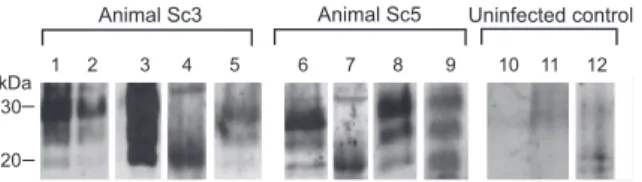
In a follow-up proof-of-concept study, which aimed to validate and expand our findings from the hamster experi-ments, five sheep naturally infected with scrapie in the field were analyzed for the presence of PrPSc in skin specimens from different body regions (head, snout, hindlimb, forelimb, perianal, axillar, and inguinal). PrPSc was visualized by Western blotting after extraction in the form of PrP27–30 from tissue samples using the anti-ovine PrP monoclonal antibodies ICSM-18 or P4. Positive specimens were found in two out of the five tested sheep: In sheep Sc3, PrP27–30 was present in a sample from the inguinal region (Figure 5, lane 5). Sheep Sc5 showed PrP27–30 in a sample from the perianal region, which was a scratching site of this animal (Figure 5, lane 6). For control purposes, a deglycosylation of a PrP27–30 extract from the same skin region of this animal was performed (Figure 5, lane 7). Furthermore, a skin sample from the snout was found positive for PrP27–30 in animal Sc5 upon detection with the ICSM-18 antibody (Figure 5, lane 8) and the anti-PrP antibody P4 (Figure 5, lane 9). All other analyzed skin specimens of the five sheep did not show specific Western blot signals for PrP27–30. Specimens from the same skin regions of four uninfected sheep served as controls and consistently produced negative results (Figure 5, lanes 10–12).
Discussion
In this study we have shown that the skin provides a reservoir for PrPSc, the biochemical marker of prion infectivity, in five different hamster TSE models,
independ-ently of whether the animals were challenged with scrapie via the p.o., i.c., or f.p. route, cerebral implantation of scrapie-contaminated s.w., or i.c. inoculation of a hamster-adapted BSE agent. Furthermore, PrPSccould be demonstrated for the first time in skin specimens from sheep naturally infected with scrapie, though in a limited number of sites investigated and at low amounts. In a time-course study using hamsters fed with scrapie agent, we were able to detect PrPScin the skin before the onset of clinical symptoms, but the bulk of skin-associated PrPSc accumulated in the clinical phase of the disease. From our Western blot findings, the final concen-tration of PrPSc in the skin of hamsters seems to be approximately 5,000–10,000 times lower than that found in the brain. This would correspond to an infectivity titre of;1 3105to 2
310550% i.c. infective doses (ID
50i.c.) per gram of skin tissue. A similar infectivity titre was previously estimated from Western blot findings for skeletal muscle tissue of clinically ill hamsters perorally challenged with 263K scrapie [23].
Pathophysiology of PrPScDeposition in the Skin
Our immunohistochemical and PET blot studies, per-formed on skin specimens from hamsters orally infected with scrapie in order to elucidate the topographical location of PrPScin the skin, revealed PrPScin small nerve fibres within the dermis but not in keratinocytes. Keratinocytes have been shown to express PrPC [24,25], and it remains to be established in future studies why this cell type—other than, for example, myocytes—does not support a detectable formation of PrPSc. Irrespective of this uncertainty, the topographical dermal location of PrPSc, which is essentially restricted to neural structures together with the late occurrence of the PrPScin the skin, point to an invasion via centrifugal spread of infection along peripheral nerves. The time-course of PrPSc accumulation in the skin, and the putative neural spreading pathways used by prions to target this organ, are strongly reminiscent of what has been previously observed for muscle tissue in the same animal model [11]. In order to further examine whether pathways other than the nervous system may be involved in the spread Figure 4.Western Blot Detection of PrP27–30 in Skin Specimens of
Hamsters Intracerebrally Challenged with BSE-H Agent
Lanes 1 and 2: skin samples from hindlimb and forelimb of a BSE-infected hamster. Amounts of tissue represented in lanes: 1, 43 mg; 2, 58 mg. doi:10.1371/journal.ppat.0030066.g004
Figure 5.PrPSc in Skin Samples from Different Body Areas of Sheep
Naturally Infected with Scrapie
Western blot detection of PrP27–30, the protease-resistant core of PrPSc,
of prions to the skin, a group of hamsters was intracerebrally infected by implantation of s.w. contaminated with scrapie agent. In this experimental paradigm, lymphotropic spread of scrapie agent through the body, as evidenced by testing the spleen and selected lymph nodes for PrPSc, was not detectable in five out of six animals. Only in the spleen of one hamster from the s.w. group was a weak signal for PrPScfound. This is indicative of a splenic infection, which occurred only relatively late in the incubation period, possibly via the peripheral nervous system. Despite the striking absence of detectable lymphotropic spread in s.w.-infected hamsters, these animals produced a practically indistinguishable skin-associated accumulation of PrPScfrom that observed for p.o.-or i.c.-challenged hamsters. Thus, propagation of infection to the skin did not show a crucial dependence on lymphotropic spreading pathways. Whether a blood-borne dissemination of agent contributes to the PrPSc contamination of skin additonally to neurally mediated invasion remains to be established.
Skin-Associated PrPSc Deposition in Sheep Naturally Infected with Scrapie
The Western blot examination of skin specimens from five sheep clinically affected with natural scrapie in the field revealed the presence of PrPScin a sample from the inguinal region of one animal, and in two samples from the snout and perianal region of another sheep. The perianal region was a scratching site of this animal but did not show macroscopi-cally visible skin alterations. Although the exact anatomical location of skin-associated PrPScin ovine scrapie remains to be determined, our findings clearly demonstrate that the skin of sheep naturally infeceted with scrapie can provide a reservoir for prions at least at late stages of incubation. The results with skin samples from scrapie-infected sheep are in good accordance with the Western blot findings in hamsters, and the anatomical organisation of hamster and sheep skin shows considerable similarities. However, because of possible anatomical differences, histological findings in hamsters cannot be extended directly to sheep without further immunohistochemical and/or PET blot examinations in ovines. Also, for a more precise assessment of potential risks possibly emanating from prions in the skin of scrapie-infected sheep, multiple tissue sites and larger numbers of animals, including those pre-clinically incubating the disease, and random case control studies are required.
Our findings raise the question of whether prions present in specific components of the skin such as peripheral nerve fibres may be involved in the natural transmission of scrapie. Dissemination of infectivity from the skin into the environ-ment could theoretically take place at skin lesions such as scrapie-induced chafing sites and other wounds or ulcers, and vectors found to be able to harbour infectivity (e.g., mites [26] or larvae and pupae [27]) may possibly take up prions from the skin of affected sheep. In this context, it has to be noted that inflammatory processes [25,28] may enhance the load of prions in infected skin regions, and that an increase of cellular prion protein expression was observed in keratino-cytes of human patients with inflammatory skin diseases [24]. Furthermore, practises of sheep shearing may account for transmission of scrapie from reservoirs in the skin. Shearing causes skin wounds in up to a third of the sheared animals, and the skin may not only provide a reservoir of prions but
also an efficient portal of entry for scrapie agent into the body [29,30]. However, previous studies on the presence of infectivity in which various tissues from scrapie-infected sheep or goats were tested did not point to the skin as a highly relevant reservoir of prions in small ruminants [31]. Fur-thermore, a substantial body of evidence suggests that many, if not the majority of cases of ovine scrapie, are caused by peroral uptake of TSE agents and subsequent invasion of the organism via the alimentary tract [32–35]. Contaminated placenta [31,36], saliva (as recently reported in the context of CWD) [37], or possibly feces or urine [38] appear to provide more relevant candidate sources of infection than the skin in the horizontal or vertical transmission of scrapie. Thus, whether and to what degree the skin plays a role in the spread of contagious scrapie in the field, and whether BSE-infected cattle, CWD-infected deer or vCJD-infected humans may also accumulate PrPSc in the skin, remains to be established in future studies.
Materials and Methods
Experiments in laboratory animals. All animal experiments were performed in accordance with European and German legal and ethical regulations and approved by the responsible review boards and authorities.
p.o. infection with scrapie brain homogenate. Outbred Syrian hamsters were fed individual food pellets doused with 100ll of a 10% (w/v)
hamster brain homogenate (corresponding to 10 mg of tissue) from terminally ill 263K scrapie–infected donors or uninfected controls as previously described [11]. Scrapie-infected animals were humanely sacrificed by exposure to CO2at 70 dpi (n¼5), 100 dpi (n¼5), and 130 dpi (n¼5), at the onset of at least two clinical signs for scrapie such as tremor of head or whole body, incoordination of gait, or difficulty in rising up from a supine position (n¼5; 138 dpi, 138 dpi, 139 dpi, 142 dpi, and 146 dpi), and at the terminal stage of disease (n¼5; 157 dpi, 158 dpi, 165 dpi, 169 dpi, and 171 dpi). Control animals, i.e., mock-challenged age-matched hamsters (three for each time-point), were sacrificed at corresponding time points.
i.c. infection with scrapie brain homogenate. Five outbred Syrian hamsters were intracerebrally infected with 50ll aliquots of 1% (w/
v) hamster brain homogenates in Tris-buffered saline ([TBS] 10 mM Tris HCl, 133 mM NaCl [pH 7.4]) from terminally ill donors challenged with 263K or BSE-H agent, or from uninfected control hamsters. The BSE-H agent was isolated in our laboratory after one passage of BSE agent from cattle in mice and subsequent trans-mission to hamsters [39]. The recipients (n¼5 for 263K,n¼6 for BSE-H) were humanely sacrificed at the end stage of clinical disease at the following dpi: 263K—83, 84, 87, 90, and 93 dpi; BSE-H —296, 296, 300, 310, 317, and 324 dpi.
i.c. infection by implantation of s.w. contaminated with scrapie agent.s.w. contaminated with 10% (w/v) brain homogenate from 263K scrapie hamsters were prepared as previously described [40] and intra-cerebrally implanted under anesthesia into reporter animals using a stereotaxic apparatus (K. Lemmer , M. Mielke, G. Pauli, and M. Beekes, unpublished data). Animals (n¼4) were humanely sacrificed at the terminal stage of scrapie at the following dpi: 91, 91, 91, and 106.
f.p. infections with scrapie brain homogenate. Outbred Syrian hamsters were challenged peripherally via the f.p. as described elsewhere [41] by inoculation of 50ll of 1% (w/v) hamster brain homogenates in
TBS from terminally ill 263K scrapie hamsters. Sacrification was performed humanely at the terminal stage of disease at the following dpi (n¼4): 117, 117, 131, and 147.
i.c.-inoculated with BSE-H agent, skin samples were dissected from the forelimb and hindlimb.
Preparation of hamster tissue samples for PET blot and immunohistochem-ical analyses.For morphological PET blot analyses on the distribution and location of PrPScin the skin, specimens from the head, the snout,
the abdomen, and the foreleg were collected. Samples were taken from hamsters that developed terminal symptoms of scrapie after peroral or s.w. infection and from age-matched perorally mock-infected controls sacrificed at corresponding time-points. The speci-mens were either immediately snap frozen or fixed in paraformalde-hyde (4% [w/v] in PBS) or Carnoy’s solution containing 60% isopropanol, 30% dichloromethane, and 10% acetic acid for up to 48 h and embedded in paraffin. Sections were placed on glass slides and a subset was stained with haematoxylin and eosin (H&E).
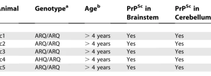
Sample preparation from sheep naturally infected with scrapie. Skin samples from field cases of ovine scrapie were collected during autopsies of cohort animals that were eradicated after identification of an index scrapie case in the respective flock. The samples analysed in this study originated from five scrapie cases (Table 1, Sc1–Sc5) and four scrapie-free control sheep. All scrapie cases were older than 4 years as determined by their dental status and showed loss of wool due to scrubbing their coat. They also nibbled off their fleece on the legs over the carpal and tarsal joints where the straight coat becomes wooly. In all scrapie-infected animals, a marked weight loss was present. The diagnosis of scrapie was confirmed according to Office International des Epizooties standards by histopathology and by immunohistochemical detection of PrPSc in the brain stem and cerebellum. Apart from animal Sc4, PrPSccould also be detected in
the tonsils from all scrapie sheep (not shown). At the polymorphic codons 136, 154, and 171 of the prion protein gene, four of them were ARQ/ARQ and one animal (Sc4) was AHQ/ARQ. According to the National Scrapie Plan for Great Britain [42], the genotypes ARQ/ ARQ and AHQ/ARQ belong to risk group 3, which refers to‘‘sheep that genetically have little resistance to scrapie and will need careful selection when used for further breeding’’. It has to be noted, however, that ARQ/ARQ and AHQ/ARQ do not provide the most prevelant genotypes in scrapie-affected sheep.
Western blot examinations. For Western blot analyses, samples were washed three times in TBS (10 mM Tris HCl, 133 mM NaCl [pH 7.4]) and incubated in a rocking device at 378C for 4 h in 900ll of
TBS containing 2 mM CaCl2and 0.25% (w/v) collagenase A (Roche,
http://www.roche.com). For positive controls, skin tissue from orally mock-infected control donors was spiked by adding 5ll of a 0.1 % (w/
v) 263K scrapie hamster brain homogenate (i.e., 5lg of brain tissue)
from i.c.-infected donors containing approximately 0.5 ng of PrPSc[3]
prior to collagenase digestion. After ultrasonification to disrupt remaining tissue structures, the samples were centrifuged for 3 min at 500g.The supernatant was carefully transferred to a new cup, whereas the pellet consisting of cell debris and the rest of the fur was removed. Subsequently, PrPScwas extracted in the form of PrP27–30 from the tissue homogenates following a previously published protocol [23]. Proteinase K–digested homogenate from 263K scrapie hamster brains, used as a PrP27–30 reference in the Western blotting analyses, was prepared as outlined previously [23]. For deglycosylation [23], extracted pellets were dissolved in 20ll of a. bidest and one fourth of
the aliquots was digested using PNGase F (New England Biolabs,
http://www.neb.com) according to the instructions of the manufac-turer prior to Western blotting. Sodium dodecyl sulfate-polyacryla-mide gel electrophoresis (SDS-PAGE) and Western blot analyses of samples from hamsters were performed as described elsewhere [23]. Western blot testing for PrPScin skin samples from scrapie-infected sheep was similarly performed using the primary antibodies ICSM-18 and P4 to label the ovine prion protein. PrP signals were visualized on an X-OMAT AR (Kodak, http://www.kodak.com) film. Films were exposed for 5–30 min.
PET blot examinations.The PET blot detection of proteinase K– resistant PrP deposits in sections of skin samples was performed on paraformaldehyde- and Carnoy-fixed specimens. For PET blot examinations of skin tissue, modifications of the original protocol [43] were necessary to remove connective tissue. After prewetting blots with TBST (10 mM TrisHCl [pH 7.8], 100 mM NaCl, 0.05 % [w/v] Tween 20), sections were digested with 1.5 mg/ml collagenase A (Roche) in a buffer containing 10 mM TrisHCl (pH 7.8), 100 mM NaCl, 100 mM CaCl2, and 0.1% (w/v) Brij 35 for 30 min at 60 8C,
followed by digestion using 250 lg/ml proteinase K (Roche) in PK
digestion buffer (10 mM TrisHCl [pH 7.8], 100 mM NaCl, 0.1% Brij 35) for 8 h at 558C. After this step, the membrane-attached proteins were fixated to the membrane. The proteins on the membranes were denatured with 3 M guanidine isothiocyanate in 10 mM TrisHCl (pH 7.8) for 20 min. Immunodetection was performed after preincubation in blocking solution (0.2% [w/v] casein in TBST) for 30 min. The monoclonal antibody 3F4 ([44]; diluted 1:3,000) was used as primary antibody and an alkaline phosphatase-coupled rabbit anti-mouse antibody (Dako, http://www.dako.com) at a dilution of 1:500 as secondary antibody. Visualization of antibody binding was achieved by using NBT/BCIP. Blots were examined using an Olympus dissecting microscope.
Immunohistochemical examinations.Frozen tissue sections as well as Carnoy-fixed sections of about 4lm were postfixed in
parafor-maldehyde (4% [w/v] in PBS) for 1 h and washed in tap water to avoid pigment deposition. Antigen retrieval was done using 4 M guanidine hydrochloride for 25 min and by microwaving five times for 3 min at 700 watts in 1 M citric acid at pH 6.0. After blocking with 0.2% casein in TBST, the primary antibody 3F4 (diluted 1:200) was used for PrP detection. Antibodies against S-100 protein (rabbit polyclonal, 1:200 in PBS; Dako,) and neurofilament (SMI31 mouse monoclonal IgG1, 1:20,000 in PBS; Sternberger Monoclonals, http://www.crpinc.com) were applied to detect small nerve fibres. As secondary antibody, a HRP-conjugated goat anti-mouse IgG1 antibody (1:400 in PBS; Dianova, http://www.dianova.de) was used for detecting the antibody SMI31. The S-100 antibody was detected using a polyvalent HRP-conjugated goat anti-rabbit antibody (EnVision, Dako). For 3F4 doublestaining, we used a biotinylated goat anti-mouse IgG2a antibody (1:400 in PBS; Dianova) or a goat anti-mouse IgG antibody without restriction to Ig-subclasses (1:100; Dako). Visualization of the different epitopes was performed using Cy2-conjugated streptavidine (green fluorescence, 1:500; Dianova) and a Cy3-conjugated goat anti-HRP antibody (red fluorescence, 1:100; Dianova). Slides were examined on an Olympus fluorescent microscope using analysis software, and Adobe Photoshop software was used for picture processing.
Acknowledgments
The skillful technical assistance of Marion Joncic, Andrea Ma¨nnel, Tatjana Pfander, and Nadine Rupprecht and the helpful discussion of veterinary infectiological aspects with Wiebke Wemheuer is gratefully acknowledged. MB would like to thank the German Bundesministe-rium fu¨r Bildung und Forschung, the German BundesministeBundesministe-rium fu¨r Gesundheit, and the European Network of Excellence‘‘NeuroPrion’’
for their sustained support.
Author contributions. AT and MB conceived and designed the experiments. AT, WSS, and AW performed the experiments. AT, WSS, AW, and MB analyzed the data. AT, WSS, AW, WW, BB, CK, and KL contributed reagents/materials/analysis tools. AT, WSS and MB wrote the paper.
Funding. This work was supported in part by grants from the European Union (QLG3-CT-2002–81030, Food-CT-2004–506579), and the Volkswagen Foundation (ZN 1294).
Competing interests.The authors have declared that no competing interests exist.
Table 1.Scrapie-Infected Sheep Tested for PrPScin the Skin
Animal Genotypea Ageb PrPScin
Brainstem
PrPScin Cerebellum
Sc1 ARQ/ARQ .4 years Yes Yes
Sc2 ARQ/ARQ .4 years Yes Yes
Sc3 ARQ/ARQ .4 years Yes Yes
Sc4 AHQ/ARQ .4 years Yes Yes
Sc5 ARQ/ARQ .4 years Yes Yes
a
The genotype is specified for the polymorphic codons 136, 154, and 171 of the ovine prion protein gene.
b
References
1. Prusiner SB (1998) Prions. Proc Natl Acad Sci U S A 95: 13363–13383. 2. Beekes M, McBride P (2007) The spread of prions through the body in
naturally acquired transmissible spongiform encephalopathies. FEBS J 274: 588–605.
3. Beekes M, Baldauf E, Diringer H (1996) Sequential appearance and accumulation of pathognomonic markers in the central nervous system of hamsters orally infected with scrapie. J Gen Virol 77: 1925–1934. 4. van Keulen LJ, Vromans ME, van Zijderveld FG (2002) Early and late
pathogenesis of natural scrapie infection in sheep. APMIS 110: 23–32. 5. Ironside JW (2000) Pathology of variant Creutzfeldt-Jakob disease. Arch
Virol Suppl: 143–151.
6. Wadsworth JD, Joiner S, Hill AF, Campbell TA, Desbruslais M, et al. (2001) Tissue distribution of protease resistant prion protein in variant Creutzfeldt-Jakob disease using a highly sensitive immunoblotting assay. Lancet 358: 171–180.
7. Mabbott NA, MacPherson GG (2006) Prions and their lethal journey to the brain. Nat Rev Microbiol 4: 201–211.
8. Bruce ME, Will RG, Ironside JW, McConnell I, Drummond D, et al. (1997) Transmissions to mice indicate that ‘new variant’ CJD is caused by the BSE agent. Nature 389: 498–501.
9. McBride PA, Schulz-Schaeffer WJ, Donaldson M, Bruce M, Diringer H, et al. (2001) Early spread of scrapie from the gastrointestinal tract to the central nervous system involves autonomic fibers of the splanchnic and vagus nerves. J Virol 75: 9320–9327.
10. Beekes M, McBride PA, Baldauf E (1998) Cerebral targeting indicates vagal spread of infection in hamsters fed with scrapie. J Gen Virol 79: 601–607. 11. Thomzig A, Schulz-Schaeffer W, Kratzel C, Mai J, Beekes M (2004) Preclinical deposition of pathological prion protein PrPSc in muscles of hamsters orally exposed to scrapie. J Clin Invest 113: 1465–1472. 12. Andre´oletti O, Simon S, Lacroux C, Morel N, Tabouret G, et al. (2004)
PrPSc accumulation in myocytes from sheep incubating natural scrapie. Nat Med 10: 591–593.
13. Peden AH, Ritchie DL, Head MW, Ironside JW (2006) Detection and localization of PrPSc in the skeletal muscle of patients with variant, iatrogenic, and sporadic forms of Creutzfeldt-Jakob disease. Am J Pathol 168: 927–935.
14. Herzog C, Riviere J, Lescoutra-Etchegaray N, Charbonnier A, Leblanc V, et al. (2005) PrPTSE distribution in a primate model of variant, sporadic, and iatrogenic Creutzfeldt-Jakob disease. J Virol 79: 14339–14345.
15. Paus R, Peters EM, Eichmuller S, Botchkarev VA (1997) Neural mechanisms of hair growth control. J Investig Dermatol Symp Proc 2: 61–68. 16. Zanusso G, Ferrari S, Cardone F, Zampieri P, Gelati M, et al. (2003)
Detection of pathologic prion protein in the olfactory epithelium in sporadic Creutzfeldt-Jakob disease. N Engl J Med 348: 711–719.
17. Gonzalez L, Jeffrey M, Siso S, Martin S, Bellworthy SJ, et al. (2005) Diagnosis of preclinical scrapie in samples of rectal mucosa. Vet Rec 156: 846–847. 18. Cunningham AA, Kirkwood JK, Dawson M, Spencer YI, Green RB, et al.
(2004) Bovine spongiform encephalopathy infectivity in greater kudu (Tragelaphus strepsiceros). Emerg Infect Dis 10: 1044–1049.
19. Safar J, Wille H, Itri V, Groth D, Serban H, Torchia M, Cohen FE, Prusiner SB (1998) Eight prion strains have PrP(Sc) molecules with different conformations. Nat Med 4: 1157–1165.
20. Lauria G, Borgna M, Morbin M, Lombardi R, Mazzoleni G, et al. (2004) Tubule and neurofilament immunoreactivity in human hairy skin: Markers for intraepidermal nerve fibers. Muscle Nerve 30: 310–316.
21. Botchkarev VA, Eichmuller S, Johansson O, Paus R (1997) Hair cycle-dependent plasticity of skin and hair follicle innervation in normal murine skin. J Comp Neurol 386: 379–395.
22. Herzog C, Sales N, Etchegaray N, Charbonnier A, Freire S, Dormont D, Deslys JP, Lasmezas CI (2004) Tissue distribution of bovine spongiform encephalopathy agent in primates after intravenous or oral infection. Lancet 363: 422–428.
23. Thomzig A, Kratzel C, Lenz G, Kru¨ger D, Beekes M (2003) Widespread
PrPSc accumulation in muscles of hamsters orally infected with scrapie. EMBO Rep 4: 530–533.
24. Pammer J, Weninger W, Tschachler E (1998) Human keratinocytes express cellular prion-related protein in vitro and during inflammatory skin diseases. Am J Pathol 153: 1353–1358.
25. Pammer J, Tschachler E (2002) A possible role of keratinocytes of skin and mucous membranes in prion propagation and transmission. J Investig Dermatol Symp Proc 7: 59–63.
26. Wisniewski HM, Sigurdarson S, Rubenstein R, Kascsak RJ, Carp RI (1996) Mites as vectors for scrapie. Lancet 347: 1114.
27. Post K, Riesner D, Walldorf V, Mehlhorn H (1999) Fly larvae and pupae as vectors for scrapie. Lancet 354: 1969–1970.
28. Heikenwalder M, Zeller N, Seeger H, Prinz M, Klohn PC, et al. (2005) Chronic lymphocytic inflammation specifies the organ tropism of prions. Science 307: 1107–1110.
29. Taylor DM, McConnell I, Fraser H (1996) Scrapie infection can be established readily through skin scarification in immunocompetent but not immunodeficient mice. J Gen Virol 77: 1595–1599.
30. Mohan J, Brown KL, Farquhar CF, Bruce ME, Mabbott NA (2004) Scrapie transmission following exposure through the skin is dependent on follicular dendritic cells in lymphoid tissues. J Dermatol Sci 35: 101–111. 31. World Health Organization (2006) Annex 1. In: Guidelines on tissue
infectivity distribution in transmissible spongiform encephalopathies. Geneva: WHO Press. pp. 22–32.
32. Pattison IH, Hoare MN, Jebbett JN, Watson WA (1974) Further observa-tions on the production of scrapie in sheep by oral dosing with foetal membranes from scrapie-affected sheep. Br Vet J 130: lxv–lxvii. 33. Hadlow WJ, Kennedy RC, Race RE (1982) Natural infection of Suffolk sheep
with scrapie virus. J Infect Dis 146: 657–664.
34. Andreoletti O, Berthon P, Marc D, Sarradin P, Grosclaude J, van KL, Schelcher F, Elsen JM, Lantier F (2000) Early accumulation of PrP(Sc) in gut-associated lymphoid and nervous tissues of susceptible sheep from a Romanov flock with natural scrapie. J Gen Virol 81: 3115–3126. 35. van Keulen LJ, Schreuder BE, Vromans ME, Langeveld JP, Smits MA (2000)
Pathogenesis of natural scrapie in sheep. Arch Virol Suppl 145: 57–71. 36. Race R, Jenny A, Sutton D (1998) Scrapie infectivity and proteinase
K-resistant prion protein in sheep placenta, brain, spleen, and lymph node: Implications for transmission and antemortem diagnosis. J Infect Dis 178: 949–953.
37. Mathiason CK, Powers JG, Dahmes SJ, Osborn DA, Miller KV, et al. (2006) Infectious prions in the saliva and blood of deer with chronic wasting disease. Science 314: 133–136.
38. Siso S, Gonzalez L, Jeffrey M, Martin S, Chianini F, et al. (2006) Prion protein in kidneys of scrapie-infected sheep. Vet Rec 159: 327–328. 39. Thomzig A, Spassov S, Friedrich M, Naumann D, Beekes M (2004)
Discriminating scrapie and bovine spongiform encephalopathy isolates by infrared spectroscopy of pathological prion protein. J Biol Chem 279: 33847–33854.
40. Lemmer K, Mielke M, Pauli G, Beekes M (2004) Decontamination of surgical instruments from prion proteins: In vitro studies on the detach-ment, destabilization and degradation of PrPSc bound to steel surfaces. J Gen Virol 85: 3805–3816.
41. Kratzel C, Mai J, Madela K, Beekes M, Kru¨ger D (2007) Propagation of scrapie in peripheral nerves after footpad infection in normal and neurotoxin exposed hamsters. Vet Res 38: 127–39.
42. Department for Environment Food and Rural Affairs (2005)Annex B.editor In: National scrapie plan for Great Britain. Available: http://www.defra.gov. uk/corporate/regulat/forms/ahealth/nsp/nsp76.pdf. Accessed 23 April 2007. 43. Schulz-Schaeffer WJ, Tscho¨ke S, Kranefuss N, Dro¨se W, Hause-Reitner D, et al. (2000) The paraffin-embedded tissue blot detects PrP(Sc) early in the incubation time in prion diseases. Am J Pathol 156: 51–56.