Myeloid Leukemia Risk: A Meta-Analysis
Yu-Tao Qin., Yong Zhang., Fang Wu., Yan Su, Ge-Ning Lu, Ren-Sheng Wang*
Department of Radiotherapy, the First Affiliated Hospital of Guangxi Medical University, Nanning, Guangxi, People’s Republic of China
Abstract
Previous observational studies investigating the association between methylenetetrahydrofolate reductase (MTHFR) polymorphisms and acute myeloid leukemia risk (AML) have yielded inconsistent results. The aim of this study is to derive a more precise estimation of the association between MTHFR (C677T and A1298C) polymorphisms and acute myeloid leukemia risk. PubMed and Embase databases were systematically searched to identify relevant studies from their inception to August 2013. Odds ratios (ORs) with 95% confidence intervals (CIs) were the metric of choice. Thirteen studies were selected for C677T polymorphism (1838 cases and 5318 controls) and 9 studies (1335 patients and 4295 controls) for A1298C polymorphism. Overall, pooled results showed that C677T polymorphism was not significant associated with AML risk(OR, 0.98–1.04; 95% CI, 0.86–0.92 to 1.09–1.25). Similar results were observed for the A1298C polymorphism and in subgroup analysis. All comparisons revealed no substantial heterogeneity nor did we detect evidence of publication bias. In summary, this meta-analysis provides evidence thatMTHFRpolymorphisms were not associated with AML risk. Further investigations are needed to offer better insight into the role of these polymorphisms in AML carcinogenesis.
Citation:Qin Y-T, Zhang Y, Wu F, Su Y, Lu G-N, et al. (2014) Association betweenMTHFRPolymorphisms and Acute Myeloid Leukemia Risk: A Meta-Analysis. PLoS ONE 9(2): e88823. doi:10.1371/journal.pone.0088823
Editor:Masaru Katoh, National Cancer Center, Japan
ReceivedOctober 19, 2013;AcceptedJanuary 10, 2014;PublishedFebruary 20, 2014
Copyright:ß2014 Qin et al. This is an open-access article distributed under the terms of the Creative Commons Attribution License, which permits unrestricted use, distribution, and reproduction in any medium, provided the original author and source are credited.
Funding:The authors have no support or funding to report.
Competing Interests:The authors have declared that no competing interests exist. * E-mail: [email protected]
.These authors contributed equally to this work.
Introduction
Worldwide, an estimated 57 000 cases of leukemia occur every year [1] and acute myeloid leukemia (AML) is the most common acute leukemia (AL). The highest incidence rate is found in males of all age groups, the fact remains to be explained [2–4]. The etiology of most types of leukemia is still unknown. Leukemia is likely to be associated with certain environmental agents, such as ionizing radiation, benzene, and cancer chemotherapy. The increase risk factors for leukemia may be both quantity and quality changes in folic acid metabolism [5–7].
The folate metabolites of carcinogens can influence the gene expression and DNA instability. DNA translocations, inversions or deletions in haematopoietic progenitor cells will lead to leukemia. Be short of folate can result in a lot of cellular disorders [8,9]. Folate metabolism participates in processes of DNA methylation, as well as involves in the synthesis and repair of DNA. That is a mechanism to prevent and repair damaged DNA [10]. The 5, 10-methylenetetrahydrofolate reductase (MTHFR) gene is found at the end of the short arm of chromosome one at locus 1p36.3. The complementary DNA sequence of this gene is approximately 2.2 kb, made up of 11exons (103–432 bp). The major product of MTHFRlocus in human is a 77-kilodaltonprotein [11].MTHFR
plays a pivotal role in the folate metabolism, it can catalyze the irreversible conversion of 5, 10-methylenetrahydrofolate to 5-methylenetrahydrofolate, which participates in the remethylation of homocysteine to methionine [12]. Two common polymor-phisms in MTHFR, C677T and A1298C, have been associated with reduced enzyme activity of MTHFR, which lead to an
accumulation of 5, 10-methylenetetrahydrofolate and DNA hypomethylation. The 5,10-methylenetetrahydrofolate donates a methyl group, which converts dUMP to dTMP and repairs DNA damages [11]. C677T polymorphism occurs in exon4, which leads alanine to be substituted by valine at codon222. People with the homozygousMTHFR677TT genotype have 30 percent enzyme activity compared with those having wild-type allele, while those with heterozygousMTHFR677 CT allele have 60 percent enzyme activity [11]. This polymorphism promotes the separation of enzyme from its co-factor, which results in the enzyme activity decrease [13]. Recently, another important polymorphism in the MTHFRgene is A1298C in exon7, which leads to a glutamate-to-alanine (A.C) change and reduced enzyme activity ofMTHFR.
To date, several studies have investigated the association between MTHFR polymorphisms and AML risk [7,11,14–30], but results from those studies remain inconsistent. Therefore, we conducted a meta-analysis of previously published studies to assess the relationship between theMTHFR polymorphisms and AML risk.
Methods
Search Strategy and Selection Criteria
hand searched for additional studies. Studies were included if they met the following inclusion criteria: (1) explored the association of MTHFR (C677T and A1298C) polymorphisms with AML risk; (2) used a case-control design; (3) provided available genotype or allele frequency of the cases and control to calculate ORs with 95% CIs. The exclusion criteria also applied: the data from study were repeated or overlapped; there was no available genotype or allele frequency; the patients were about therapy-related AML; the studies were review, case report, or comment.
Data Extraction
Two investigators (YTQ and FW) independently extracted data using a standardized data-collection form. Study characteristics extracted from each article were as follows: first author, year of publication, country of origin, racial decent, participant age, number of participants, source of controls, genotype studied, and available genotype frequency information forMTHFRC677T and A1298C. Any disagreements were resolved by consensus and a third author (YZ). All data were extracted from the published studies and no authors were contacted to require further information.
Statistical Analysis
The strength of the association betweenMTHFR(C677T and A1298C) polymorphisms and AML risk was measured by using crude odds ratio (OR) with 95% confidence interval (CI). The pooled ORs were estimated in following models: allele contrast (T vs. C), codominant model (CT vs. CC; TT vs. CC), dominant model (TT+CT vs.CC), and recessive model (TT vs. CT+CC), respectively. For MTHFR A1298C polymorphism, we assessed the same association. The Cochran Q test was used to test statistical heterogeneity. TheI2statistics [31] was also calculated to quantify the proportion of the variations across studies. AP value of less than 0.1 for the Q statistic was considered as heterogeneity across studies, allowing for the use of a random-effects model (DerSimonian and Laird method [32]. Otherwise, a fixed-effects model (Mantel–Haenszel method [33]) was applied.
Subgroup analysis based on ethnicity (Caucasian, Asian, and Brazilian), sample size (large sample size$100, and small sample size, 100), and HWE was performed to assess the source of heterogeneity. We also assessed the influence of individual studies on the combined risk estimate by sequentially omitting one study each time.
Potential publication bias was assessed both by visually inspecting of the Begg funnel plot and statistically via Egger’s unweighted regression tests [34]. All statistical analyses were conducted using Stata version 11.0 (Stata Corporation, College Station, TX). AllP values are tailed where 0.05 was considered statistically significant except in the test for heterogeneity.
Results
Identification of Eligible Studies
The search strategy yielded 35 potential studies from PubMed and Embase databases. However, most of them were excluded after reviewing titles and abstracts, leaving 19 for full-text review. The literature search and detailed study selection procedures were presented in Figure S1. Six studies were excluded (two studies [26,27] were conference articles, and two [28,29] with patients were about therapy-related AML, one [11] was review article, and one [30] was supplementary material). Finally, 13 studies [7,14–25] were included in this meta-analysis.
Study Characteristics
The main characteristics of the included studies were shown in
Table 1. These studies were published between 1999 and 2012. Sample size ranged from 27 to 1,700 (including 1,838 patients with AML and 5,318 healthy controls). Among these, five studies were in Caucasian descent [17,19–22], five studies of Asian descent [14,18,23–25] and three studies of Brazilian descent [7,15,16]. Thirteen studies including 1838 cases and 5318 controls had examined the association ofMTHFRC677T polymorphism with AML risk, and 9 studies with a total of 1335 patients and 4295 controls investigated the association between MTHFR A1298C Table 1.Characteristics of studies included in this meta-analysis.
First author Year Country Racial decent Cases, n
Controls, n
Source of
controls HWE StudiedMTHFRgenotypes
Hussain [14] 2012 India Asian 112 251 Population yes C677T
Lightfoot [19] 2010 United Kingdom Caucasian 89 824 Population yes C677T and A1299C
Vahid [20] 2010 Iran Caucasian 106 97 Population yes C677T and A1299C
Amorim [15] 2008 Brazil Brazilian 50 248 Population yes C677T and A1299C
Kim [24] 2008 Korea Asian 389 1700 Population yes C677T and A1299C
Barbosa [7] 2008 Brazil Brazilian 27 100 Population yes C677T and A1299C
Bolufer [22] 2007 Spain Caucasian 302 454 Population yes C677T
Moon [23] 2007 South Korea Asian 200 434 Population yes C677T and A1299C
Chen [25] 2006 China Asian 40 157 Population yes C677T
Costa Ramos [16] 2006 Brazil Brazilian 182 315 Population yes C677T and A1299C
Hur [18] 2006 Korea Asian 55 200 Population no C677T and A1299C
Deligezer [17] 2003 Turkey Caucasian 49 161 Population yes C677T
Skibola [21] 1999 United Kingdom Caucasian 237 377 Hospital yes C677T and A1299C
HWE, Hardy-Weinberg equilibrium;MTHFR, Methylenetetrahydrofolate reductase. doi:10.1371/journal.pone.0088823.t001
polymorphism and AML risk. Of these, 12 studies were population-based and one was hospital-based.
MTHFR C677T
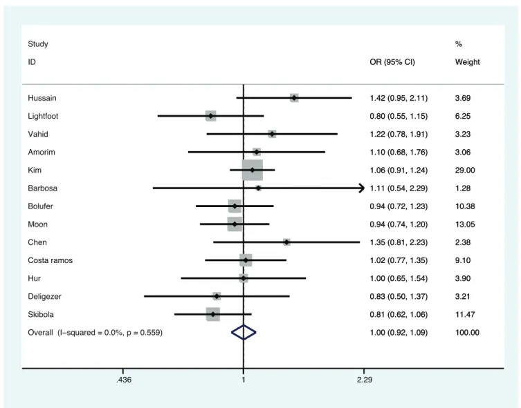
Figure 1 showed the results from a fixed-effects model combining the ORs for the association of MTHFR C677T polymorphism and AML risk. Overall, the pooled results showed that the MTHFR C677T polymorphism was not associated with the development of AML (OR, 0.98–1.04; 95% CI, 0.86–0.92 to 1.09–1.25; P, 0.750–0.976), without statistically significant be-tween-study heterogeneity (I2, 0.0%–26.4%; P for heterogeneity, 0.178–0.573). Table 2 showed that the Asian and Brazilian subgroups were at increased risk in some genetic models. Caucasians may even have some low-level protection in some models (OR 0.81–0.89).
MTHFRA1298C
Figure 2 presented the results from a fixed-effects model combining the ORs for the association of MTHFR A1298C polymorphism and AML risk. Overall, the estimate results indicated non-significant increased risk association of MTHFR A1298C polymorphism with AML risk in some genetic models
(OR, 1.11–1.13), without zero heterogeneity (Pfor heterogeneity, 0.562–0.955). Table 3 shows that the Brazilian subgroup are at increased risk in all genetic models (OR, 1.1–1.4), and in two genetic models, so are the Asians (OR, 1.23–1.25) as well as the HWE studies (OR, 1.11) and even small sample size studies (OR, 1.36–1.50).
Publication Bias
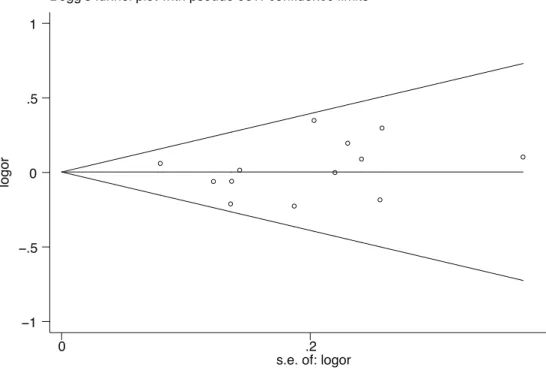
The Begg rank correlation test and Egger linear regression tests for publication bias in the meta-analysis indicated no obvious publication bias among studies (Begg’s test, P = 0.360; Egger’s test, P = 0.659; Figure 3).
Discussion
To the best of our knowledge, this is the first meta-analysis to assess the association betweenMTHFRpolymorphisms and AML risk. Thirteen studies (1838 cases and 5318 controls) and 9 studies (1335 patients and 4295 controls) explored the association between the C677T and A1298C polymorphisms and AML risk, respectively. Results of this study suggested thatMTHFR(C677T and A1298C) polymorphisms were not significantly associated
Figure 1. Meta-analysis for the association of acute myeloid leukemia risk withMTHFRC677T polymorphism (Tvs.C).
with AML risk. Moreover, similar results were observed in subgroup analyses based on ethnicity, sample size, and HWE in controls.
Nowadays, several meta-analyses have been performed to clarify the association between MTHFR (C677T and A1298C) polymorphisms and risk of several cancers. For instance, You et al have demonstrated that the MTHFRC677T and A1298C polymorphisms were associated with bladder cancer risk [35]. Wei et al provided evidence that theMTHFRC677T polymor-phism increased the risk for developing colorectal cancer [36]. However, a meta-analysis by Ding et al indicated that no
significant association was observed between MTHFR C677T polymorphism and susceptibility to ovarian cancer [37]. Besides, Niu et al suggested that no significant association between MTHFR A1298C polymorphism head and neck cancer [38], which were consistent with our results. These inconsistent and confusing conclusions can be attributed to several factors. Different selection criteria and selection bias might account for the diversity of the results. In addition, the reason might be the complexity of the folate metabolic pathway because MTHFR is only one of many enzymes involved in the pathway. Moreover, the studies with small sample size will have a lower statistical
Table 2.Distribution ofMTHFRC677T genotypes and allelic frequencies in acute myeloid leukemia patients.
Genetic comparisons
Population and
subgroups under analysis Studies Fixed-effects model
OR (95% CI) p-value I2,% pfor heterogeneity
T vs. C All 13 1.00 (0.92–1.09) 0.976 0.0 0.559
Caucasian 5 0.89 (0.76–1.03) 0.119 0.0 0.573
Asian 5 1.07 (0.95–1.20) 0.279 0.0 0.417
Brazilian 3 1.04 (0.83–1.31) 0.720 0.0 0.951
Large sample size 7 1.01 (0.92–1.11) 0.862 15.4 0.312
Small sample size 6 0.97 (0.80–1.18) 0.776 0.0 0.629
All in HWE 12 1.00 (0.92–1.09) 0.976 0.0 0.473
CT vs. CC All 13 0.98 (0.86–1.11) 0.750 10.5 0.340
Caucasian 5 0.81 (0.66–1.01) 0.056 26.0 0.248
Asian 5 1.14 (0.95–1.36) 0.169 0.0 0.680
Brazilian 3 0.94 (0.69–1.30) 0.722 0.0 0.824
Large sample size 7 0.99 (0.86–1.14) 0.873 0.0 0.578
Small sample size 6 0.95 (0.73–1.24) 0.704 42.1 0.125
All in HWE 12 0.96 (0.84–1.09) 0.530 0.0 0.455
TT vs. CC All 13 1.04 (0.87–1.25) 0.648 2.9 0.417
Caucasian 5 0.88 (0.64–1.21) 0.427 0.0 0.411
Asian 5 1.12 (0.88–1.42) 0.370 41.7 0.143
Brazilian 3 1.20 (0.72–1.97) 0.484 0.0 0.997
Large sample size 7 1.05 (0.86–1.29) 0.606 28.8 0.209
Small sample size 6 1.00 (0.66–1.51) 0.985 0.0 0.553
All in HWE 12 1.05 (0.88–1.27) 0.570 7.9 0.367
TT+CT vs. CC All 13 0.99 (0.88–1.12) 0.913 0.0 0.573
Caucasian 5 0.83 (0.68–1.01) 0.061 0.0 0.433
Asian 5 1.14 (0.96–1.35) 0.143 0.0 0.933
Brazilian 3 0.99 (0.74–1.34) 0.965 0.0 0.875
Large sample size 7 1.00 (0.88–1.15) 0.956 0.0 0.585
Small sample size 6 0.96 (0.74–1.23) 0.737 12.3 0.336
All in HWE 12 0.98 (0.87–1.11) 0.762 0.0 0.580
TT vs. CT+CC All 13 1.02 (0.86–1.20) 0.836 26.4 0.178
Caucasian 5 0.95 (0.71–1.29) 0.748 15.4 0.316
Asian 5 1.01 (0.82–1.26) 0.892 63.3 0.028
Brazilian 3 1.23 (0.76–1.99) 0.398 0.0 0.985
Large sample size 7 1.02 (0.86–1.23) 0.797 42.2 0.110
Small sample size 6 0.99 (0.67–1.46) 0.950 16.6 0.306
All in HWE 12 1.04 (0.88–1.23) 0.631 24.3 0.205
MTHFR, methylenetetrahydrofolate reductase; OR, odds ratio; CI, confidence interval; vs., versus; HWE, Hardy-Weinberg equilibrium. doi:10.1371/journal.pone.0088823.t002
power than those with large sample size. Furthermore, the different mechanisms of carcinogenesis of different cancers might due to gene–variant associations vary in different kinds of diseases.
Several studies have demonstrated that individuals with MTHFR 677 TT genotype, lack of vitamins B6 and B12, methionine and folate, and high consumption of alcohol are at increased risk of developing colorectal tumors [39–42]. How-ever, no studies have reported these gene-nutrient interactions with the risk of AML. The present study was lack of data to estimate the association of gene-nutrient and risk of AML. These interesting clues may be useful for future research. Dietary intake of several nutrients could influence the distribu-tion of intracellular folate metabolites. Vitamins B6 and B12 may affect DNA synthesis and MTHFR enzyme activity. Moreover, high consumption of alcohol might take place of more nutritious foods, which may lead to the intake deficiency of folate and B vitamins [43]. Deficiency of folate is associated with carcinogenesis mainly in two ways [8]: (1) The conversion of dUMP to dTMP, using for DNA synthesis and repair, demands methyl group donated by 5, 10-methyleneTHF, so lack of folate can intervene thymidylate biosynthesis and then lead to leads to errors in DNA synthesis, strand breakage, and chromosomal repair. (2) Low-level 5-methylTHFmay result in
DNA hypomethylation and cause proto-oncogene expression due to cellular S-adenosylmethionine used up. Thus, cohort studies are needed to focus on gene-nutrient interactions in the future.
In order to better estimate the association of MTHFR (C677T and A1298C) polymorphisms with AML risk, subgroup analysis based on ethnicity, sample size and HWE, was performed. Although Asian and Brazilian subgroups were at increased risk in some genetic models, no significant associations between MTHFR (C677T and A1298C) polymorphisms and AML risk were found in sample size subgroups or all in HWE, which indicated that the results of our analysis was reliable and stable. The real effect of MTHFR (C677T and A1298C) polymorphisms may be concealed by the causal genes in AML. Moreover, different ethnicity of genotypic milieu and living surroundings might have an effect on AML risk, which may led to an effect in our results.
Several limitations might be acknowledged in this meta-analysis. First, we only selected the published articles to acquire data for analyses, and the unpublished article’s effect was unknown. Thus, it is necessary to conduct a system review to avoid the potential effect in analysis. Second, our study was based on single-factor estimate, which explained the effects of two polymorphisms on AML risk respectively and lack of combination of two
Figure 2. Meta-analysis for the association of acute myeloid leukemia risk withMTHFRA1299C polymorphism (Cvs.A).
polymorphisms analysis. So, conducting a meta-analysis to investigate the combination of these two functional polymorphisms may offer better insight into MTHFR (C677T and A1298C) polymorphisms on AML risk. Third, there were no significant effects for both polymorphisms. Fourth, gene and gene-environment interactions might also be considered in future studies. In spite of these, our meta-analysis also has two advantages as follows: (1) there was no significant absence of evidence of publication bias in the present study, which highlighted further, ensured the reliability of association analysis our findings. (2) There was no evidence of statistical heterogeneity between the
analyses of two polymorphisms and AML risk underpins the combinability of the component studies.
In conclusion, our meta-analysis indicates thatMTHFRC677T polymorphism is not associated with AML risk, as well as A1298C polymorphism. Future well-design study is warranted to estimate the effect of combination of two polymorphisms and gene-environment interactions. If epidemiologic study confirms the role of gene-environment interactions, additional studies will be needed to further elucidate the potential biological mecha-nisms involved.
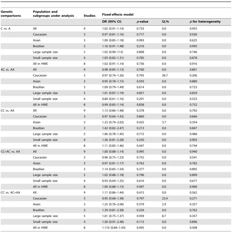
Table 3.Distribution ofMTHFRA1298C genotypes and allelic frequencies in acute myeloid leukemia patients.
Genetic comparisons
Population and
subgroups under analysis Studies Fixed-effects model
OR (95% CI) p-value I2,% pfor heterogeneity
C vs. A All 9 1.02 (0.91–1.14) 0.733 0.0 0.955
Caucasian 3 0.97 (0.81–1.16) 0.717 0.0 0.926
Asian 3 1.00 (0.85–1.18) 0.993 0.0 0.625
Brazilian 3 1.16 (0.91–1.48) 0.216 0.0 0.995
Large sample size 5 1.02 (0.90–115) 0.808 0.0 0.746
Small sample size 4 1.03 (0.82–1.31) 0.785 0.0 0.878
All in HWE 8 1.02 (0.91–1.14) 0.736 0.0 0.916
AC vs. AA All 9 0.98 (0.85–1.13) 0.760 0.0 0.801
Caucasian 3 0.97 (0.74–1.26) 0.795 36.7 0.206
Asian 3 0.95 (0.78–1.15) 0.593 0.0 0.880
Brazilian 3 1.09 (0.79–1.49) 0.614 0.0 0.723
Large sample size 5 1.01 (0.87–1.19) 0.857 0.0 0.859
Small sample size 4 0.84 (0.61–1.16) 0.291 0.0 0.523
All in HWE 8 0.99 (0.85–1.14) 0.838 0.0 0.732
CC vs. AA All 9 1.13 (0.86–1.48) 0.378 0.0 0.792
Caucasian 3 0.97 (0.66–1.42) 0.860 0.0 0.666
Asian 3 1.23 (0.74–2.02) 0.425 3.7 0.354
Brazilian 3 1.42 (0.82–2.47) 0.213 0.0 0.847
Large sample size 5 1.06 (0.78–1.45) 0.715 0.0 0.486
Small sample size 4 1.36 (0.81–2.28) 0.250 0.0 0.903
All in HWE 8 1.11 (0.85–1.46) 0.447 0.0 0.744
CC+AC vs. AA All 9 1.00 (0.88–1.14) 0.995 0.0 0.940
Caucasian 3 0.96 (0.75–1.23) 0.752 0.0 0.541
Asian 3 0.97 (0.81–1.17) 0.762 0.0 0.782
Brazilian 3 1.14 (0.85–1.54) 0.377 0.0 0.895
Large sample size 5 1.02 (0.88–1.18) 0.796 0.0 0.899
Small sample size 4 0.93 (0.69–1.25) 0.616 0.0 0.677
All in HWE 8 1.00 (0.88–1.15) 0.947 0.0 0.900
CC vs. AC+AA All 9 1.11 (0.86–1.44) 0.415 0.0 0.562
Caucasian 3 0.95 (0.68–1.38) 0.797 23.4 0.271
Asian 3 1.25 (0.76–2.06) 0.379 2.9 0.357
Brazilian 3 1.39 (0.81–2.38) 0.234 0.0 0.762
Large sample size 5 1.01 (0.75–1.37) 0.939 8.7 0.357
Small sample size 4 1.50 (0.91–2.48) 0.113 0.0 0.896
All in HWE 8 1.110 (0.84–1.43) 0.495 0.0 0.508
MTHFR, methylenetetrahydrofolate reductase; OR, odds ratio; CI, confidence interval; vs., versus; HWE, Hardy-Weinberg equilibrium. doi:10.1371/journal.pone.0088823.t003
Supporting Information Figure S1 Flow chart.
(DOC)
Checklist S1 PRISMA checklist.
(DOC)
Author Contributions
Conceived and designed the experiments: R-SW. Performed the experiments: YS G-NL FW. Analyzed the data: YZ. Contributed reagents/materials/analysis tools: Y-TQ YZ YS. Wrote the paper: Y-TQ.
References
1. Pui C (2006) Childhood Leukaemias, 2nd ed. Cam-bridge, United Kingdom: Cambridge University Press.
2. Ross JA, Davies SM, Potter JD, Robison LL (1994) Epidemiology of childhood leukemia, with a focus on infants. Epidemiol Rev 16: 243–72.
3. Pui CH (2000) Acute lymphoblastic leukemia in children. Curr Opin Oncol 12: 3–12.
4. Henderson ES (1990) Acute leukemia: gen-eral considerations. In: Williams WJ, Beutler E, Erslev AJ, Lichtman MA, eds. Hematology, 4 th ed. New York: McGraw-Hill. p. 237.
5. Greaves MF (1997) Aetiology of acute leukaemia. Lancet 349: 344–9. 6. Smith MT, Zhang L (1998) Biomarkers of leukemia risk: benzene as a model.
Environ Health Perspect 106 Suppl 4: 937–46.
7. Barbosa CG, Souza CL, de Moura Neto JP (2008) Methylenetetrahydrofolate reductase polymorphisms in myeloid leukemia patients from Northeastern Brazil. Genetics and Molecular Biology 31: 1 (29–32).
8. Duthie SJ, Narayanan S, Brand GM, Pirie L, Grant G (2002) Impact of folate deficiency on DNA stability. J Nutr 132: 2444S–2449S.
9. Giovannucci E (2002) Epidemiologic studies of folate and colorectal neoplasia: a review. J Nutr 132: 2350S–2355S.
10. Yamada K, Chen Z, Rozen R, Matthews RG (2001) Effects of common polymorphisms on the properties of recombinant human methylenetetrahydro-folate reductase. Proc Natl Acad Sci U S A 98: 14853–8.
11. Robien K, Ulrich CM (2003) 5,10-Methylenetetrahydrofolate reductase polymorphisms and leukemia risk: a HuGE minireview. Am J Epidemiol 157: 571–82.
12. Blount BC, Mack MM, Wehr CM, MacGregor JT, Hiatt RA, et al. (1997) Folate deficiency causes uracil misincorporation into human DNA and chromosome breakage: implications for cancer and neuronal damage. Proc Natl Acad Sci U S A 94: 3290–5.
13. Guenther BD, Sheppard CA, Tran P, Rozen R, Matthews RG, et al. (1999) The structure and properties of methylenetetrahydrofolate reductase from Esche-richia coli suggest how folate ameliorates human hyperhomocysteinemia. Nat Struct Biol 6: 359–65.
14. Hussain SR, Naqvi H, Raza ST, Ahmed F, Babu SG, et al. (2012) Methylenetetrahydrofolate reductase C677T genetic polymorphisms and risk
of leukaemia among the North Indian population. Cancer Epidemiol 36: e227– 31.
15. Amorim MR, Zanrosso CW, Magalhaes IQ, Pereira SC, Figueiredo A, et al. (2008) MTHFR 677C–.T and 1298A–.C polymorphisms in children with Down syndrome and acute myeloid leukemia in Brazil. Pediatr Hematol Oncol 25: 744–50.
16. da Costa Ramos FJ, Cartaxo Muniz MT, Silva VC, Araujo M, Leite EP, et al. (2006) Association between the MTHFR A1298C polymorphism and increased risk of acute myeloid leukemia in Brazilian children. Leuk Lymphoma 47: 2070– 5.
17. Deligezer U, Akisik E, Dalay N (2003) Genotyping of the MTHFR gene polymorphism, C677T in patients with leukemia by melting curve analysis. Mol Diagn 7: 181–5.
18. Hur M, Park JY, Cho HC, Lee KM, Shin HY, et al. (2006) Methylenetetrahy-drofolate reductase A1298C genotypes are associated with the risks of acute lymphoblastic leukaemia and chronic myelogenous leukaemia in the Korean population. Clin Lab Haematol 28: 154–9.
19. Lightfoot TJ, Johnston WT, Painter D, Simpson J, Roman E, et al. (2010) Genetic variation in the folate metabolic pathway and risk of childhood leukemia. Blood 115: 3923–9.
20. Vahid P, Farnaz R, Zaker F, Farzaneh A, Parisa R (2010) Methylenetetrahy-drofolate reductase gene polymorphisms and risk of myeloid leukemia. Laboratory Medicine 41: 490–4.
21. Skibola CF, Smith MT, Kane E, Roman E, Rollinson S, et al. (1999) Polymorphisms in the methylenetetrahydrofolate reductase gene are associated with susceptibility to acute leukemia in adults. Proceedings of the National Academy of Sciences of the United States of America 96: 12810–5. 22. Bolufer P, Collado M, Barragan E, Cervera J, Calasanz MJ, et al. (2007) The
potential effect of gender in combination with common genetic polymorphisms of drug-metabolizing enzymes on the risk of developing acute leukemia. Haematologica 92: 308–14.
23. Moon HW, Kim TY, Oh BR, Min HC, Cho HI, et al. (2007) MTHFR 677CC/ 1298CC genotypes are highly associated with chronic myelogenous leukemia: a case-control study in Korea. Leuk Res 31: 1213–7.
Figure 3. Publication bias test (MTHFRC677T: Tvs.C).
24. Kim HN, Kim YK, Lee IK, Yang DH, Lee JJ, et al. (2009) Association between polymorphisms of folate-metabolizing enzymes and hematological malignancies. Leuk Res 33: 82–7.
25. Chen BA, Jiang N, Ji MJ, Hou P, Lu ZH, et al. (2006) A new method for 5, 10-methylenetetrahydrofolate reductase single nucleotide polymorphisms genotyp-ing used to study susceptibility of hematological malignancy. Zhongguo Shi Yan Xue Ye Xue Za Zhi 14: 1069–73.
26. Sazawal S, Bohra B, Kaur P, Chikkara S, Chaubey R, et al. (2010) MTHFR genetic polymorphisms and susceptibility to acute myeloid Leukemia in India. Indian Journal of Hematology and Blood Transfusion 26: 148–9.
27. Ann Bishop L, Dengel DR, Kelly A, Sinaiko AR, Petryk A, et al. (2009) The C677T methylenetetrahydrofolate reductase polymorphism and insulin resis-tance in childhood cancer survivors. Blood 422.
28. Guillem VM, Collado M, Terol MJ, Calasanz MJ, Esteve J, et al. (2007) Role of MTHFR (677, 1298) haplotype in the risk of developing secondary leukemia after treatment of breast cancer and hematological malignancies. Leukemia 21: 1413–22.
29. Bolufer P, Collado M, Barragan E, Calasanz MJ, Colomer D, et al. (2007) Profile of polymorphisms of drug-metabolising enzymes and the risk of therapy-related leukaemia. Br J Haematol 136: 590–6.
30. Savitskaya T, Lipay N, Krivko M, Petina O, Kokarava M (2009) Polymorphisms of genes MDR1 and MTHRF in children with acute leukemia. European Journal of Cancer, Supplement 7: 571–572.
31. Deeks JJ, Higgins JPT, Altman DG (2008) Analysing data and undertaking meta-analyses. In:Higgins, J. P. T Green, S. editors. Cochrane handbook for systematic reviews of interventions. Version 5.0.1. Chapter 9. The Cochrane Collaboration.
32. DerSimonian R, Laird N (1986) Meta-analysis in clinical trials. Control Clin Trials 7: 177–88.
33. Sutton AJ, Abrams KR, Jones DR (2000) Methods for meta-analysis in medical research. Chichester: Wiley.
34. Egger M, Davey Smith G, Schneider M, Minder C (1997) Bias in meta-analysis detected by a simple, graphical test. BMJ 315: 629–34.
35. You W, Li Z, Jing C, Qian-Wei X, Yu-Ping Z, et al. (2013) MTHFR C677T and A1298C Polymorphisms Were Associated with Bladder Cancer Risk and Disease Progression: A Meta-Analysis. DNA Cell Biol 32: 260–7.
36. Teng Z, Wang L, Cai S, Yu P, Wang J, et al. (2013) The 677C.T (rs1801133) polymorphism in the MTHFR gene contributes to colorectal cancer risk: a meta-analysis based on 71 research studies. PLoS One 8: e55332.
37. Ding XP, Feng L, Ma L (2012) MTHFR C677T polymorphism and ovarian cancer risk: a meta-analysis. Asian Pac J Cancer Prev 13: 3937–42. 38. Niu YM, Shen M, Li H, Ni XB, Zhou J, et al. (2012) No association between
MTHFR A1298C gene polymorphism and head and neck cancer risk: a meta-analysis based on 9,952 subjects. Asian Pac J Cancer Prev 13: 3943–7. 39. Chen J, Giovannucci EL, Hunter DJ (1999) MTHFRpolymorphism,
methyl-replete diets and the risk of colorectal carcinoma and adenoma among U.S. men and women: an example of geneenvironment interactions in colorectal tumorigenesis. J Nutr 129(suppl): 560S–4S.
40. Ulrich CM, Kampman E, Bigler J, Schwartz SM, Chen C, et al. (1999) Colorectal adenomas and the C677T MTHFRpolymorphism: evidence for geneenvironment interaction? Cancer Epidemiol Biomarkers Prev 8: 659–68. 41. Slattery ML, Potter JD, Samowitz W, Schaffer D, Leppert M (1999)
Methylenetetrahydrofolate reductase, diet, and risk of colon cancer. Cancer Epidemiol Biomarkers Prev 8: 513–18.
42. Levine AJ, Siegmund KD, Ervin CM, Diep A, Lee ER, et al. (2000) The methylenetetrahydrofolate reductase 677C–.Tpolymorphism and distal colo-rectal adenoma risk. Cancer Epidemiol Biomarkers Prev 9: 657–63. 43. Halsted CH (1995) Alcohol and folate intereactions: clinical impli-cations. In:
Bailey LB, ed. Folate in health and disease. New York, NY: Marcel Dekk er, Inc, 313–27.