Correspondence to:
Zora PAVLOVIĆ-POPOVIĆ Institute for Pulmonary Diseases of Vojvodina
Department for Sarcoidosis and Difuse Pulmonary Diseases Put dr Goldmana 4 21204 Sremska Kamenica Serbia
zora.pavlovic-popovic@institut.rs
SUMMARY
Introduction The splenic involvement is common in sarcoidosis, but its real frequency is still obscure, depending doubtless on the method of splenomegaly detection. Splenomegaly may be accompanied with pain or anemia, leucopenia and thrombocytopenia.
Objective The aim of this study was to investigate the frequency of splenomegaly related to clinical characteristics of sarcoidosis and to solve the dilemma – whether to introduce medicaments, and when to perform splenectomy.
Methods The method of the study is a retrospective and prospective analysis of the patients’ material. Results The study included 540 patients with sarcoidosis in a 20-year period. Of them, 26% had splenom-egaly detected by computerized tomography screening. Splenomsplenom-egaly was more frequently registered in the patients with a longer history of sarcoidosis (38%), as compared to those with a shorter history of the disease (23%) (p<0.05). Splenomegaly was more frequently registered in the patients with other extrapulmonary lesions detected (33%) than in those who had no extrapulmonary manifestations of sarcoidosis (17%) (p<0.01). Indications, possible benefits and complications of splenectomy were ana-lysed in 11 sarcoidosis patients undergoing this intervention for various reasons, of which the follow-up period ranged from one to 20 years.
Conclusion Splenomegaly was more frequent in chronic cases or in the patients with established sarcoid lesions of other extrapulmonary organs. The primary treatment of uncomplicated symptomatic splenic sarcoidosis includes medicamentous therapy. Occasionally, splenectomy is required. Prognostically, splenomegaly indicates an unfavorable course of the disease.
Keywords: sarcoidosis; clinical characteristics; splenomegaly
Splenomegaly in Sarcoidosis: Frequency, Treatment,
Prognosis and Long-term Follow-up
Zora Pavlović-Popović1,2, Bojan Zarić2, Zdravko Kosjerina2, Dragana Petrović3
1University of Novi Sad, Faculty of Medicine, Novi Sad, Serbia;
2Institute for Pulmonary Diseases of Vojvodina, Sremska Kamenica, Serbia; 3Institute of Oncology of Vojvodina, Sremska Kamenica, Serbia
INTRODUCTION
Sarcoidosis is a multisystem granulomatous disease of unknown etiology. As sarcoidosis may affect any organ, the abdominal organs may be involved as well. The spleen is frequent-ly affected in sarcoidosis, but it is not always particularly investigated.
The normal human spleen weighs approxi-mately 150 to 250 grams. The organ must dou-ble in size before it can be palpated beyond the costal margin. The term ‘massive splenom-egaly’ denotes the splenic weight greater than 1,000 grams, or 4 to 6 times the normal weight, when the spleen is palpable more than 4 cm bellow the costal margin. Spleens involved with sarcoidosis rarely exceed two kilograms in weight, but may also be normal in size.
A slightly enlarged spleen is often asymp-tomatic. The patients with sarcoidosis of the spleen develop symptoms which are usually as-sociated with a massive splenomegaly. These symptoms include early satiety, left upper-quadrant fullness, and arch, while extremely severe pain may be due to splenic infarct sec-ondary to gastric compression, which may be the first symptom of the spleen involved with sarcoidosis [1]. Functional disorders in patients
with splenic infiltration include pancytopenia, anemia, leukopenia and thrombocytopenia, in-ducing fatigue and petechiae.
The splenic involvement with sarcoidosis is probable if the spleen is enlarged on the palpa-tion or radiography in patients with established sarcoidosis. As the definite histological diagno-sis may be established by splenectomy when necessary, it is important to differentiate be-tween the splenic involvement with sarcoidosis and the sarcoid-like reaction, the latter associ-ated with lymphoma and malignant tumors of other organs [2].
In patients with sarcoidosis, CT imaging re-veals heterogeneous splenomegaly with multi-ple hypovascular nodules. Being hypovascular, the nodules are mostly discrete, but they may coalesce and become bigger [3, 4].
280
Pavlović-Popović Z. et al. Splenomegaly in Sarcoidosis: Frequency, Treatment, Prognosis and Long-term Follow-up
tients failing to adequately respond to the applied medi-camentous treatment, in severe hypersplenism and pain, prophylaxis for splenic rupture, and neoplastic exclusion.
OBJECTIVE
The aim of this study was to investigate the frequency of splenomegaly related to clinical characteristics of sarcoido-sis and to solve the dilemma – whether to introduce medi-caments, and when to perform splenectomy.
METHODS
The investigated group included patients with the diagno-sis of sarcoidodiagno-sis established in the Institute for Pulmonary Diseases of Vojvodina, Serbia, in the period from 1992 to 2012. The patients selected for the study were submitted to control examinations and their available medical files were reviewed. With regard to a long course of the disease, the collected data were used in two methodological proce-dures – the retrospective and the prospective one. On the basis of the retrospective analyses of the patients’ material, including the medical files (the initial diagnoses and the findings on control examinations) of a certain number of the patients with sarcoidosis, an insight into the former course of the disease was obtained. The diagnosis of sar-coidosis was based on the consistent clinical and radio-logical picture, the historadio-logical finding of non-caseating granulomas in one or more of the affected tissue systems, and exclusion of other granulomatous conditions mimick-ing sarcoidosis. The prospective analysis of the patients’ material included further monitoring of their disease through control examinations of the registered patients. The comparative analysis of the former condition and the findings at control examinations have shed light on the evolution of the disease and effects of the treatment on splenomegaly in the patients with sarcoidosis.
RESULTS
There were 540 patients with sarcoidosis (212 males and 328 females) registered over the period from 1992 to 2012. These patients were submitted to the spleen size assess-ment by ultrasound of the abdomen. In this group, the patients with splenomegaly were additionally submitted to computerized tomography of the abdomen, which con-firmed splenomegaly in 140 (26%) of them. No difference in the frequency of splenomegaly has been registered be-tween the sexes. Severe pancytopenia was registered in two patients, who were submitted to splenectomy. In the group of sarcoidosis patients with a longer follow-up (>2 years) due to length of the disease, splenomegaly was more fre-quently registered (38%), compared to the patients with a shorter (≤2 years) disease history (23%) (p<0.05) (Table 1).
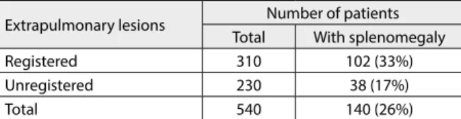
In the patients with other extrapulmonary lesions registered, the frequency of splenomegaly was 33%, as
compared to 17% in sarcoidosis patients with no other extrapulmonary lesions registered (p<0.01) (Table 2).
Hemoglobin, hematocrit and leucocyte levels were similar in sarcoid patients with either normal or enlarged spleen. Thrombocyte levels were statistically significantly lower in female sarcoid patients with splenomegaly than in those with a normal-sized spleen. This difference was not registered in male patients. A negative correlation was registered in female patients between hemoglobin, hemat-ocrit and thrombocyte values and the spleen size, but this correlation was not found in male patients. Two of 140 patients with splenomegaly had anemia, while neutropenia and thrombocytopenia were registered in 8 and 14 of 140 patients, respectively. Pancytopenia was registered in 2 pa-tients. Anemia accompanied with thrombocytopenia was registered in 2 female patients as well. Generally, 28 of 140 patients with splenomegaly (20%) had altered peripheral blood values suggesting hypersplenism.
In the further course of the study, 11 patients with sar-coidosis undergoing splenectomy for a variety of reasons were followed over the period of 1 to 20 years. This group included 4 males and 7 females, ranging from 20 to 54 years of age at the time of establishing the initial diagno-sis. In this group of sarcoid patients with splenomegaly, the predominant symptoms were the abdominal pain, weakness and fatigue. Anemia, leukopenia and thrombo-cytopenia were registered in 2 patients. The immediate reasons for coming to see a doctor were skin lesions, joint pains and enlarged lymph nodes. To establish the diagno-sis of sarcoidodiagno-sis, biopsy samples of the skin lesions were taken, and the lungs were also sampled by transbronchial biopsy in 3 patients. This was due to the fact that 50% of these patients had a normal chest X-ray finding. In the patients with splenomegaly, splenectomy was often indi-cated in order to exclude lymphoma or other malignant hematological disease, or in cases of massive splenomegaly with a threatening rupture, or a severe pancytopenia re-sponding poorly to the applied medicamentous therapy. The 1–20-year follow-ups of 11 patients with sarcoidosis submitted to splenectomy has revealed that 55% of the
Table 1. Computerized tomography established splenomegaly in patients with sarcoidosis
Disease history (years) Number of patients
Total With splenomegaly
≤2 440 102 (23%)
>2 100 38 (38%)
Total 540 140 (26%)
The diference between patients with the disease history of ≤2 and >2 years: p<0.05
Table 2. Splenomegaly and other extrapulmonary manifestations in patients with sarcoidosis
Extrapulmonary lesions Number of patients
Total With splenomegaly
Registered 310 102 (33%)
Unregistered 230 38 (17%)
Total 540 140 (26%)
patients required no further medicamentous treatment be-cause sarcoidosis was in remission, or no medicamentous treatment was recommended either. One fatal outcome was due to neither sarcoidosis nor splenectomy, but the patient had a car accident. In other patients of this group, 45% of sarcoidosis cases were resolved by corticosteroids.
DISCUSSION
The recent data suggest the frequency of splenic involve-ment with sarcoidosis ranges from l0% to over 50% [6]. The palpable spleen is registered in 2–42% of the patients with sarcoidosis [7]. The study in Serbia reported a palpa-ble spleen in 1%, and splenomegaly in 4% of the patients with sarcoidosis [8], while the study in Finland reported a palpable spleen in 6% of their examined patients with sarcoidosis [9]. Splenomegaly is more frequently registered at autopsy 54%, and granulomas in the spleen are found at autopsy in 38–77% of the patients with sarcoidosis [7, 9, 10]. Longcope and Freiman [10] reported an enlarged spleen in 65% of the autopsied subjects with sarcoidosis. In another study of 111 sarcoidosis patients, the spleen was found to be involved in 53% of the cases [11]. A review of the literature, comprising 6,074 patients from 29 stud-ies, shows that splenomegaly is common, but a massive splenomegaly is rare (approximately 3%) in sarcoidosis patients with splenic involvement [12]. The age, sex and race of the patients with splenic sarcoidosis correlate to other sarcoidosis patients [13]. Splenomegaly due to sar-coidosis is associated with multiple organs involvement that almost always includes the lungs, and very often the liver [13, 14]. The radiological finding of the chest is rarely normal in sarcoidosis patients with splenic involvement [15]. The isolated cases of sarcoidosis of the spleen, with no extrasplenic involvement, have been reported in the literature, but rarely so [16].
The frequency of radiological abnormalities of the spleen is unknown, as all examined series are either retro-spective, or they have a biased selection. The retrospective analysis of abdominal CT findings in 49 patients with the tissue-proven sarcoidosis revealed spleen abnormalities in 26 (53%) patients [17]. In their recent studies, Ebert et al. found nodules in the spleen in 15% of the patients with sar-coidosis who underwent CT scanning of the abdomen [4]. The nodular lesions correlated with the activity of the dis-ease [4].
Comparing the contrast CT and MR imaging, the latter appears to be an equally good, or a slightly better imaging procedure to detect the nodules in the spleen and liver [18].
Ultrasound of the abdomen has also been applied in the diagnostic assessment of splenomegaly in 108 patients with sarcoidosis, detecting the sarcoid involvement of the spleen in 22 (20.3%) of them [19].
In general, the treatment of splenic sarcoidosis includes the medicamentous therapy with prednisone, methotrex-ate and/or antimalarial drugs, providing a good treatment response. In study of 24 patients with sarcoid
splenom-egaly, a good control of splenomegaly was achieved by corticosteroid therapy in 17 patients. Methotrexate and azathioprine are used to treat the patients with intolerance to corticosteroids, or those with a refractory disease. In some cases, splenectomy should be indicated. Baughman et al. [20] detected splenomegaly in 32 of 235 patients with sarcoidosis; in 11 of 32 patients, the enlarged spleen was palpable 4 cm below the costal margin and 9 of these pa-tients had a good response to the applied medicamentous treatment while 2 patients required splenectomy. Patel [21] reported a patient intolerant to corticosteroids with a sys-temic sarcoidosis which also involved the bone marrow, who responded well to TNF antagonist adalimumab, with hemoglobin normalization and splenomegaly reduction to normal levels in a two-year period.
Sarcoidosis of the spleen may cause hypersplenism, which induces hematological disorders including anemia, leukopenia, thrombocytopenia or any of their combina-tions, including pancytopenia [12]. In sarcoidosis, throm-bocytopenia with splenomegaly is not always the result of the splenic sequestration of thrombocytes; autoimmune thrombocytopenia may result from the development of thrombocyte-associated immunoglobulins. However, a low frequency of hypersplenism in the patients with sar-coidosis (range 0–22%) has been reported in the literature [22], despite the common splenic involvement. Pham et al. [12] reported 1 case of giant sarcoid splenomegaly treated by splenectomy in order to control the hematological ab-normalities. A case with giant splenomegaly, pancytopenia and hypercalcemia has also been reported. The spleen was 15 cm below the costal margin [23].
Splenectomy is quite rarely indicated by hypersplen-ism or the symptoms (uneasiness) resulting from the compression in the abdomen or infarction of the spleen. It is however unavoidable in cases of the rupture of the spleen or the splenic artery, which are fortunately rare in sarcoidosis, with just one report each [24]. On the other hand, splenectomy should be indicated as a precaution against the splenic rupture, in cases of a gross enlarge-ment, severe hypersplenism and pain, indefinite diagnosis requiring exclusion of a lymphoma or other hematological malignancies, when the medicamentous therapy failed to produce a satisfactory response, or it was contraindicated [6]. In these cases splenectomy is the last treatment option to be applied. When assessing the indications for it, long-term benefits and post-splenectomy complications should be considered. These cases are few and their long-term follow-up is often difficult [25].
282
CONCLUSION
In our study, the CT screening of the abdomen revealed the spleen involvement in one fourth of the examined pa-tients with sarcoidosis. Splenomegaly was more frequent in chronic cases, or in the patients with established sarcoid lesions of other extrapulmonary organs. Hypersplenism
due to the peripheral blood alterations occurs exception-ally rarely. The symptoms produced by the compression of the enlarged spleen in the abdomen, as well as hypersplen-ism, require a medicamentous treatment. In our study, splenectomy was rarely performed for splenic sarcoidosis. Prognostically, the disease in these cases takes an unfavo-rable course due to its long duration.
1. Liu Y. Clinical significance of diffusely increased splenic uptake on FDG-PET. Nucl Med Commun. 2009; 30:763-69.
2. Kojima M, Nakamura S, Fujisaki M, Hasegawa H, Maeda D, Suito T, et al. Sarcoid-like reaction in the regional lymph nodes and spleen in gastric carcinoma: a clinicopathologic study of five cases. Gen Diagn Pathol. 1996/1997; 142:347-52.
3. Judson MA, Baughman RP, Teirstein AS, Yeager H; ACCESS Research Group. Defining organ involvement in sarcoidosis: the ACCESS proposed instrument. Sarcoidosis Vasc Diff Lung Dis. 1999; 16:75-86.
4. Porter JC, Matutes E, Michell DN. The spleen, bone marrow and blood. In: Mitchell DN, Wells A, Spiro SG, Moller DR, editors. Sarcoidosis. London: Hodder & Stoughton; 2012. p.250-5. 5. Aly Y, Popescu A, Woodlock TJ. Extrapulmonary sarcoidosis: rapid
spontaneous remission of marked splenomegaly. J Natl Med Assoc. 1996; 88:714-6.
6. Kurosaki F, Bando M, Nakayama M, Mato N, Yamasawa H, Higashizawa T, et al. A patient with sarcoidosis who developed heterochronic involvements in different organs from initial organs during 7 years. Respir. Res. 2014; 52(1):71-4.
7. Hunninghake GW, Costabel U, Ando M, Baughman RP, Cordier JF, du Bois R, et al. ATS/ERS/WASOG statement on sarcoidosis. Sarcoidosis Vasc Diff Lung Dis. 1999; 16:149-73.
8. Pavlovic-Popovic Z, Djuric B. Relevant prognostic predictors of sarcoidosis. Sarcoidosis Vasc Diffuse Lung Dis. 2001; 18:53. 9. Selroos O. Fine-needle aspiration biopsy of the spleen in diagnosis
of sarcoidosis. Ann N Y Acad Sci. 1976; 278:517-21.
10. Logcope W, Freiman D. A study of sarcoidosis based on a combined investigation of 160 cases including 30 autopsies from the Johns Hopkins and Massachusetts General Hospital. Medicine. 1952; 31:1-132.
11. Sharma O, Chan K. Treatment of sarcoidosis: a practical guide. Biodrugs. 1999; 12:201-65.
12. Pham A, Grayson G, Rodriguez M, Zreik R. Massive splenomegaly and pancytopenia: a rare presentation of sarcoidosis in an 8-year-old female. Pediatr Blood Cancer. 2013; 60(S2):S57.
13. Baughman RP, Teirstein AS, Judson MA, Rossman MD, Yeager H Jr, Bresnitz EA, et al. Clinical characteristics of patients in a case control study of sarcoidosis. Am J Respir. Crit Care Med. 2001; 164:1885-9.
14. Kawano S, Kato J, Kawano N, Yoshimura Y, Masuyama H, Fukunaga T, et al. Sarcoidosis manifesting as cardiac sarcoidosis and massive splenomegaly. Internal Med. 2012; 51:65-69.
15. Thanos L, Zormpala A, Brountzos E, Nikita A, Kelekis D. Nodular hepatic and splenic sarcoidosis in a patient with normal chest radiograph. Eur J Radiol. 2002; 41:10-1.
16. Zia H, Zeman H, Brody F. Laparoscopic splenectomy for isolated sarcoidosis of the spleen. J Laparoendosc Adv Surg Tech A. 2005; 15:160-2.
17. Folz SJ, Johnson CD, Swensen S. Abdominal manifestations of sarcoidosis in CT studies. J Comput Assist Tomogr. 1995; 19:573-9. 18. Warshauer DM, Semelka RC, Ascher SM. Nodular sarcoidosis of
the liver and spleen: Appearance on MR images. J Mag Resonance Imag. 1994; 4:553-7.
19. Siniluoto TMJ, Tikkakoski TA, Lähde ST, Päivänsalo MJ, Koivisto MJ. Ultrasound or CT in splenic diseases? Acta Radiol. 1994; 37:597-605. 20. Baughman RP, Ohmichi M, Lover EE. Combination therapy for
sarcoidosis. Sarcoidosis Vasc Diffuse Lung Dis. 2001; 18:133-7. 21. Patel SR. Systemic sarcoidosis with bone marrow involvement
responding to therapy with adalimumab: a case report. J Med Case Report. 2009; 3:8573.
22. Lawrence HJ, Greenberg BR. Autoimmune thrombocytopenia in sarcoidosis. Am J Med. 1985; 79(6):761-4.
23. Kruthoff K, Gyetko M, Scheiman J. Giant splenomegaly and refractory hypercalcemia due to extrapulmonary sarcoidosis. Successful treatment by splenectomy. Arch Intern Med. 1993; 153:2793-6.
24. Barton JH, Tavora F, Farb A, Li L, Burke AP. Unusual cardiovascular manifestations of sarcoidosis, a report of the cases: coronary artery aneurysm with myocardial infarction, symptomatic mitral valvular disease, and sudden death from ruptured splenic artery. Cardiovasc Pathol. 2010; 19(4):119-23.
25. Shiber J, Fontane E, Prisk D. A traumatic splenic rupture: dreaded complication of splenomegaly. Trop Med Surg. 2014; 2(1):162. 26. Mana J, Salazar A, Manresa F. Clinical factors predicting persistence
of activity in sarcoidosis: a multivariate analysis of 193 cases. Respiration. 1994; 61:219-25.
27. Neville E, Walker AN, James DG. Prognostic factors predicting the outcome of sarcoidosis: an analysis of 818 patients. Quart J Med. 1983; 208:525-33.
REFERENCES
КРАТАК САДРЖАЈ
Увод Код осо ба обо ле лих од сар ко и до зе сле зи на је че сто за хва ће на, али ствар на уче ста лост ни је по зна та, већ за ви си од ме то де от кри ва ња. Спле но ме га ли ја мо же би ти пра ће на бо лом или ане ми јом, ле у ко пе ни јом и тром бо ци то пе ни јом. Циљ ра да Циљ ис тра жи ва ња је био да се утвр ди уче ста лост спле но ме га ли је у по гле ду кли нич ких од ли ка сар ко и до зе и раз ре ши те ра пиј ска ди ле ма: да ли је по треб но ме ди ка мент-но ле че ње и ка да тре ба ура ди ти спле нек то ми ју.
Ме то де ра да Бо ле снич ки ма те ри јал је об ра ђен ре тро спек-тив но и про спек спек-тив но.
Ре зул та ти То ком два де се то го ди шњег пе ри о да ис пи та но је 540 бо ле сни ка са сар ко и до зом. Код 26% ис пи та ни ка ком пју-тер ском то мо гра фи јом уста но вље на је спле но ме га ли ја. Она је би ла че шћа код ис пи та ни ка ко ји су ду го бо ло ва ли и ду го кли нич ки пра ће ни (38%) у од но су на бо ле сни ке чи ја је
бо-лест тра ја ла кра ће (23%) (p<0,05). Та ко ђе је би ла че шћа код бо ле сни ка са дру гим екс тра то ра кал ним ле зи ја ма (33%) у од но су на бо ле сни ке код ко јих се ни су зна ле дру ге ло ка ци је сар ко и до зних ле зи ја (17%) (p<0,01). Ин ди ка ци је, ду го роч не ко ри сти и ком пли ка ци је ана ли зи ра не су код 11 сар ко и до-зних бо ле сни ка ко ји ма је ура ђе на спле нек то ми ја из ра до-зних раз ло га пра ће них у пе ри о ду од јед не до 20 го ди на. За кљу чак Спле но ме га ли ја је би ла че шћа у хро нич ним слу ча је ви ма, као и код обо ле лих где су сар ко и до зним ле-зи ја ма би ли за хва ће ни и дру ги екс тра пул мо нал ни ор га ни. При мар на те ра пи ја не ком пли ко ва них сле зин ских сар ко и-до за са симп то ми ма са сто ји се од ме ди ка мент ног ле че ња. По не кад је по треб но при ме ни ти спле нек то ми ју. Прог но за спле но ме га ли је је ло ша.
Кључ не ре чи: сар ко и до за; кли нич ке од ли ке; спле но ме га-ли ја
Учесталост, лечење, прогноза и дугогодишње клиничко праћење
спленомегалије у саркоидози
Зора Павловић-Поповић1,2, Бојан Зарић2, Здравко Косјерина2, Драгана Петровић3
1Универзитет у Новом Саду, Медицински факултет, Нови Сад, Србија; 2Институт за плућне болести Војводине, Сремска Каменица, Србија; 3Институт за онкологију Војводине, Сремска Каменица, Србија