_________________________________________________________ Journal of Experimental Biology and Agricultural Sciences http://www.jebas.org
KEYWORDS
Pyrin
FMF
In Silico
Molecular modeling
ΔΔG
ABSTRACT
Present study was carried out for the molecular modeling of the pyrin protein. Tertiary structure of pyrin protein was developed by de novo modeling and treading methods. Subsequent evaluation of the developed model was also carried out and found it stereochemical correct. Furthermore, influence of the mutation on the stability of the pyrin tertiary structure and development of Familial Mediterranean Fever was also studied in the present study. Total 66 mutations were localized at B30.2 domain of pyrin protein and this domain is responsible for manifestation of Familial Mediterranean Fever. It was also reported that among 66 localized mutations 24 mutations affects the stability of pyrin structure while 25 mutations have neutral effect on the stability and rest 17 mutations have stabilizing effect on the tertiary structure of pyrin.
All the article published by Journal of Experimental Biology and Agricultural Sciences is licensed under a Creative Commons Attribution-NonCommercial 4.0 International License Based on a work at www.jebas.org.
Arakelov G G
Russian - Armenian (Slavonic) University, 123 HovsepEminst., Yerevan, 0051, Armenia.. Institute of Molecular Biology of the National Academy of Sciences of the Republic of Armenia, 7 Hasratyanst., 0014, Yerevan, Armenia
Received – April 05, 2015; Revision – April 15, 2015; Accepted – April 25, 2015 Available Online – April 30, 2015
DOI: http://dx.doi.org10.18006/2015.3(2).220.225
INFLUENCE OF THE MUTATION ON THE STABILITY OF PYRIN PROTEIN AND
DEVELOPMENT OF FAMILIAL MEDITERRANEAN FEVER
E-mail: [email protected] (Arakelov G G)
Peer review under responsibility of Journal of Experimental Biology and Agricultural Sciences.
* Corresponding author
Journal of Experimental Biology and Agricultural Sciences, April - 2015; Volume – 3(2)
Journal of Experimental Biology and Agricultural Sciences
http://www.jebas.org
ISSN No. 2320 – 8694
_________________________________________________________ Journal of Experimental Biology and Agricultural Sciences http://www.jebas.org
1 Introduction
The Familial Mediterranean Fever (FMF) also known by Armenian disease is a hereditary autoinflammatory disease. It is the most widely spread autosomal recessive disease caused by mutation in the MEFV (Mediterranean FeVer) gene which located on the short arm of the sixteenth chromosomes
(16р13.3) and responsible for the synthesis of pyrin. More than
100000 patients suffered from this disease worldwide (Pras et al., 1992; The French FMF Consortium, 1997; Drenth & van der Meer, 2001; Kastner, 2005). The disease predominantly occurs in the group of people originating from around the Mediterranean basin and most frequently affect the Armenians, Sephardic Jews, Arabs and Turks ethnic groups (Kuijk et al., 2008). The FMF develops in the homozygous of MEFV. This gene comprises 10 exons and 781 codons among these 87 are located on the 10th exon of MEFV gene (Centola et al., 1998), encoding C-terminal domain of B30.2. In such case the cause
of disease for 80% of patients is М694V-mutation. The carrier
state of М694V-mutation is considered to be the factor of severe acute FMF. Other spread mutation is M680I which accompanied by a mild course of FMF (Touitou, 2001; Gershoni-Baruch et al., 2001; Ergüven et al., 2008). Total seven types of attacks were reported for this disease but ninety percent of patients have abdominal attack with acute peritoneal inflammation before the age of 18. With this patient frequently face the high risk of joint destruction, chest attack includes pleuritis, scrotal attack, Myalgia, Erysipeloid and fever.
Molecular genetic studies has suggested that the MEFV gene expressed in granulocytes, monocytes, dendritic, skin fibroblasts, peritoneal and in synovial membrane cells resulting of this expression synthesis of pyrin occurred which consisting of 781 amino acid residues (Mansfield et al., 2001). The secondary structure of the pyrin can be represented with three domains and two motifs (Chae et al.,2009 ). N-terminal domain of pyrin contains DAPIN (located from 1 to 92 amino acids; bZIP basic domain (266-280 amino acids); B-box type zinc-finger motif (370-412 amino acids) and putative NLS motif (420-437 amino acids) consisting of two overlapping nuclear localization signals while the C-terminal contains B30.2 domain ( 580-775 amino acids), which can be divided into two subdomains: N-terminal Pry and C-terminal Spry. From above mentioned domains in PDB (Protein Data Bank) is the structure of only two of domains: DAPIN (PDB id:
2MPC) and В30.2 (PDB id: 2WL1) (Weinert et al., 2009). The aim of present study was to develop in silico tertiary structure of pyrin and to investigate the effects of FMF mutations at B30.2 domain on the stability of pyrin protein structure.
2 Materials and Methods
In silico molecular modeling of pyrin tertiary structure was carried out by using software package ROSETTA 3.5 (Leaver-Fay et al., 2011). For determining the accuracy and resolution of the obtained model VADAR (Willard et al., 2003) and RESPROX (Berjanskii et al., 2012) programs were used. For
studied the effect of mutations on the stability of the pyrin tertiary structure SDM (Worth et al., 2011) software was used, and the same software was used for the estimation of Gibbs
free energy (ΔG) for the native pyrin and its mutated forms,
which was followed by calculation of the difference (ΔΔG) between ΔG native and ΔG mutated structures (ΔΔG = ΔG native- ΔGmutated), which is an indicator of the effect of mutations
on the stability of the proteins tertiary structure. Models visualization and analysis were performed using VMD 1.9 program (Humphrey et al., 1996).
These software packages have been used in the operating system Linux, by 24-nood computer cluster of IMB NAS RA (Hakobyan & Nazaryan, 2010) and HPC of M.V. Lomonosov Moscow State University (Sadovnichy et al., 2013).
3 Results and Discussion
Total 1000000 models of the pyrin protein tertiary structure were constructed with the help of de novo and threading modeling, among these model represented in figure 1 was chosen on the basis of the lowest Gibbs energy and higher number of occurrences. Selection of best molecular model was followed by the process of reliability and resolution verification of the pyrin structure; it was carried out by estimating the degree of stereochemical correctness. Verification of distribution of torsion angles rotation in the
backbone φ and ψ, which is the main index of the
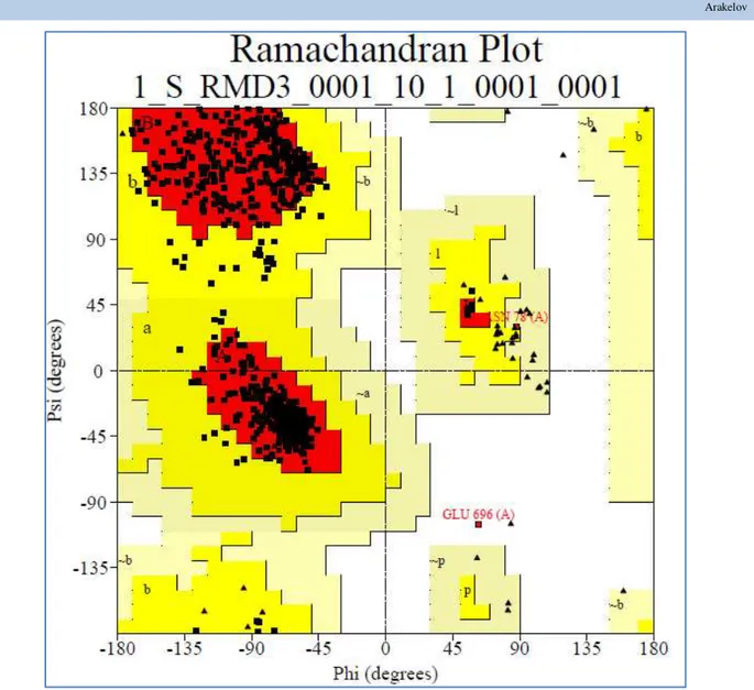
stereochemical correctness of protein, was carried out and the result of this test was visualized as a Ramachandran plot (Figure. 2). This figure presented torsion angles for all amino acids of pyrin. Lysine and proline residues are shown separately as triangles, because they are not tied to any definite region of the map. Painted regions on the map show its main favorable field while the darker region favors the combination
of φ and ψ angles. Furthermore, unpainted regions of the figure
2 represent the unfavorable regions of the map. Location of unfavorable regions is only allowed for glycine and proline residues, as they have other favorable and unfavorable regions because of its special stereochemistry.
Analysis of the Ramachandran plot of selected pyrin molecular models showed that 603 (91.6%) amino acids of pyrin were located in the most favored regions of map (A, B, L), in addition (a, b, l, p) 53 (8.1%) are available in allowable and less allowable regions (~ a, ~ b, ~ l, ~ p) -1 (0.2%) and only one amino acid (0.2%) ASN78 was located in the unacceptable regions of the map. Thus, 99.7% amino acids of the developed molecular structure are localized in the permissible regions of Ramachandran plot and it is quite enough to assess the quality and stereochemical correctness of the developed pyrin molecular structure. It has been experimentally established that if the favorable region contains higher than 90% amino acids the developed tertiary structure is stereochemically correct. .
221 Arakelov
_________________________________________________________ Journal of Experimental Biology and Agricultural Sciences http://www.jebas.org
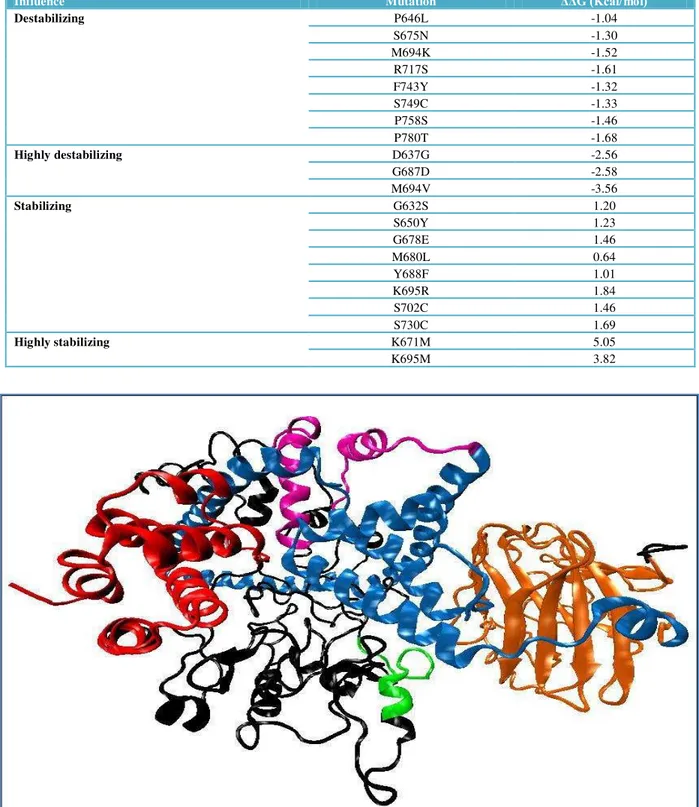
Table 1 Influence of mutations on the stability of pyrin tertiary structure.
Figure 1 Molecular Structure of pyrin developed by de novo and threading modeling technique DAPIN domain is shown by red color, bZIP – green, B-box type Zn-fingers - purple, α-Helix – blue, B30.2 – orange, and black color shows undescribed regions.
Influence Mutation ΔΔG (Kcal/mol)
Destabilizing P646L -1.04
S675N -1.30
M694K -1.52
R717S -1.61
F743Y -1.32
S749C -1.33
P758S -1.46
P780T -1.68
Highly destabilizing D637G -2.56
G687D -2.58
M694V -3.56
Stabilizing G632S 1.20
S650Y 1.23
G678E 1.46
M680L 0.64
Y688F 1.01
K695R 1.84
S702C 1.46
S730C 1.69
Highly stabilizing K671M 5.05
K695M 3.82
_________________________________________________________ Journal of Experimental Biology and Agricultural Sciences http://www.jebas.org
Figure 2 Ramachandran plot for the developed pyrin tertiary structure.
Furthermore, the stereochemical correctness of the obtained structure was also checked by distribution of rotation angles of the side chain. This is made possible by its resolution. As a result of validation it was reported that obtained model has a resolution of 1.6 Å and this is the highest resolution for such a large protein.
After molecular modeling of pyrin tertiary structure and assessment of its accuracy; effect of mutation on the stability of pyrin tertiary structure was also conducted with help of
SDM program. For this calculated ΔΔG was estimated by the
difference between ΔG native and ΔG mutated structures of
pyrin. Based on the obtained value a measure of change in protein stability under the influence of mutations was tested. In this study all the significant mutations (66) which were localized on the domain B30.2 of pyrin protein was studied.
Table 1 shows the values of ΔΔG for destabilizing, highly
destabilizing, stabilizing and highly stabilizing mutations.
Results of the study revealed that total 13 mutations (R628K, D661N, M680I, I720M, V722M, I729V, R737K, A744S, A744T, I755V, R761C, I772V, Q778L) have slightly destabilizing effect on the pyrin structure while 8 mutations (P646L, S675N, M694K, R717S, F743Y, S749C, P758S, P780T) have destabilizing and 3 mutations showed highly destabilizing (D637G, G687D,M694V) effect on the pyrin structure. Out of 66 reported mutations, 25 mutations are not showing any effect on the stability of the pyrin structure and considered as neutral mutation and not effecting the stability of the pyrin (N599D, I640M, I641F, R652C, R652H, R653H, E656A, I666V, M680V, T681I, M693I, L709R, M694L, K695N, V704I, P705S, R708C, R717H, V726A, N733S, F743L, Q753H, P754R, R761H, P769A).
Among the reported mutations, total 17 mutations shows stabilizing effect on the structure of pyrin, among these stabilizing mutations 7 were considered as slightly stabilizing
223 Arakelov
_________________________________________________________ Journal of Experimental Biology and Agricultural Sciences http://www.jebas.org
(G632A, L649P, V659F, Y688C, M694I, R717L, N766H) - while 8 were stabilizing (G632S, S650Y, G678E, M680L, Y688F, K695R, S702C, S730C) and rest 2 were considered as highly stabilizing (K671M, K695M). Effect of mutation on the structural stability and expression of pyrin was also reported by various researchers (Mansfield et al., 2001; Tchernitchko et al., 2004; Papin et al., 2007). In present study a correlation between the influence of the most common mutations and stability of the pyrin tertiary structure was also reported. These mutations affect the degree of severity of FMF. It has been also reported that the mutation M694V is a factor of severe flow of
FMF and has highly destabilizing effect (ΔΔG = -3.56) on the stability of the pyrin tertiary structure, while the mutation M680I is a factor of milder flow of FMF as compared to M694V and has slightly destabilizing effect (ΔΔG = -0.98).
Conclusions
The developed pyrin molecular model was found it stereochemical correct with resolution 1.6 Å. Furthermore, it has been reported that mutations those are localized on the B30.2 domain of pyrin having destabilizing or stabilizing effect on the protein tertiary structure and a correlation was also reported between the severity of mutations and stability of the pyrin tertiary structure.
Conflict of Interest
Author has not declared any conflict of interest.
Acknowledgements
I would like to express my sincere gratitude to my scientific adviser Ph.D., Professor K.B. Nazaryan.
References
Berjanskii M, Zhou J, Liang Y, Lin G, Wishart DS (2012) Resolution-by-Proxy: A Simple Measure for Assessing and Comparing the Overall Quality of NMR Protein Structures. Journal of Biomolecular NMR 53: 167-80. doi: 10.1007/s10858-012-9637-2.
Centola M, Aksentijevich I, Kastner DL (1998) The hereditary periodic fever syndromes: molecular analysis of a new family of inflammatory diseases. Human Molecular Genetics 7: 1581-1588. doi: 10.1093/hmg/7.10.1581.
Chae JJ, Aksentijevich I, Kastner DL (2009) Advances in the understanding of familial Mediterranean fever and possibilities for targeted therapy. British Journal of Haematology 146: 467–
478. doi: 10.1111/j.1365-2141.2009.07733.
Drenth JPH, van der Meer JWM (2001) Hereditary Periodic fever. The New England Journal of Medicine 345: 1748-1757. doi: 10.1056/NEJMra010200.
Ergüven M, Emeksiz C, Deveci M, Ozlü SG (2008) Relation between microalbuminuria and gene mutations in familial Mediterranean fever. The Turkish Journal of Pediatrics 50: 326-330.
Gershoni-Baruch R, Brik R, Shinawi M, Livneh A (2002) The differential contribution of MEFV mutant alleles to the clinical profile of familial Mediterranean fever. European Journal of Human Genetics 10: 145 – 149. doi: 10.1038/sj/ejhg/5200776.
Hakobyan D, Nazaryan K (2010) Molecular dynamics study of interaction and substrate channeling between neuron-specific enolase and B-type phosphoglycerate mutase. Proteins 78: 1691–1704. doi: 10.1002/prot.22686.
Humphrey W, Dalke A, Schulten K (1996) VMD: visual molecular dynamics. Journal of Molecular Graphics 14: 33-38 doi:10.1016/0263-7855(96)00018-5.
Kastner DL (2005) Hereditary periodic fever syndromes. Hematology / the Education Program of the American Society of Hematology 1: 74-81. doi: 10.1182/asheducation-2005.1.74.
Kuijk LM, Hoffman HM, Neven B, Frenkel J (2008) Episodic autoinflammatory disorders in children. In Handbook of systemic autoimmune diseases 1st ed. Amsterdam: Elsevier Pp. 119-35.
Leaver-Fay A, Tyka M, Lewis SM, Lange OF, Thompson J, Jacak R, Kaufman K, Renfrew PD, Smith CA, Sheffler W, Davis IW, Cooper S, Treuille A, Mandell DJ, Richter F, Ban YE, Fleishman SJ, Corn JE, Kim DE, Lyskov S, Berrondo M, Mentzer S, Popović Z, Havranek JJ, Karanicolas J, Das R, Meiler J, Kortemme T, Gray JJ, Kuhlman B, Baker D, Bradley P (2011) ROSETTA3: an object-oriented software suite for the simulation and design of macromolecules. Methods in Enzymology 487: 545-574. doi:10.1016/B978-0-12-381270-4.00019-6.
Mansfield E, Chae JJ, Komarow HD, Brotz TM, Frucht DM, Aksentijevich I, Kastner DL (2001) The familial Mediterranean fever protein, pyrin, associates with microtubules and colocalizes with actin filaments. Blood 98: 851- 859. doi: http://dx.doi.org/10.1182/blood.V98.3.851.
Pras E, Aksentijevich I, Gruberg L, Balow JE Jr, Prosen L, Dean M, Steinberg AD, Pras M, Kastner DL (1992) Mapping of a gene causing familial Mediterranean fever to the short arm of chromosome 16. The New England Journal of Medicine 326: 1509-1513. doi: 10.1056/NEJM199206043262301.
Sadovnichy V, Tikhonravov A, Voevodin VI, Opanasenko V (2013) "Lomonosov": Supercomputing at Moscow State University. In Contemporary High Performance Computing: From Petascale toward Exascale (Chapman & Hall/CRC Computational Science), Boca Raton, USA, CRC Press Pp.283-307.
_________________________________________________________ Journal of Experimental Biology and Agricultural Sciences http://www.jebas.org
The French FMF Consortium (1997) A candidate gene for familial Mediterranean fever. Nature Genetics 17: 25—31. doi:10.1038/ng0997-25.
Touitou I (2001) The spectrum of Familial Mediterranean Fever (FMF) mutations. European Journal of Human Genetics 9: 473- 483.
Weinert C, Grütter C, Roschitzki-Voser H, Mittl PR, Grütter MG (2009) The Crystal Structure of Human Pyrin B30.2 Domain: Implication for Mutations Associated with Familial
Mediterranean Fever. Journal of Molecular Biology 394: 226-236. doi:10.1016/j.jmb.2009.08.059.
Willard L, Ranjan A, Zhang H, Monzavi H, Boyko RF, Sykes BD, Wishart DS VADAR: a web server for quantitative evaluation of protein structure quality. Nucleic Acids Research 31 : 3316-3319. doi: 10.1093/nar/gkg565.
Worth CL, Preissner R, Blundell TL (2011) SDM - a server for predicting effects of mutations on protein stability and malfunction. Nucleic Acids Research 39: 215-222. doi: 10.1093/nar/gkr363.