Bovine Gamma Delta T Cells Contribute to
Exacerbated IL-17 Production in Response to
Co-Infection with Bovine RSV and
Mannheimia haemolytica
Jodi L. McGill1*, Rachel A. Rusk2, Mariana Guerra-Maupome2, Robert E. Briggs3, Randy E. Sacco3
1Department of Diagnostic Medicine/Pathobiology, College of Veterinary Medicine, Kansas State
University, Manhattan, Kansas, United States of America,2Pathobiology Graduate Program, Department of Diagnostic Medicine/Pathobiology, College of Veterinary Medicine, Kansas State University, Manhattan, Kansas, United States of America,3Ruminant Diseases and Immunology Research Unit, National Animal Disease Center, Agricultural Research Service, Ames, Iowa, United States of America
*jlmcgill@vet.k-state.edu
Abstract
Human respiratory syncytial virus (HRSV) is a leading cause of severe lower respiratory tract infection in children under five years of age. IL-17 and Th17 responses are increased in children infected with HRSV and have been implicated in both protective and pathogenic roles during infection. Bovine RSV (BRSV) is genetically closely related to HRSV and is a leading cause of severe respiratory infections in young cattle. While BRSV infection in the calf parallels many aspects of human infection with HRSV, IL-17 and Th17 responses have not been studied in the bovine. Here we demonstrate that calves infected with BRSV express significant levels of IL-17, IL-21 and IL-22; and both CD4 T cells andγδT cells con-tribute to this response. In addition to causing significant morbidity from uncomplicated infections, BRSV infection also contributes to the development of bovine respiratory dis-ease complex (BRDC), a leading cause of morbidity in both beef and dairy cattle. BRDC is caused by a primary viral infection, followed by secondary bacterial pneumonia by patho-gens such asMannheimia haemolytica. Here, we demonstrate thatin vivoinfection withM.
haemolyticaresults in increased expression of IL-17, IL-21 and IL-22. We have also devel-oped anin vitromodel of BRDC and show that co-infection of PBMC with BRSV followed by
M.haemolyticaleads to significantly exacerbated IL-17 production, which is primarily medi-ated by IL-17-producingγδT cells. Together, our results demonstrate that calves, like humans, mount a robust IL-17 response during RSV infection; and suggest a previously unrecognized role for IL-17 andγδT cells in the pathogenesis of BRDC.
OPEN ACCESS
Citation:McGill JL, Rusk RA, Guerra-Maupome M, Briggs RE, Sacco RE (2016) Bovine Gamma Delta T Cells Contribute to Exacerbated IL-17 Production in Response to Co-Infection with Bovine RSV and Mannheimia haemolytica. PLoS ONE 11(3): e0151083. doi:10.1371/journal.pone.0151083
Editor:Steven M. Varga, University of Iowa, UNITED STATES
Received:December 22, 2015
Accepted:February 23, 2016
Published:March 4, 2016
Copyright:This is an open access article, free of all copyright, and may be freely reproduced, distributed, transmitted, modified, built upon, or otherwise used by anyone for any lawful purpose. The work is made available under theCreative Commons CC0public domain dedication.
Data Availability Statement:All relevant data are within the paper.
Introduction
Human respiratory syncytial virus (HRSV) is a leading cause of acute lower respiratory tract disease in human infants worldwide. It is estimated that over 33 million children under the age of 5 years old contract the disease, resulting in over 3 million hospitalizations and nearly 200,000 deaths [1]. Currently, no licensed vaccine is available for HRSV.
Bovine respiratory syncytial virus (BRSV) is genetically and antigenically closely related to HRSV. BRSV infection is among the leading causes of lower respiratory tract infections in young calves [2–4]. BRSV infection in the calf parallels many features of HRSV infection in humans, including age-dependent susceptibility, microscopic lesions and innate and adaptive immune responses [3,5,6]. BRSV infection in neonatal calves represents a fully permissive, natural host-pathogen interaction that closely mimics HRSV infection in human infants. Thus, BRSV infection is increasingly recognized as an excellent model for studying disease pathogen-esis and immunity to HRSV infection in children, as well as for pre-clinical testing of novel vac-cines and therapeutics [3,5,6].
In addition to causing morbidity in calves due to uncomplicated infections, BRSV is an important contributing factor in the development of bovine respiratory disease complex (BRDC), a multifactorial disease condition affecting cattle in all stages and categories of pro-duction [2–4,7]. The etiology of BRDC is not well understood but usually affects animals that are stressed due to weaning, shipping or comingling, and is a result of co-infections by multiple viral and bacterial agents, including BRSV,Mannheimia haemolytica,Pasteurella multocida
andHistophilus somni[8–13]. BRDC is the leading cause of morbidity and mortality among feedlot cattle and is the most common cause of weaned dairy heifer mortality [3,4]. Economic costs to the cattle industry due to BRDC have been estimated as high as $3 billion annually due to death losses, reduced performance and costs of vaccinations and treatment modalities [14]. Features of BRDC include significant pro-inflammatory cytokine production and broncho-pneumonia including severe neutrophilia in the lungs. The mechanisms of immune suppres-sion and disease pathogenesis contributing to the development of BRDC remain unclear.
Hallmarks of severe RSV infection in infants and BRSV infection in calves include rapid neutrophil infiltration, excessive mucus production and a delayed virus-specific CD8 T cell response [15–17]. Strong expression of Th2 cytokines such as IL-13 are commonly implicated in disease pathogenesis [18]. However, recent results from humans and mice have suggested that IL-17-dependent immune activation may also be a critical factor in the response to RSV infection [19–23].
IL-17 is a pro-inflammatory cytokine that is primarily produced by activatedγδT cells and CD4+T cells, termed Th17 cells [24]. The main effector cytokines associated with Th17 immunity are IL-17A, (IL-17), IL-17F, IL-21 and IL-22. IL-17, like IFNγ, is a“master regula-tor”of downstream cytokine and chemokine responses. Expression of IL-17 stimulates stro-mal and epithelial cells to produce chemokines such as CXCL1, CCL20, IL-6, and most importantly, IL-8, leading to recruitment and activation of neutrophils and monocytes [25]. While critical for protection from several types of extracellular bacterial and fungal infections, the role of IL-17 and Th17 cells in immunity against intracellular pathogens such as RSV remains less clear. Faberet al. reported significantly lower levels of IL-17 in the bronchoalveo-lar lavage (BAL) of severely ill, pediatric RSV patients compared to less severe cases [19]. Sim-ilarly, a study by Larranagaet al. revealed higher plasma levels of IL-17 in infants with moderate RSV infection compared to those with severe disease [26]. In contrast, Mukherjee
et al. observed increased levels of IL-17 in tracheal aspirates from severely ill infants compared to healthy controls [21]. Mice deficient in STAT1 [27] or CCR7 [28] display increased pathol-ogy in the lungs that is attributed to increased production of IL-17; and animals deficient in Competing Interests:The authors have declared
IL-17 display significantly reduced mucus production and increased CD8 T cell responses in the lungs during primary RSV infection [21]. The role of IL-17 in RSV-associated allergic sen-sitization appears similarly complex, with reports that this cytokine both prevents [22] and promotes [21] airway reactivity.
Here, we report for the first time that calves infected with BRSV express increased levels of IL-17 in the lungs, and develop virus-specific IL-17 producing lymphocytes in the blood by day 7 after infection. Similarly, calves vaccinated with a live-attenuated strain of BRSV mount virus-specific IL-17 responses in the blood. Both CD4 andγδT lymphocytes have the capacity to secrete IL-17 in response to viral infection. Using anin vitroco-infection model of BRSV andM.haemolyticato model BRDC, we further demonstrate that BRSV-induced expression of IL-17 is significantly exacerbated in the presence of secondary bacterial exposure toM. haemo-lytica, suggesting a mechanism that may be contributing to neutrophilia in the lungs of calves with BRDC. Interestingly, this exacerbated IL-17 production appears primarily mediated byγδ
T cells, as depletion of these cells from culture ablates the effect. Together, our results demon-strate that calves, like humans and mice, mount a significant IL-17 response during acute BRSV infection and suggest a possible role forγδT lymphocytes in contributing to BRDC-associated immunopathology in the calf.
Materials and Methods
Animals
BRSV infected animals. Lung samples and peripheral blood samples were collected over 3
experiments from a total of 16, 4–6 week old Holstein calves. Eight calves were infected with BRSV Strain 375 via aerosol inoculation as previously described [29,30]. Eight uninfected, age-matched calves served as negative controls. All animals were housed in a climate-controlled, biosafety level-2 facility at the National Animal Disease Center in Ames, IA. Peripheral blood was collected via the jugular vein on day 0 prior to infection, and on day 3 and 7 post infection. Calves were monitored daily following experimental infection. Clinical signs were evaluated including rectal temperature, evidence of ocular or nasal discharge, cough, dyspnea, and appe-tite. The animal care protocol included provisions for a humane endpoint as determined by the discretion of the attending clinical veterinarian. No animals died or required euthanasia prior to the experimental endpoint. Methods to minimize pain and distress included the avoidance of prolonged restraint and the inclusion of euthanasia as an intervention strategy. Fundamental to the relief of pain and distress is the ability to recognize clinical signs of RSV in calves. The investigator provides assurance that any unrelieved pain and distress occurred only for the minimum time necessary to achieve study objectives and was based on sound scientific ratio-nale. Calves were euthanized by barbiturate overdose on day 7-post infection. Lungs were removed and samples from representative gross lesion and nonlesion tissue collected from each calf and stored in RNAlater(Invitrogen, Life Technologies).
M.haemolyticainfected animals. Lung samples were collected from a total of 8, 6-month
attending clinical veterinarian. Methods to minimize pain and distress included the avoidance of prolonged restraint and the inclusion of euthanasia as an intervention strategy. No animals died or required euthanasia prior to the experimental endpoint. Calves were euthanized by cap-tive bolt on day 3-post infection. Lungs were removed and samples from representacap-tive gross lesion and non-lesion tissue collected from each calf and stored in RNAlater.
BRSV vaccinated animals. PBMC were collected from a total of 16 female adult Holstein
cows housed at the dairy facility at the National Animal Disease Center in Ames, IA (n = 8) or the Kansas State Dairy in Manhattan, KS (n = 8). All cows were on an annual vaccination program and received a multivalent commercial vaccine containing live attenuated BRSV (BoviShield Gold FP5) within 4–12 weeks of inclusion in the study. No experimental end-point was established for these animals, as they were healthy and were not experimentally infected. All provisions to minimize pain and distress were used including phlebotomy by experienced handlers and avoidance of prolonged restraint. All animals tested positive for antibodies to BRSV (titer of 1:32–1:128), and positive for BRSV-specific CD4 T cell responses prior to enrollment into the study. Six cows that were not on the annual vaccination program due to inclusion in another study were used as nonvaccinated controls. Control cows were also housed at the Kansas State University Dairy in Manhattan, KS. These animals had a BRSV titer of 1:8–1:64, but had no detectable BRSV-specific CD4 T cell responses as mea-sured by T cell proliferation assays.
All animal studies were conducted in strict accordance with federal and institutional guide-lines and were approved by the National Animal Disease Center Institutional Animal Care and Use Committee or the Kansas State University Institutional Animal Care and Use Committee, as appropriate.
Peripheral blood collection and mononuclear cell preparation
Mononuclear cells were isolated by density centrifugation of peripheral blood collected from the jugular vein into 2x acid citrate dextrose. Contaminating red blood cells were removed using hypotonic lysis. Cells were washed and resuspended in complete RPMI (cRPMI) as previ-ously described [30].
CD4 T cells andγδT cells were enriched from PBMC using Magnetic Activated Cell Sorting (MACS) as previously published [33]. Briefly, PBMC were labeled with 10μg/mL mouse anti-bovine CD4 (clone ILA11A) or 10μg/mL mouse anti-bovineγδT cell receptor (Clone GB21A), both from Washington State Monoclonal Antibody Center (Pullman, WA). Cells were then labeled with anti-mouse IgG2A+B magnetic beads (Miltenyi Biotech) and purified by positive sorting per manufacturer’s instructions.
WC1 expressing subsets ofγδT cells were isolated by FACS sorting as previously described [30,33]. PBMC were labeled with mouse bovine WC1.1 (Clone BAG25A), mouse anti-bovine WC1.2 (Clone CACTB32A) and mouse anti-anti-bovineγδT cell receptor (Clone
GB21A), all from Washington State Monoclonal Antibody Center (Pullman, WA). Cells were then labeled with anti-mouse IgG1-AF488, anti-mouse IgM-PE and anti-mouse IgG2b-AF647 secondary antibodies (Invitrogen, Life Technologies). Cells were sort purified based upon their expression of theγδT cell receptor and WC1.1, WC1.2 or lack of WC1 expression. Subsets were sorted to>90% purity using a BD FACS Aria Cell Sorting System (BD
Biosci-ences, San Jose, CA).
In vitro
cell stimulation
PBMC or MACS purified T lymphocytes were plated at a concentration of 4x10⁶cells/mL in cRPMI (100μl/well) in sterile, round-bottom, 96-well tissue-culture-treated plates (BD Biosci-ences). For experiments requiring antigen-presenting cells (APC), monocytes were plated at a ratio of 1:5 with purified CD4 orγδT cells. In some experiments, cells were labeled with Cell Trace Violet per manufacturer’s recommendations (Invitrogen, Life Technologies) prior to being placed in culture. Forin vitrore-stimulation with BRSV, cells were incubated at a 0.1 multiplicity of infection (MOI) with BRSV Strain 375 for 90 min at 37°, washed once and resuspended in cRPMI for the remaining incubation period. For experiments using heat-killed BRSV, virus stock was inactivated for 30 minutes at 56°C, then 50μl added to the cultures (final volume of 200μl/well).
Forin vitroBRDC experiments, cells were infected with BRSV Strain 375 as above. Four hours later,M.haemolyticastrain D153, serotype A1 orPastuerella multocidastrain P1062, serotype A3 was suspended in antibiotic-free RPMI and added to the cell cultures at ~0.1 MOI as determined by spectrophotometric growth curves. After 3–4 hours, cells were resuspended in antibiotic-containing cRPMI to prevent bacterial overgrowth. Importantly, we performed dose titrations to determine the optimal concentration ofM.haemolyticato add to our cul-tures. The addition of higher concentrations ofM.haemolyticaresulted in significant cell death, likely a result of the action of leukotoxin [34–36], while an MOI of 0.5 or less did not induce significantly increased cell death over uninfected cultures (not shown). Heat-inactivated
M.haemolyticaD153 was prepared by suspending bacteria in antibiotic-free RPMI at approxi-mately 0.1 MOI, then boiling for 15 minutes. InactivatedM.haemolyticawas then added to PBMC cultures for 3–4 hours, followed by centrifugation and a media change to antibiotic-containing cRPMI as above.
At the indicated times (24 hours or 6 days of culture), PBMC, CD4 orγδT cells were pel-leted by centrifugation and cell culture supernatants were collected and stored at−80°C. Cell
pellets were then lysed with Buffer RLT (Qiagen, Valencia, CA) containing 2-mercaptoethanol and stored at−80°C.
Real-time PCR
RNA was isolated from lung tissue samples using Trizol Reagent (Invitrogen, Life Technolo-gies) according to manufacturer’s instructions. The RNA concentration in each sample was determined using a NanoDrop 2000 spectrophotometer (Thermo Scientific, Wilmington, DE). RNA (300 ng per sample) was DNase-treated and cDNA synthesized using random primers and Superscript III Reverse Transcriptase according to the manufacturer's instructions (Invi-trogen, Life Technologies).
For PBMC, purified T cell populations and bovine turbinates (BT), RNA was extracted using the RNeasy Mini RNA Isolation kit (Qiagen) according to manufacturer's instructions. Contaminating genomic DNA was removed using the RNase-Free DNase digestion set (Qia-gen) as per manufacturer's instructions. Total eluted RNA was reverse transcribed into cDNA using Superscript III Reverse Transcriptase and Random Primers following the manufacturer's instructions.
1 min at 60°, 15 seconds at 95°, 15 seconds at 60°). Relative gene expression was determined using the 2-ΔΔCtmethod with RPS9 as the reference housekeeping gene.
IL-17 ELISA
The Bovine IL-17A VetSet ELISA Development kit was purchased from Kingfisher Biotech, Inc. (St. Paul, MN). ELISAs were performed using cell culture supernatants according to manu-facturer’s instructions.
Stimulation of BT
BT cells were cultured in cMEM, plated in 96-well, flat-bottom tissue-culture-treated plates and grown to confluence overnight at 37°C. Cell culture supernatants from day 6 stimulated PBMC, CD4 orγδT cell culture supernatants were diluted 1:2 with cMEM, and then added to the BT cells. Cells were incubated overnight, then lysed with Buffer RLT (Qiagen, Valencia, CA) containing 2-mercaptoethanol and stored at−80°C until RNA extraction.
Statistical Analysis
ΔΔCt values were used in the statistical analysis of relative gene expression.ΔΔCt values were transformed (2-ΔΔCt) and are shown as expression relative to uninfected or unstimulated
con-trol samples, as appropriate. [40]
Statistical analyses were performed using Prism v6.0f software (Graphpad Software, Inc.) Statistical comparisons were performed using a two-tailed Student’st-test when comparing control to infected or stimulated samples; or a one-way ANOVA with Bonferri post-test analy-sis, when comparing multiple treatment conditions (in vitroBRDC studies).
Results
IL-17 response in the lungs and peripheral blood of calves infected with
bovine RSV
IL-17 expression is increased in mice and humans with acute RSV infection [20–23,41], but has not been studied in calves with acute BRSV infection. To this end, neonatal Holstein calves
Table 1. Primers used for qPCR.
Gene (alternate name) Accession number Strand Sequence (5’-3’) Reference
IL17 NM_001008412 Forward CACAGCATGTGAGGGTCAAAC [37]
Reverse GGTGGAGCGCTTGTGATAAT
IL-21 NM_198832 Forward GTGGCCCATAAGTCAAGCTC [38]
Reverse CGCTCACAGTGTCTCTTTAC
IL-22 NM_001098379 Forward GACTGTGGAGTTTGGCTCCCCCTTTTC [38]
Reverse GAGATTAAAGTCATTGGAGAACTGAAC
IL-8 X78306 Forward AAGCTGGCTGTTGCTCTC [29]
Reverse GGCATCAGAAGTTCTGTACTC
MUC5AC BN001491 Forward CAGACCCTCCACCTTCTTCA [39]
Reverse GGTCCTCGAAGCTGTTCTTG
MUC5B BN0011492 Forward TCTACCTGACCGTGGAGACC [39]
Reverse GTTGATGATGCTGCACTGCT
RPS9 NM_001101152.1 Forward CGCCTCGACCAAGAGCTGAAG [29]
Reverse CCTCCAGACCTCACGTTTGTTCC
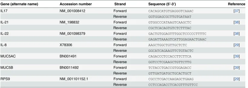
were infected with BRSV strain 375 via aerosol inoculation. On day 7-post infection, lung sam-ples were analyzed by qPCR for expression of IL-17 and the Th17-associated cytokines IL-21 and IL-22. As seen inFig 1, BRSV infection resulted in significantly increased expression of IL-17 and IL-22 in the lungs of infected calves (Fig 1A). IL-21 expression was not significantly increased in pneumonic lung at this time.
IL-17 promotes increased production of the pro-inflammatory chemokine, IL-8. IL-8 is increased in humans with RSV [42,43] and we have previously demonstrated that IL-8 is upre-gulated in the lungs of calves with BRSV [29]. In agreement with these previous reports, IL-8 expression was significantly increased in the lung lesions of calves infected with BRSV com-pared to lungs from uninfected control calves (Fig 1B).
To determine if BRSV infection induced the development of systemic Th17 responses, peripheral blood was collected from control and BRSV-infected calves on day 7-post infection. PBMC were restimulated with heat-killed BRSV antigen for 24 hours (Fig 1C) and examined by qPCR for expression of IL-17. PBMC from calves with acute BRSV infection expressed sig-nificantly increased levels of IL-17 in recall response to BRSV infection. Importantly, signifi-cant IL-17 expression was not observed in PBMC collected from the calves on day 0 or 3-post infection (data not shown). PBMC were also stimulated with heat-killed BRSV antigen for 6 days and culture supernatants analyzed by ELISA. In agreement with the qPCR results, PBMC
Fig 1. IL-17 and Th17 responses in the lungs and peripheral blood of calves infected with BRSV.Calves (n = 8) were infected via aerosol inoculation with BRSV Strain 375 (RSV) as described in Materials and Methods. Control calves remained uninfected (n = 8). On day 7 post-infection, the animals were sacrificed and the lungs analyzed by qPCR for expression of IL-17, IL-21 and IL-22 (A) and IL-8 (B). In separate experiments, PBMC were isolated from control (n = 8) and BRSV infected calves (n = 8) on day 7-post infection. PBMC were stimulated with BRSV for 24 hours and then analyzed for expression of IL-17 by qPCR (C); or for 6 days and then cell culture supernatants were analyzed by ELISA for expression of IL-17 (D). For qPCR analysis, results were normalized to the housekeeping gene RPS-9, and expressed relative to samples from uninfected control calves. Results were pooled from a total of three independent experiments. Data represent means±SEM.
from infected, but not control calves secreted significantly increased concentrations of IL-17 in recall response to BRSV antigen (Fig 1D).
IL-17 response in the peripheral blood of calves vaccinated with
live-attenuated BRSV
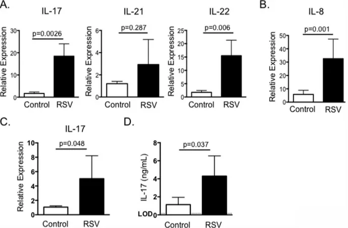
We next chose to determine if BRSV vaccination also induces the development of virus-specific IL-17 responses. PBMC were collected from adult Holstein cows within 4–12 weeks of
Fig 2. IL-17 and Th17 responses from BRSV vaccinated cattle.Peripheral blood was collected from cows receiving annual vaccinations with a multivalent vaccine containing live-attenuated BRSV (n = 8), or from control cows that were not included in the vaccination program due to inclusion in another study (n = 6). PMBC were isolated and stimulated with BRSV for 24 hours or 6 days, as inFig 1. RNA was isolated from the cells and analyzed by qPCR for expression of IL-17 (A, left panel), IL-21 (B) and IL-22 (C). Cell culture supernatants were also analyzed by ELISA for IL-17 (A, right panel). For qPCR analysis, results were normalized to the housekeeping gene RPS-9, and expressed relative to unstimulated control samples. Results were pooled from two independent experiments. Data represent means±SEM.
receiving an annual booster of live, attenuated BRSV vaccine. Peripheral blood was also col-lected from control cows that were not included in the annual vaccination program due to inclusion in another study. PBMC from both groups of animals were stimulated with heat-killed BRSV strain 375 and assessed by qPCR 24 hours later for expression of IL-17. In agree-ment with our results from calves with acute BRSV infection, cells from BRSV vaccinated ani-mals produced virus-specific IL-17 as measured by qPCR (Fig 2A) and by ELISA (Fig 2B). Similar to our results from BRSV infected calves, PBMC from vaccinated cows also expressed increased levels of IL-21 and IL-22 in recall responses to BRSV antigen (Fig 2).
Severe RSV infection in humans is associated with increased neutrophil infiltration and increased mucus production. IL-17 reportedly increases expression of IL-8, a critical neutrophil chemoattractant, and the genes encoding mucins, MUC5AC and MUC5B [24]. To confirm that IL-17 and the Th17 associated cytokines induced by BRSV displayed similar activity in the bovine, we treated BT cells for 24 hours with supernatants collected from BRSV antigen-stimu-lated cultures. BT cells were then analyzed by qPCR for expression of IL-8 (Fig 3A), MUC5AC (Fig 3B) and MUC5B (Fig 3C). As seen inFig 3, cell culture supernatants from non-vaccinated cows that were stimulated with BRSV induced only minor expression of IL-8, MUC5B and MUC5AC by BT, while stimulated supernatants from vaccinated calves induced increased expression of all three target genes by BT cells.
CD4 and
γδ
T cells produce IL-17 in response to BRSV
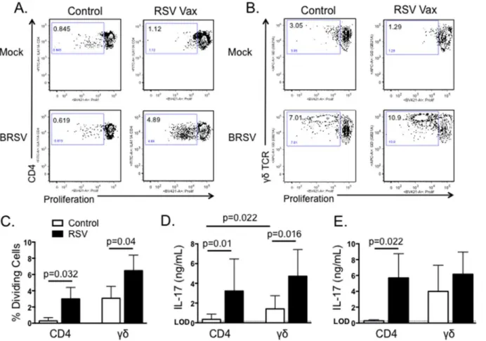
Both CD4 T cells andγδT cells have the capacity to produce IL-17 in response to RSV infection in humans and mice [24]. Therefore, we next chose to determine which T cell subsets from cat-tle produce IL-17 in recall response to BRSV stimulation. PBMC from BRSV vaccinated catcat-tle were labeled with CellTrace Violet, then cultured for 6 days in the presence or absence of heat-killed BRSV. CD4 T cells andγδT cells were then analyzed by flow cytometry for BRSV-specific proliferation as measured by CellTrace Violet dilution. As seen inFig 4A–4C, both CD4 T cells andγδT cells from vaccinated cows proliferate in specific response to stimulation with BRSV antigen. Not surprisingly, we observed higher levels of non-specific proliferation byγδT cells from control, non-vaccinated cows; however, BRSV-induced proliferation was still significantly
Fig 3. Cell culture supernatants from BRSV stimulated PBMC induce expression of IL-8, MUC5B and MUC5AC by BT cells.PBMC from control (nonvaccinated) and BRSV vaccinated cows were isolated and stimulated with heat-killed BRSV for 6 days as inFig 2. Cell culture supernatants were then diluted 1:1 in cMEM and added to confluent BT cells in 96 well plates for 24 hours. RNA was the isolated from the BT and analyzed by qPCR for expression of IL-8 (A), MUC5B (B) and MUC5AC (C). For qPCR analysis, results were normalized to the housekeeping gene RPS-9, and expressed relative to
unstimulated control samples. Results were pooled from two independent experiments. Data represent means±SEM.
increased above background levels (Fig 4C). Unfortunately, an antibody is not available for intracellular cytokine staining of bovine IL-17. Therefore, CD4 T cells andγδT cells were puri-fied by MACS and cultured with autologous APC in the presence or absence of BRSV antigen for 6 days. Cell culture supernatants were then analyzed by ELISA to determine concentrations of IL-17. As seen inFig 4D, both CD4 T cells andγδT cells secrete IL-17 in specific response to BRSV antigen.γδT cell production of non-specific IL-17 was increased compared to CD4 T cells, but stimulation with BRSV antigen resulted in a significant increase in virus-specific IL-17 production by purifiedγδT cells. Importantly, we observed similar IL-17 production when ana-lyzing CD4 andγδT cells isolated from peripheral blood of BRSV infected calves (Fig 4E).
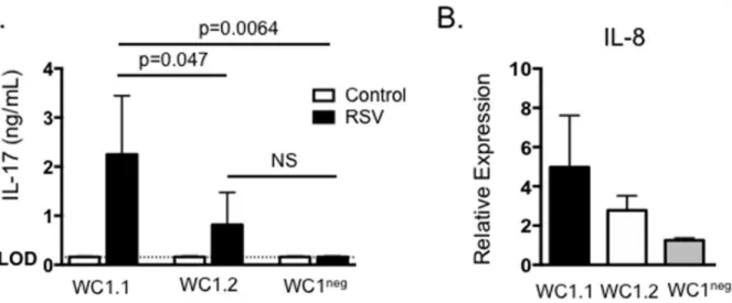
In cattle,γδT cells are phenotypically and functionally divided into subsets based upon their expression of WC1. We have previously demonstrated that WC1neg, WC1.1+and WC1.2+γδT cell subsets respond to BRSV infection, and the subsets differ in their response to virus with regards to inflammatory chemokine expression, IFNγproduction and production of regulatory cytokines such as IL-10 [30]. We next chose to determine the contribution of indi-vidual WC1-expressing subsets in production of IL-17 during the response to BRSV.γδT cells
Fig 4. Both CD4 T cells andγδT cells produce IL-17 in response to BRSV.PBMC were isolated from control or BRSV vaccinated cows and labeled with Cell Trace Violet. Cells were then cultured for 6 days with BRSV. On day 6, CD4 T cells (A) andγδT cells (B) were analyzed for virus-specific proliferation as measured by Cell Trace Violet dilution. Representative flow plots are shown in A and B. Aggregate results are shown in C. (D) CD4 T cells andγδT cells from BRSV vaccinated or nonvaccinated animals were isolated by MACS and cultured in the presence of autologous APC±BRSV. After 6 days, cell culture supernatants were analyzed by ELISA for IL-17. (E) CD4 T cells andγδT cells were MACS purified from peripheral blood of calves infected or not with BRSV strain 375 for 7 days. Purified cells were cultured in the presence of autologous APC±BRSV. After 6 days, cell culture supernatants were analyzed by ELISA for IL-17. For A-C, background levels of proliferation were subtracted and results are presented as change over mock. Results are pooled from two
independent experiments. Data represent means±SEM.
were FACS purified into subsets based upon their expression of theγδT cell receptor and WC1.1, WC1.2 or lack of WC1 as previously described [30,33]. Cells were cultured as inFig 4
and then supernatants were assessed for concentrations of IL-17 by ELISA. As seen inFig 5A, WC1.1+γδT cells secreted significant levels of IL-17 in response to BRSV antigen, while nei-ther WC1.2+nor WC1negsubsets produced IL-17, suggesting the WC1.1 subset may be the pri-maryγδT cell-derived source of IL-17 during BRSV infection in the calf. Stimulation of BT cells with culture supernatants from all three subsets of purifiedγδT cell subsets resulted in an increase in IL-8 expression (Fig 5B).
IL-17 expression in the lungs during
M.
haemolytica
infection
M.haemolyticais the predominant bacterial isolate recovered from cases of bovine pneumonia and the leading cause of direct economic loss from BRDC in the United States [44]. IL-8 is upregulated in the lungs ofM.haemolyticainfected cattle [45,46], and significant numbers of neutrophils are recruited to the lungs following infection, resulting in the exacerbated inflam-mation and tissue damage typical of BRDC [8,34]. We next determined if IL-17 is contributing to this increase in IL-8 production and neutrophil recruitment duringM.haemolytica infec-tion. To this end, cattle were infected withM.haemolyticavia endoscope-guided inoculation. On day 3 after inoculation, animals were sacrificed and their lungs examined by qPCR for expression of IL-17. As seen inFig 6A,M.haemolyticainfection results in increased expression of IL-17, IL-22 and IL-23 in the lungs by day 3 after infection. In agreement with previous stud-ies [45,46], we also observed significantly increased expression of IL-8 in lung tissue fromM.
haemolyticainfected calves compared to controls (Fig 6B).
In vitro
infection with BRSV and
M.
haemolytica
induces exacerbated
production of IL-17
Primary BRSV infection is a significant factor promoting the development of secondary bacterial pneumonia caused by pathogens such asM.haemolytica. We are interested in determining
Fig 5. WC1.1+γδT cells produce IL-17 in response to BRSV.γδT cells were purified from the peripheral blood of BRSV vaccinated or control animals by FACS based upon their expression of theγδT cell receptor and either expression of WC1.1, WC1.2 or lack of WC1. Cells were cultured in the presence of autologous APC±BRSV for 6 days. Cell culture supernatants were then analyzed by ELISA for IL-17. (B) Cell culture supernatants from (A) were also diluted 1:1 and added to BT cells for 24 hours. After 24 hours, BT were analyzed by qPCR for expression of IL-8. For qPCR analysis, results were normalized to the housekeeping gene RPS-9, and expressed relative to unstimulated control samples. Results are pooled from two independent experiments. Data represent means±SEM.
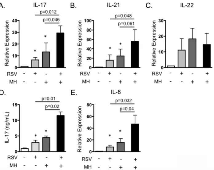
mechanisms of immunity that predispose an animal to the development of BRDC. IL-17 plays a critical role in neutrophil recruitment and inflammation, and neutrophilia in the lungs is a trait characteristic of severe pasteruellosis in calves with BRDC [45]. Therefore, we chose to deter-mine the effect of secondary bacterial exposure on IL-17 expression and its possible role in the development of exacerbated bacterial pneumonia. To this end, we developed anin vitromodel of BRDC. PBMC were infected with BRSV, followed by exposure to liveM.haemolytica, mim-icking a situation that may occur in the lungs, where T lymphocytes recruited to control BRSV infection are exposed toM.haemolyticaduring secondary bacterial infection. InFig 7, PBMC were infected with 0.1 MOI of BRSV strain 375 for 6 hours, thenM.haemolyticastrain D153 was added to the cultures at an MOI of 0.1. Cells and bacteria were incubated for 4 hours in anti-biotic free media, then antianti-biotics were added to control possible bacterial overgrowth. Cells were stimulated for 18 hours and then analyzed by rtPCR, and were also cultured for 6 days and supernatants measured by ELISA.M.haemolyticaproduces a potent leukotoxin that can induce rapid cell death in activated bovine PBMC [34–36]. Thus, dose titration experiments were per-formed to determine the optimal dose ofM.haemolyticato add that did not induce significantly increased cell death in our PBMC cultures. Doses ofM.haemolyticaabove 0.5 MOI induced sig-nificantly increased cell death in our PBMC coinfection cultures (data not shown), thus we selected an MOI of 0.1. As seen inFig 7A, cells exposed to either BRSV alone orM.haemolytica
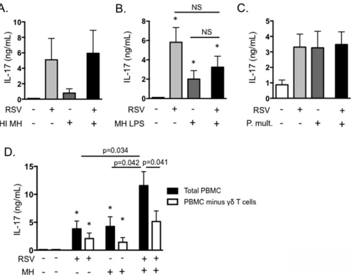
alone secreted IL-17. However, IL-17 was significantly increased in the presence of both BRSV andM.haemolytica. A similar exacerbation of IL-21 expression was also observed in the pres-ence of both BRSV andM.haemolytica, but not following stimulation with either pathogen sin-gly (Fig 7B). In contrast, expression of IL-22 was not increased following exposure to both BRSV andM.haemolytica(Fig 7C). ELISA for IL-17 protein in day 6 culture supernatants confirmed our qPCR results, demonstrating increased production of IL-17 in response to BRSV orM. hae-molyticaalone, but significantly exacerbated production in response to the pathogens together (Fig 7D). Addition of the stimulated cell supernatants to BT cells as inFig 3, also resulted in sig-nificantly increased production of IL-8 (Fig 7E). This suggests that the increased production of IL-17 during co-infections may significantly alter inflammatory cytokine production by epithe-lial and stromal cells in the lungs, leading to the severe neutrophilia that is a hallmark of the BRDC phenotype (Fig 7). Interestingly, a similar result is not observed in response to heat-inac-tivatedM.haemolytica(Fig 8A) or in response to purifiedM.haemolyticaLPS (Fig 8B).
Fig 6.M.haemolyticainfection induces IL-17 and Th17 cytokine production.Calves (n = 4) were infected with a field strain ofM.haemolyticavia endoscope-guided inoculation as described in Materials and Methods. Control calves were sham inoculated (n = 4). On day 3 after infection, animals were sacrificed and their lungs analyzed by qPCR for expression of IL-17, IL-21 and IL-22 (A); and for expression of IL-8 (B). Results were normalized to the housekeeping gene RPS-9, and expressed relative to samples from uninfected control calves. Results are pooled from one experiment. Data represent means±SEM.
Pasteruerella multocidais also a known cause of BRDC in young calves. LikeM.haemolytica
infection, pathogenesis ofP.multocidapneumonia includes severe neutrophilia in the lungs [9]. We next determined if co-infection withP.multocidaresults in a similar exacerbation of IL-17. Interestingly, althoughP.multocidaalone promotes IL-17 production by bovine PBMC, co-culturing of PBMC withP.multocidaand BRSV together did not result in the same enhanced IL-17 phenotype that was observed in response toM.haemolytica(Fig 8B).
To determine which cells were responsible for the exacerbated production of IL-17, we next conducted depletion experiments.γδT cells were depleted from the cultures by MACS, prior to BRSV/M.haemolyticaco-infection. As seen inFig 8D, depletion ofγδT cells resulted in slight, but not significant reductions in IL-17 production in cultures exposed to BRSV orM.
haemolyticaindividually. However, depletion ofγδT cells significantly reduced the exacer-bated production of IL-17 in co-infected cultures (Fig 8D). Depletion of CD4 T cells from the cultures yielded an overall reduction in IL-17 production in both singly and co-infected
Fig 7.In vitroinfection with BRSV andM.haemolyticaresults in exacerbated IL-17 production.PBMC from BRSV vaccinated cows were stimulated with BRSV for 6 hours, then cultured with 0.1 MOIM.haemolyticain antibiotic free media for 4 hours. After four hours, antibiotics were added, and then incubations were continued for 18 hours (A-C) or 6 days (D and E). Control cultures were included that received neither pathogen, BRSV alone orM.
haemolyticaalone. (A-C) Cells were analyzed by qPCR for expression of IL-17 (A), IL-21 (B) and IL-22 (C). Cell culture supernatants from day 6 were analyzed by ELISA for IL-17 (D) and were diluted 1:1 and added to BT for 24 hours (E). BT were analyzed by qPCR for expression of IL-8 after overnight stimulation as inFig 3. Results in A-C are pooled from three independent experiments with n = 12. Results in D and E are from two experiments with n = 8. Data represent means±SEM.
cultures, but did not alter the exacerbated phenotype observed following exposure to both pathogens (not shown). Together, our results suggest thatγδT cells are a significant source of IL-17 duringin vitrovirus/bacterial coinfections, suggestingγδT cell derived IL-17 may also be important in the pathogenesis of BRDC in the calf.
Discussion
We demonstrate here for the first time that neonatal calves infected with BRSV mount a robust IL-17 response in the lungs and peripheral blood. Both CD4 andγδT cells are a significant source of IL-17 in recall responses to BRSV antigen. Similarly, both CD4 T cells andγδT cells in rodents and humans contribute to IL-17 production in the lungs during RSV infection [19–
23,27,28]. Previous results have suggested thatγδT cells may contribute to early IL-17
Fig 8.γδT cells are the primary source of exacerbated IL-17 production in anin vitromodel of BRDC.(A and B) PBMC from BRSV vaccinated cows were stimulated with BRSV for 6 hours, then with 0.1 MOI of heat-inactivatedM.haemolytica(A) or 1μg/mL purified LPS fromM.haemolytica(B). Control cells remained unstimulated, were stimulated with only BRSV or were stimulated with only heat-inactivatedM.haemolyticaLPS. After six days, cell culture supernatants were analyzed by ELISA for IL-17. (C) PBMC from BRSV vaccinated cows were stimulated with BRSV for 6 hours, then cultured with 0.1 MOI
P.multocidain antibiotic free media for 4 hours. After four hours, antibiotics were added, and then incubations were continued for 6 days. Control cultures were included that received neither pathogen, BRSV alone orP.multocidaalone. Cell culture supernatants were analyzed by ELISA for IL-17. (D) PBMC were depleted ofγδT cells using MACS. Total PBMC andγδT cell-depleted PBMC were then cultured in the presence of BRSV andM.haemolyticaas above. After 6 days, cell culture supernatants were analyzed by ELISA. Results in A-C are from one experiment with n = 4. Results in D are pooled from 2 independent experiments with n = 8. Data represent means±SEM.
production, while Th17 cells are more important later in infection. On day 7 post-infection in the calf, our results indicate that both cell subsets have the capacity for significant virus-specific IL-17 production when isolated from peripheral blood. We were unable to detect BRSV-spe-cific IL-17 production by peripheral blood lymphocytes at earlier time points after infection, although possible IL-17 expression in the lungs at early time points was not examined. In future studies, a more detailed analysis of the kinetics of IL-17 production by both cell types in the blood and at the site of virus infection in the lungs will be valuable.
Currently, the extent ofγδT cell involvement in RSV disease in humans is poorly defined. We have previously described potent chemokine and cytokine production by bovineγδT cells responding toin vitroandin vivoBRSV infection [30]; and we demonstrate here thatγδT cells may play a critical role in IL-17 production during RSV infection in the calf. We suggest that the bovine represents an excellent model for further delineating the role ofγδT cells in protec-tion or immunopathology in the lungs during RSV infecprotec-tion.γδT cells are conserved amongst all vertebrate species, and are found in large proportions in a number of animal species includ-ing cattle, sheep, pigs and poultry [47–50]. In young cattle,γδT cells can comprise as high as 60% of circulating blood mononuclear cells, while the percentage in adult animals declines to around 8–18% [47,51]. The percentage ofγδT cells in humans is much smaller, ranging from 2–5% in healthy adults [52]. However, the frequency ofγδT cells circulating in human patients with certain bacterial and protozoal infections can increase to as high as 57% of circulating CD3+T cells [52].γδT cells in both humans and cattle are hypothesized to play an immune sentinel role, and large proportions are also found in the respiratory tract, mucosa and skin of both species [53]. With the exception of their increased abundance in the bovine immune sys-tem, literature suggests many similarities between bovine and humanγδT cells. Amongst these, the ability to process and present antigens via MHC class II [54,55], lack of MHC restriction [53,56], potent inflammatory chemokine and cytokine production [57], and innate recognition of conserved pathogen associated molecular patterns via pattern recognition recep-tors such as TLR and NLR [30,57–61].
Studies in mice and humans have demonstrated production of IL-17 during acute infection and recovery from BRSV. However, it remains debated if this response has positive or negative consequences on the host. Some reports in children have suggested increased levels of IL-17 in the BAL and plasma correlate with less severe RSV disease [19,26]; while others have reported the opposite [21]. In mice, it appears that IL-17 primarily contributes to immunopathology, increased production of IL-13 and increased mucus production in the lungs [21,23,27,28,41]; although these effects vary depending upon the virus strain used. Importantly, conclusions drawn from rodent models of RSV must be interpreted with caution, as it is clear that mice are only semi-permissive to virus infection and do not reflect many aspects of disease pathogenesis and immunity observed in RSV-infected children [3,5,6]. As a fully-permissive host for BRSV, the calf model may be ideal for more clearly determining the role of IL-17 in immunity to RSV infection, as well as elucidating the circumstances that contribute to a protective or pathogenic role for IL-17.
production. In contrast, however, we observed significantly increased production of IL-17 in cell cultures exposed to both BRSV andM.haemolyticatogether, compared to either pathogen singly. It is important to note that ourin vitroBRDC experiments were all carried out using lymphocytes isolated from peripheral blood. It is currently unknown if similarly exacerbated IL-17 production would be observed using cells isolated from the lungs, or duringin vivo
BRSV/M.haemolyticaco-infection. It is possible that lung-resident subsets of CD4 andγδT cells may respond differently to co-infection than cells from the blood, or that effects of thein vivolung microenvironment may alter IL-17 production and the inflammatory response. How-ever, given the significant neutrophilia that is observed in calves with BRDC, ourin vitro exper-iments support the possibility that exacerbated IL-17 production, and hence exacerbated neutrophil recruitment, may be occurring duringin vivoinfection. Thus, based upon results from ourin vitrocoinfection model, we hypothesize that increased and exacerbated production of IL-17 in the lungs may contribute to the unbalanced immune response and disease observed in calves with BRDC, and suggest thatγδT cells may be critical to this outcome. In future experiments, we plan to test this hypothesis and determine the contribution of IL-17 produc-tion on disease pathogenesisin vivoin the BRDC affected calf.
M.haemolyticaandP.multocidaare closely related members of the family Pasteurellaceae and common causes of the secondary bacterial pneumonia observed in calves with BRDC [9,62]. Using ourin vitroco-infection model of BRDC, we observed thatM.haemolytica infec-tion resulted in exacerbated IL-17 producinfec-tion, while secondary infecinfec-tion withP.multocidadid not. Importantly, althoughM.haemolyticaandP.multocidaare in the same family, they are genetically quite different and induce a different pathologic outcome in the infected host [8,9]. In particular,M.haemolyticainduces a more acute infection, while pneumonia caused byP.
multocidaprogresses more slowly. At this time we do not know the mechanism contributing to exacerbated IL-17 during BRSV/M.haemolyticacoinfection, nor is it clear whyP.multocida
does not induce a similar response. One possible mechanism may be the action ofM. haemoly-tica’sleukotoxin. Leukotoxin binds CD18 on ruminant leukocytes causing rapid, necrotic cell death, and is hypothesized to play a role in the inflammation and neutrophilia observed in cat-tle withM.haemolyticapneumonia [34].P.multocidadoes not express leukotoxin [8,9]. Fur-ther analysis of differential immune responses induced by these two pathogensin vitroandin vivomay contribute to our knowledge of host immunity and disease pathogenesis during BRDC in the calf.
In conclusion, with this report, we have demonstrated for the first time the production of IL-17, IL-21 and IL-22 in the calf with BRSV infection. We have also described a possible mechanism for IL-17 leading to exacerbated inflammation that is commonly observed during BRDC in the calf. Our results have implications for both children infected with RSV, and neo-natal calves affected by respiratory disease. A more detailed analysis of thein vivorole of IL-17 in both contexts is critical to increase our understanding of disease pathogenesis for both RSV and BRDC. Further studies may also implicate IL-17 and Th17 responses as a critical target for therapeutic intervention in both children and cattle with RSV.
Acknowledgments
Author Contributions
Conceived and designed the experiments: JLM REB RES. Performed the experiments: JLM RAR MGM. Analyzed the data: JLM RAR MGM. Contributed reagents/materials/analysis tools: JLM RES REB. Wrote the paper: JLM RES.
References
1. Nair H, Nokes DJ, Gessner BD, Dherani M, Madhi SA, Singleton RJ, et al. Global burden of acute lower respiratory infections due to respiratory syncytial virus in young children: a systematic review and meta-analysis. Lancet. 2010; 375(9725):1545–55. Epub 2010/04/20. doi:10.1016/S0140-6736(10)60206-1 PMID:20399493; PubMed Central PMCID: PMC2864404.
2. Gershwin LJ. Bovine respiratory syncytial virus infection: immunopathogenic mechanisms. Anim Health Res Rev. 2007; 8(2):207–13. doi:10.1017/S1466252307001405PMID:18218161.
3. Sacco RE, McGill JL, Palmer MV, Lippolis JD, Reinhardt TA, Nonnecke BJ. Neonatal calf infection with respiratory syncytial virus: drawing parallels to the disease in human infants. Viruses. 2012; 4 (12):3731–53. PMID:23342375; PubMed Central PMCID: PMC3528288.
4. Sacco RE, McGill JL, Pillatzki AE, Palmer MV, Ackermann MR. Respiratory syncytial virus infection in cattle. Vet Pathol. 2014; 51(2):427–36. doi:10.1177/0300985813501341PMID:24009269.
5. Bem RA, Domachowske JB, Rosenberg HF. Animal models of human respiratory syncytial virus dis-ease. Am J Physiol Lung Cell Mol Physiol. 2011; 301(2):L148–56. Epub 2011/05/17. doi:10.1152/ ajplung.00065.2011PMID:21571908; PubMed Central PMCID: PMC3154630.
6. Sacco RE, Durbin RK, Durbin JE. Animal models of respiratory syncytial virus infection and disease. Curr Opin Virol. 2015; 13:117–22. doi:10.1016/j.coviro.2015.06.003PMID:26176495.
7. Snowder GD, Van Vleck LD, Cundiff LV, Bennett GL. Bovine respiratory disease in feedlot cattle: envi-ronmental, genetic, and economic factors. J Anim Sci. 2006; 84(8):1999–2008. doi: 10.2527/jas.2006-046PMID:16864858.
8. Singh K, Ritchey JW, Confer AW. Mannheimia haemolytica: bacterial-host interactions in bovine pneu-monia. Vet Pathol. 2011; 48(2):338–48. doi:10.1177/0300985810377182PMID:20685916.
9. Dabo SM, Taylor JD, Confer AW. Pasteurella multocida and bovine respiratory disease. Anim Health Res Rev. 2007; 8(2):129–50. doi:10.1017/S1466252307001399PMID:18218157.
10. Yates WD, Babiuk LA, Jericho KW. Viral-bacterial pneumonia in calves: duration of the interaction between bovine herpesvirus 1 and Pasteurella haemolytica. Can J Comp Med. 1983; 47(3):257–64. PMID:6315196; PubMed Central PMCID: PMC1235935.
11. Agnes JT, Zekarias B, Shao M, Anderson ML, Gershwin LJ, Corbeil LB. Bovine respiratory syncytial virus and Histophilus somni interaction at the alveolar barrier. Infect Immun. 2013; 81(7):2592–7. doi: 10.1128/IAI.00108-13PMID:23649093; PubMed Central PMCID: PMC3697614.
12. Fulton RW. Bovine respiratory disease research (1983–2009). Anim Health Res Rev. 2009; 10(2):131– 9. Epub 2009/12/17. doi:10.1017/S146625230999017XPMID:20003649.
13. Gershwin LJ, Berghaus LJ, Arnold K, Anderson ML, Corbeil LB. Immune mechanisms of pathogenetic synergy in concurrent bovine pulmonary infection with Haemophilus somnus and bovine respiratory syncytial virus. Vet Immunol Immunopathol. 2005; 107(1–2):119–30. doi:10.1016/j.vetimm.2005.04. 004PMID:15979157.
14. Watts JL, Sweeney MT. Antimicrobial resistance in bovine respiratory disease pathogens: measures, trends, and impact on efficacy. Vet Clin N Am Food Anim Pract. 2010; 26(1):79–88, table of contents. doi:10.1016/j.cvfa.2009.10.009PMID:20117544.
15. Blodorn K, Hagglund S, Gavier-Widen D, Eleouet JF, Riffault S, Pringle J, et al. A bovine respiratory syncytial virus model with high clinical expression in calves with specific passive immunity. BMC Vet Res. 2015; 11:76. doi:10.1186/s12917-015-0389-6PMID:25890239; PubMed Central PMCID: PMC4377052.
16. Heidema J, Lukens MV, van Maren WW, van Dijk ME, Otten HG, van Vught AJ, et al. CD8+ T cell responses in bronchoalveolar lavage fluid and peripheral blood mononuclear cells of infants with severe primary respiratory syncytial virus infections. J Immunol. 2007; 179(12):8410–7. PMID: 18056387.
18. Mukherjee S, Lukacs NW. Association of Interleukin- 13 in RSV induced pulmonary disease: Still a promising target. Expert Rev Anti Infect Ther. 2010; 8(6):617–21. doi:10.1586/eri.10.39PMC2955489. PMID:20521887
19. Faber TE, Groen H, Welfing M, Jansen KJ, Bont LJ. Specific increase in local IL-17 production during recovery from primary RSV bronchiolitis. J Med Virol. 2012; 84(7):1084–8. doi:10.1002/jmv.23291 PMID:22585726.
20. Huang H, Saravia J, You D, Shaw AJ, Cormier SA. Impaired gamma delta T cell-derived IL-17A and inflammasome activation during early respiratory syncytial virus infection in infants. Immunol Cell Biol. 2015; 93(2):126–35. doi:10.1038/icb.2014.79PMID:25267484; PubMed Central PMCID:
PMC4323740.
21. Mukherjee S, Lindell DM, Berlin AA, Morris SB, Shanley TP, Hershenson MB, et al. IL-17-induced pul-monary pathogenesis during respiratory viral infection and exacerbation of allergic disease. Am J Pathol. 2011; 179(1):248–58. doi:10.1016/j.ajpath.2011.03.003PMID:21703407; PubMed Central PMCID: PMC3123803.
22. Newcomb DC, Boswell MG, Reiss S, Zhou W, Goleniewska K, Toki S, et al. IL-17A inhibits airway reac-tivity induced by respiratory syncytial virus infection during allergic airway inflammation. Thorax. 2013; 68(8):717–23. doi:10.1136/thoraxjnl-2012-202404PMID:23422214; PubMed Central PMCID: PMC3916091.
23. Stoppelenburg AJ, Salimi V, Hennus M, Plantinga M, Huis in 't Veld R, Walk J, et al. Local IL-17A poten-tiates early neutrophil recruitment to the respiratory tract during severe RSV infection. PloS one. 2013; 8(10):e78461. doi:10.1371/journal.pone.0078461PMID:24194936; PubMed Central PMCID: PMC3806820.
24. Bystrom J, Al-Adhoubi N, Al-Bogami M, Jawad AS, Mageed RA. Th17 lymphocytes in respiratory syn-cytial virus infection. Viruses. 2013; 5(3):777–91. doi:10.3390/v5030777PMID:23462708; PubMed Central PMCID: PMC3705295.
25. Khader SA, Gopal R. IL-17 in protective immunity to intracellular pathogens. Virulence. 2010; 1(5):423– 7. doi:10.4161/viru.1.5.12862PMID:21178483; PubMed Central PMCID: PMC2953849.
26. Larranaga CL, Ampuero SL, Luchsinger VF, Carrion FA, Aguilar NV, Morales PR, et al. Impaired immune response in severe human lower tract respiratory infection by respiratory syncytial virus. Pediatr Infect Dis J. 2009; 28(10):867–73. PMID:19738511.
27. Hashimoto K, Durbin JE, Zhou W, Collins RD, Ho SB, Kolls JK, et al. Respiratory syncytial virus infec-tion in the absence of STAT 1 results in airway dysfuncinfec-tion, airway mucus, and augmented IL-17 levels. J Allergy Clin Immunol. 2005; 116(3):550–7. doi:10.1016/j.jaci.2005.03.051PMID:16159623. 28. Kallal LE, Hartigan AJ, Hogaboam CM, Schaller MA, Lukacs NW. Inefficient lymph node sensitization
during respiratory viral infection promotes IL-17-mediated lung pathology. J Immunol. 2010; 185 (7):4137–47. doi:10.4049/jimmunol.1000677PMID:20805422; PubMed Central PMCID: PMC4417502.
29. Sacco RE, Nonnecke BJ, Palmer MV, Waters WR, Lippolis JD, Reinhardt TA. Differential expression of cytokines in response to respiratory syncytial virus infection of calves with high or low circulating 25-hydroxyvitamin D3. PloS one. 2012; 7(3):e33074. Epub 2012/03/14. doi:10.1371/journal.pone. 0033074PMID:22412984; PubMed Central PMCID: PMC3297628.
30. McGill JL, Nonnecke BJ, Lippolis JD, Reinhardt TA, Sacco RE. Differential chemokine and cytokine production by neonatal bovine gammadelta T-cell subsets in response to viral toll-like receptor agonists and in vivo respiratory syncytial virus infection. Immunology. 2013; 139(2):227–44. doi:10.1111/imm. 12075PMID:23368631; PubMed Central PMCID: PMC3647189.
31. Amrine DE, White BJ, Larson RL, Mosier DA. Pulmonary lesions and clinical disease response to Man-nheimia haemolytica challenge 10 days following administration of tildipirosin or tulathromycin. J Anim Sci. 2014; 92(1):311–9. doi:10.2527/jas.2013-6577PMID:24243906.
32. Capik SF, White BJ, Lubbers BV, Apley MD, Mosier DA, Larson RL, et al. Characterization of Mannhei-mia haemolytica in beef calves via nasopharyngeal culture and pulsed-field gel electrophoresis. J Vet Diagn Invest. 2015; 27(5):568–75. doi:10.1177/1040638715597724PMID:26330399.
33. McGill JL, Sacco RE, Baldwin CL, Telfer JC, Palmer MV, Waters WR. Specific recognition of mycobac-terial protein and peptide antigens by gammadelta T cell subsets following infection with virulent Myco-bacterium bovis. J Immunol. 2014; 192(6):2756–69. doi:10.4049/jimmunol.1302567PMID:24532582. 34. Jeyaseelan S, Sreevatsan S, Maheswaran SK. Role of Mannheimia haemolytica leukotoxin in the
path-ogenesis of bovine pneumonic pasteurellosis. Anim Health Res Rev. 2002; 3(2):69–82. PMID: 12665107.
36. Singh K, Confer AW, Step DL, Rizzi T, Wyckoff JH 3rd, Weng HY, et al. Cytokine expression by pulmo-nary leukocytes from calves challenged with wild-type and leukotoxin-deficient Mannheimia haemoly-tica. Vet J. 2012; 192(1):112–9. doi:10.1016/j.tvjl.2011.05.015PMID:21696986.
37. Thacker TC, Palmer MV, Waters WR. T-cell mRNA expression in response to Mycobacterium bovis BCG vaccination and Mycobacterium bovis infection of white-tailed deer. Clin Vacc Immunol. 2009; 16 (8):1139–45. doi:10.1128/CVI.00424-08PMID:19515866; PubMed Central PMCID: PMC2725541. 38. Rainard P, Cunha P, Bougarn S, Fromageau A, Rossignol C, Gilbert FB, et al. T helper 17-associated
cytokines are produced during antigen-specific inflammation in the mammary gland. PloS one. 2013; 8 (5):e63471. doi:10.1371/journal.pone.0063471PMID:23696826; PubMed Central PMCID:
PMC3656053.
39. Hoorens PR, Rinaldi M, Li RW, Goddeeris B, Claerebout E, Vercruysse J, et al. Genome wide analysis of the bovine mucin genes and their gastrointestinal transcription profile. BMC Genomics. 2011; 12:140. doi:10.1186/1471-2164-12-140PMID:21385362; PubMed Central PMCID: PMC3056801.
40. Livak KJ, Schmittgen TD. Analysis of relative gene expression data using real-time quantitative PCR and the 2(-Delta Delta C(T)) Method. Methods. 2001; 25(4):402–8. Epub 2002/02/16. doi:10.1006/ meth.2001.1262PMID:11846609.
41. Stoppelenburg AJ, de Roock S, Hennus MP, Bont L, Boes M. Elevated Th17 response in infants under-going respiratory viral infection. Am J Pathol. 2014; 184(5):1274–9. doi:10.1016/j.ajpath.2014.01.033 PMID:24650560.
42. Bont L, Heijnen CJ, Kavelaars A, van Aalderen WM, Brus F, Draaisma JT, et al. Peripheral blood cyto-kine responses and disease severity in respiratory syncytial virus bronchiolitis. Eur Respir J. 1999; 14 (1):144–9. PMID:10489842.
43. Noah TL, Becker S. Chemokines in nasal secretions of normal adults experimentally infected with respiratory syncytial virus. Clin Immunol. 2000; 97(1):43–9. doi:10.1006/clim.2000.4914PMID: 10998316.
44. Welsh RD, Dye LB, Payton ME, Confer AW. Isolation and antimicrobial susceptibilities of bacterial path-ogens from bovine pneumonia: 1994–2002. J Vet Diag. 2004; 16(5):426–31. PMID:15460326. 45. Caswell JL, Middleton DM, Gordon JR. The importance of interleukin-8 as a neutrophil chemoattractant
in the lungs of cattle with pneumonic pasteurellosis. Can J Vet Res. 2001; 65(4):229–32. PMID: 11768129; PubMed Central PMCID: PMC1189684.
46. Malazdrewich C, Ames TR, Abrahamsen MS, Maheswaran SK. Pulmonary expression of tumor necro-sis factor alpha, interleukin-1 beta, and interleukin-8 in the acute phase of bovine pneumonic pasteurel-losis. Vet Pathol. 2001; 38(3):297–310. PMID:11355660.
47. Mackay CR, Hein WR. A large proportion of bovine T cells express the gamma delta T cell receptor and show a distinct tissue distribution and surface phenotype. Int Immunol. 1989; 1(5):540–5. PMID: 2535142.
48. Mackay CR, Maddox JF, Brandon MR. Three distinct subpopulations of sheep T lymphocytes. E J Immunol. 1986; 16(1):19–25. doi:10.1002/eji.1830160105PMID:3081353.
49. Pieper J, Methner U, Berndt A. Characterization of avian gammadelta T-cell subsets after Salmonella enterica serovar Typhimurium infection of chicks. Infect Immunit. 2011; 79(2):822–9. doi:10.1128/IAI. 00788-10PMID:21078853; PubMed Central PMCID: PMC3028855.
50. Sinkora M, Butler JE. The ontogeny of the porcine immune system. Dev Comp Immunol. 2009; 33 (3):273–83. doi:10.1016/j.dci.2008.07.011PMID:18762210.
51. Rogers AN, Vanburen DG, Hedblom EE, Tilahun ME, Telfer JC, Baldwin CL. Gammadelta T cell func-tion varies with the expressed WC1 coreceptor. J Immunol. 2005; 174(6):3386–93. Epub 2005/03/08. PMID:15749871.
52. Morita CT, Jin C, Sarikonda G, Wang H. Nonpeptide antigens, presentation mechanisms, and immuno-logical memory of human Vgamma2Vdelta2 T cells: discriminating friend from foe through the recogni-tion of prenyl pyrophosphate antigens. Immunol Rev. 2007; 215:59–76. doi:10.1111/j.1600-065X. 2006.00479.xPMID:17291279.
53. Hayday AC. [gamma][delta] cells: a right time and a right place for a conserved third way of protection. Annu Rev Immunol. 2000; 18:975–1026. Epub 2000/06/03. doi:10.1146/annurev.immunol.18.1.975 PMID:10837080.
54. Brandes M, Willimann K, Moser B. Professional antigen-presentation function by human gammadelta T Cells. Science. 2005; 309(5732):264–8. Epub 2005/06/04. doi:10.1126/science.1110267PMID: 15933162.
56. Jutila MA, Holderness J, Graff JC, Hedges JF. Antigen-independent priming: a transitional response of bovine gammadelta T-cells to infection. Anim Health Res Rev. 2008; 9(1):47–57. Epub 2008/03/19. doi:10.1017/S1466252307001363PMID:18346297.
57. Hayday AC. Gammadelta T cells and the lymphoid stress-surveillance response. Immunity. 2009; 31 (2):184–96. Epub 2009/08/25. doi:10.1016/j.immuni.2009.08.006PMID:19699170.
58. Hedges JF, Lubick KJ, Jutila MA. Gamma delta T cells respond directly to pathogen-associated molec-ular patterns. J Immunol. 2005; 174(10):6045–53. Epub 2005/05/10. PMID:15879098.
59. Kerns HM, Jutila MA, Hedges JF. The distinct response of gammadelta T cells to the Nod2 agonist mur-amyl dipeptide. Cell Immunol. 2009; 257(1–2):38–43. Epub 2009/03/25. doi:10.1016/j.cellimm.2009. 02.004PMID:19306993; PubMed Central PMCID: PMC2688450.
60. Wesch D, Beetz S, Oberg HH, Marget M, Krengel K, Kabelitz D. Direct costimulatory effect of TLR3 ligand poly(I:C) on human gamma delta T lymphocytes. J Immunol. 2006; 176(3):1348–54. Epub 2006/ 01/21. PMID:16424161.
61. Wesch D, Peters C, Oberg HH, Pietschmann K, Kabelitz D. Modulation of gammadelta T cell responses by TLR ligands. Cel Mol Life Sci. 2011; 68(14):2357–70. Epub 2011/05/12. doi: 10.1007/s00018-011-0699-1PMID:21560072.
62. Srikumaran S, Kelling CL, Ambagala A. Immune evasion by pathogens of bovine respiratory disease complex. An Health Res Rev. 2007; 8(2):215–29. Epub 2008/01/26. doi:10.1017/
S1466252307001326PMID:18218162.