Limiting glutamate transmission in a
Vglut2
-expressing
subpopulation of the subthalamic nucleus is sufficient
to cause hyperlocomotion
Nadine Schweizera,b,1, Stéfano Pupea,b,c,1, Emma Arvidssona,b, Karin Nordenankara,b, Casey J. A. Smith-Anttilaa,b, Souha Mahmoudid, Anna Andréna,b, Sylvie Dumase, Aparna Rajagopalana,b, Daniel Lévesqued, Richardson N. Leãob,c, and Åsa Wallén-Mackenziea,b,2
Units ofaFunctional Neurobiology andbDevelopmental Genetics, Department of Neuroscience, Biomedical Center, Uppsala University, S-751 24 Uppsala, Sweden;cBrain Institute, Federal University of Rio Grande do Norte, 2155-59056-450 Natal-RN, Brazil;dFaculty of Pharmacy, Université de Montréal, Montréal, QC, Canada H3C 3J7; andeOramacell, 75006 Paris, France
Edited* by Tomas G. M. Hokfelt, Karolinska Institutet, Stockholm, Sweden, and approved April 18, 2014 (received for review December 17, 2013)
The subthalamic nucleus (STN) is a key area of the basal ganglia circuitry regulating movement. We identified a subpopulation of neurons within this structure that coexpressesVglut2andPitx2, and by conditional targeting of this subpopulation we reduced Vglut2expression levels in the STN by 40%, leavingPitx2 expres-sion intact. This reduction diminished, yet did not eliminate, glu-tamatergic transmission in the substantia nigra pars reticulata and entopeduncular nucleus, two major targets of the STN. The knock-out mice displayed hyperlocomotion and decreased latency in the initiation of movement while preserving normal gait and balance. Spatial cognition, social function, and level of impulsive choice also remained undisturbed. Furthermore, these mice showed reduced dopamine transporter binding and slower dopamine clearance in vivo, suggesting thatVglut2-expressing cells in the STN regulate dopaminergic transmission. Our results demonstrate that altering the contribution of a limited population within the STN is suffi-cient to achieve results similar to STN lesions and high-frequency stimulation, but with fewer side effects.
Parkinson disease
|
deep brain stimulation|
vesicular transporter|
optogenetics|
striatumT
he subthalamic nucleus (STN) has long been a structure ofinterest for researchers and clinicians alike. There is ample evidence that high-frequency stimulation of the STN improves symptoms such as tremor, rigidity, and slowness of movement, so called bradykinesia, in patients with Parkinson disease (see ref. 1 for review), but the mechanism through which this is achieved is still unknown. Some studies suggest that electrical stimulation causes a hyperexcitation of this structure (2), whereas others find evidence that the opposite is true (3–5). Other possible inter-pretations include the activation of the zona incerta, a neigh-boring white-matter structure (6) or of fibers coming from the motor cortex (7). Bilateral lesions of the STN improve locomo-tion (8), a result that is consistent with the inactivalocomo-tion hypoth-esis. However, previous studies have also found cognitive side effects when using high-frequency stimulation of the STN (9), findings supported by lesion studies in experimental animals, which led to abnormalities in operant tasks involving attention and impulsivity (10, 11). The projections of the STN to other regions help explain the multiple roles of this structure: It sends projections to other targets in the basal ganglia, such as the in-ternal segment of the globus pallidus [also termed the entope-duncular nucleus (EP) in rodents] and the substantia nigra pars reticulata (SNr) (12, 13). The STN is also part of a circuit that includes the prefrontal cortex and the nucleus accumbens (14). It is currently unknown, however, whether these different roles re-flect a heterogeneous population of cells, characterized by distinct gene expression. If that is the case, it would allow direct control over each cell population, facilitating the investigation of their respective roles. In rodents, the STN is believed to be composed
solely of glutamatergic neurons, characterized by expression of the
subtype 2Vesicular glutamate transporter(Vglut2), whereas the other
two subtypes (Vglut1andVglut3) have not been detected (15, 16).
Selective targeted deletion of Vglut2 expression in this nucleus
would therefore provide a specific loss-of-function model that would bypass a common problem presented by traditional lesions with pharmacological agents, which have patterns of diffusion that likely affect surrounding structures (17). It is known, however, that Vglut2 is expressed in many other parts of the brain (18), and a complete knockout in the mouse is not viable (19, 20). There is also
evidence that the promoter driving expression of the Paired-like
homeodomain 2(Pitx2) gene is strong in the mouse STN (21) but is
also not specific to this structure and a full knockout of Pitx2
ex-pression results in premature death (22). To achieve the desired level of specificity, using a conditional knockout technique pre-viously used to eliminate glutamatergic transmission in other cell
types (23), we crossed Pitx2-Cre and Vglut2-lox mice, producing
Vglut2f/f;Pitx2-Creconditional knockout (cKO) mice in which Vglut2 expression in the STN was strongly reduced in comparison with expression levels in littermate control mice. To understand the physiological contribution of the selected subpopulation of STN cells, we characterized these cKO mice with regard to anatomical,
Significance
The subthalamic nucleus (STN) has an important role in loco-motion, as evidenced by the successful use of high-frequency stimulation of this structure as treatment for Parkinson dis-ease. There is considerable uncertainty, however, regarding the mechanism through which this effect is achieved. We iden-tified a promoter diversity within this nucleus and observed a movement phenotype displayed as decreased latency in initia-tion of movement and increased locomoinitia-tion in both horizontal and vertical planes as a consequence of blunting, but not
elim-inating, expression ofVglut2in the STN of mice. In contrast to
various lesion and high-frequency-stimulation studies of the STN, our genetic approach leaves cognitive functions intact. Taken together, our findings could be important for advancing future therapeutic strategies.
Author contributions: N.S., S.P., S.D., D.L., R.N.L., and Å.W.-M. designed research; N.S., E.A., K.N., C.J.A.S.-A., S.M., A.A., S.D., A.R., D.L., R.N.L., and Å.W.-M. performed re-search; N.S., S.P., E.A., K.N., C.J.A.S.-A., S.D., D.L., R.N.L., and Å.W.-M. analyzed data; and N.S., S.P., and Å.W.-M. wrote the paper.
The authors declare no conflict of interest.
*This Direct Submission article had a prearranged editor. 1N.S. and S.P. contributed equally to this work.
2To whom correspondence should be addressed. E-mail: [email protected].
This article contains supporting information online atwww.pnas.org/lookup/suppl/doi:10. 1073/pnas.1323499111/-/DCSupplemental.
NEUROSCI
electrophysiological, and molecular properties, as well as their performance in a range of behavioral tasks.
Results
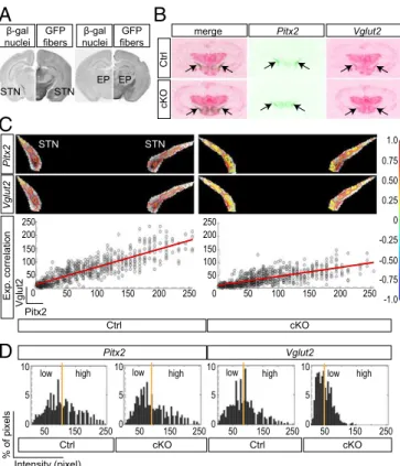
Pitx2-Cre–Mediated Targeting Shifts the Expression Profile ofVglut2 in the STN from High to Low Levels.The activity of thePitx2-Cre
transgene (21) was validated using theTau-mGFPCre-reporter
transgene (24), which showedβ-gal–immunopositive cell bodies
in the STN (Fig. 1A) and GFP in the target areas of STN
neu-rons, including the EP, corresponding to the internal segment of
globus pallidus in primates (25) (Fig. 1A).β-gal was also detected
in the mammillary nucleus (MN) and the posterior
hypothala-mus (PH), the two additional regions wherePitx2expression has
been described (21). Mice gene-targeted forVglut2specifically in
Pitx2-Cre–expressing cells were produced by breeding Pitx2-Cre
mice withVglut2f/f(19) mice, thereby producing cKO and control
littermate mice. STN expression of all Vglutsubtypes was
ana-lyzed on a single-cell level using multiplex single-cell RT-PCR analysis on freshly dissociated neurons from postnatal day (P) 1
and 20 control and cKO mice, detecting theVglut2WT allele in
38% and 40% of control cells, respectively, and the KO allele in 15% of cKO cells at P1, which increased to 28% at P20. The
STN P20 cells were also analyzed for Vglut1expression, which
was detected in both control and cKO cells, and forVglut3
ex-pression, which was not detected at all (Fig. S1). In addition,
analysis of MN and PH cells verified the expression of both Vglut1andVglut2also in these areas and showed the incidence of
the KO allele in 12% and 17% ofVglut2-expressing cells of cKO
mice. In addition, all three areas contained cells coexpressing Vglut1andVglut2(Fig. S1). Quantitative radioactive oligoprobe in situ hybridization was used to analyze the distribution of Vglut2mRNA in the STN, its overlap withPitx2, and the extent
of theVglut2deletion in the adult mouse. Serial section analysis
confirmed Vglut2 expression in the adult STN and revealed
a reduction of Vglut2expression in the STN of the cKO mice
detected upon pure visual inspection (Fig. 1B). Closer
exami-nation ofPitx2andVglut2expression in the STN by correlation
analysis in each individual (two representative cases are shown in
Fig. 1C) revealed a similar distribution of high and low
expres-sion values in control mice, whereas Vglut2expression was
se-verely diminished in cKO mice, as reflected by the horizontally
deflected correlation curve (Fig. 1C,Right). Subsequent
quanti-tative evaluation of the difference in STN Vglut2 mRNA
ex-pression between control and cKO brains showed a global ratio
of STN vs. thalamus at 1.57 (± 0.16) in the control and 0.94
(± 0.12) in the cKO, a finding indicating a 40% decrease of
Vglut2 expression in the STN of the cKO brains. A similar quantification in the MN and PH did not show a significant
decrease ofVglut2expression in either area (Fig. S1). A
distri-bution analysis ofPitx2 andVglut2 mRNA pattern in the STN
revealed that both genes show a large dynamic range from low to high expression levels in the control, a range that remained the
same forPitx2in the cKO but that was altered forVglut2, which
instead showed a more homogenousVglut2mRNA distribution
with disappearance of high labelings (Fig. 1D). Further, the
distribution of the labeling analyzed by quintiles showed that in the cKO the number of STN cells that expressed a very low level of mRNA was increased by 70%. The correlation analysis
indi-cates that thePitx2andVglut2signals are highly mutually related
in both control and cKO. The Pearson correlation ranged from 0.80 to 0.87 in control and 0.62 to 0.78 in cKO STN, the lower correlation level in the cKO being due to the change of the signal distribution for cKO and a usually higher variation, in relative terms, in weaker signal ranges. Moreover, the correlation data
indicate that the decrease ofVglut2signal is not associated with
a subregion of the STN but covers the extent of this structure. Because multiple structures are innervated by the STN (13), to
globally assess brain anatomy upon the limited deletion ofVglut2
expression in the STN introduced here we analyzed serial sections spanning the entire brain upon in situ hybridization for excitatory (Vglut1andVglut2), inhibitory (glutamic acid decarboxylase;GAD)
and dopaminergic (tyrosine hydroxylase;TH) markers, respectively,
all of which seemed normal (Fig. S2), apart from the selective
reduction ofVglut2expression in the STN reported above.
Diminished, but Not Eliminated, Glutamatergic Transmission in the Main Target Areas of the STN. To understand the physiological
consequences of the decreased Vglut2 expression levels in the
STN, we used parasagittal slices containing the STN, EP, and SNr (26) to assess synaptic currents in STN targets. Excitatory postsynaptic currents (EPSCs) in the EP and SNr were elicited
by a single 400-μs shock delivered by a concentric electrode placed
on the STN (Fig. 2A). EPSC amplitude in EP cells increased as
stimulus intensity was increased in both control and cKO mice. However, the dependency of EPSC amplitude on stimulus
in-tensity was greater in control mice (Fig. S3A). It has previously
been shown that a single stimulation pulse generates STN-dependent multiple EPSCs (compound EPSCs) in post-synaptic neurons (26), and our data in control animals
corrobo-rate this finding (Fig. 2A). However, in cKO mice, EPSCs in both
GFP fibers β-gal nuclei
STN STN
EP EP
A
B
C
merge Pitx2 Vglut2
Ctrl
cKO
Pitx2 Vglut2
Intensity (pixel)
% of pixels
GFP fibers β-gal nuclei
Ctrl cKO
Pitx2
Vglut2
Exp. correlation 0 0
Pitx2
Vglut2
50 100 150 200 250 50 100 150 200 250
50 100 150 200 250
50 100 150 200 250
-0.50 -0.25 0 0.25 0.50 0.75 1.0
-1.0 -0.75
D
0 5 10
0 5 10
0 5 10
0 5 10
50 150 250 50 150 250 50 150 250 50 150 250
low high
low high low high low high
Ctrl cKO Ctrl cKO
STN STN
Fig. 1. ThePitx2-Cretransgene is expressed in the STN and mediates a shift inVglut2mRNA levels and distribution. (A) Immunohistochemistry forβ-gal (nuclear,Left) and GFP (projections,Right) on coronal mousePitx2-CreTau-mGFP
EP and SNr consisted of a single event (Fig. 2A). Because com-pound EPSCs generate larger long-lasting currents (26), we compared EPSCs in control and cKO mice by measuring the area under the curve (current density) of EPSCs. Current densities in cKO were dramatically decreased in both EP and SNr cells (Fig.
2B,P=0.04,n=20 cells andP=0.004,n=20 cells, respectively).
In addition, cKO mice displayed smaller amplitudes of the first
EPSC peak in both nuclei (Fig. 2B,P= 0.002,n =20 andP=
0.001,n= 20, respectively), whereas stimulation threshold (Fig.
2B,P=0.03,n=20 andP=0.02,n=20, respectively) and EPSC
rise time (EP control/cKO 0.47 ±0.07ms vs. 1.04 ± 0.13, P =
0.001,n=20; SNr control/cKO 0.36±0.10ms vs. 0.91±0.11,P=
0.002,n=20) were greater in EP and SNr cells of the cKO mice.
The impact of impaired synaptic transmission between the STN and postsynaptic targets in vivo was evaluated by light stimulation of STN cells transduced by stereotaxic injection of an
adeno-associated virus vector (AAV) with Cre-dependent
Channelrho-dopsin 2(ChR2). Injection of AAV-ChR2 inPitx2-Cre–expressing control and cKO mice produced a robust expression of ChR2 and
its reporter gene encoding the YFP in the STN (Fig. 2C). Using
multisite probes with silicon substrate, we recorded single-unit
ac-tivity in the STN and EP in response to light stimulation (Fig. 2D
andE). Single 10-s-long light pulses caused a large increase in firing
frequency of STN neurons in both control and cKO mice (P=0.01,
n =6 units per two animals andP=0.003,n=7 units per two
animals, respectively; Fig. 2D). However, light stimulation of STN in
cKO mice caused no significant change in EP cell firing frequency
(n=14 cells per two animals; Fig. 2EandF), whereas in control
animals firing frequency was markedly increased (P=0.00003,n=
30 cells per two animals; Fig. 2EandF). These experiments verified
that the targeted loss ofVglut2in thePitx2-Cre–expressing cells
of the STN severely affects the ability of STN to generate postsynaptic activity.
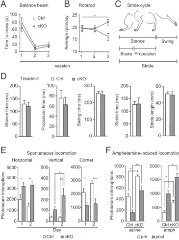
Normal Motor Coordination and Gait, but Accentuated Movement. Because the STN is heavily involved in motor behavior, we tested the Vglut2f/f,Pitx2-Cre mouse line in a battery of motoric tests.
Littermate control and cKO mice were tested on the balance beam, the rotarod, and in the treadmill to assess fine and crude motor coordination and gait, respectively. The time to cross the
Current Density (pA.ms) STN stimulation STN stimulation SNr recording SNr recording STN stimulation STN stimulation EP recording EP recording * ** ** **
B
A
Control cKO Control cKO STN EP Fiber Silicon probe Ctrl cKO STN EP STN stimulation STN stimulation STN recordingSTN recording STN stimulationSTN stimulationEP recordingEP recording
C
D
E
Ctrl cKO Before Light STN EP EPSC amplitude (pA) Current Density (pA.ms) 0 120 EPSC amplitude (pA) 0 120 0 600 0 600 Threshold (V) 45 0 Threshold (V) 30 0 * * STNF
Ctrl cKO EP Ctrl cKO Frequency (Hz) 0 40 Frequency (Hz) 0 40 0 40 0 40 * ** * 50pA 10ms ChR2-YFP DAPI STN 25mV 500ms 50mV 0.5ms Light During Before Light Light During DG ThFig. 2. Attenuation ofVglut2expression in the STN severely impairs synaptic communication postsynaptically. (A) (Left) Diagram showing stimulation and recording electrode placement in parasagittal slices containing the STN, EP, and SNr. (Right) Representative examples of synaptic currents elicited by STN stimulation (10% above threshold) in SNr and EP cells in control and cKO mice. (B) Mean EPSC amplitude, current density, and stimulation threshold for EP and SNr cells in control (black bars) and cKO (red bars) mice. (C) Photomi-crograph showing representativePitx2-Cre–driven ChR2 expression (through
its reporting protein YFP) selectively in the STN with high magnification in inset. Dentate gyrus (DG) and thalamus (Th) are indicated for reference. (D) Example of extracellular recordings in the STN of control and cKO mice be-fore and during light stimulation. (E) Single units isolated from EP recordings before and during light stimulation. (F) Summary of firing frequency in STN and EP recordings before and during light stimulation in control (black bars) and cKO (red bars) mice. *P<0.05; **P<0.001. cKO, conditional knockout; Ctrl, control.
*
session
Stance time (ms)
Propulsion time (ms) Swing time (ms) Stride time (ms) Stride length (mm)
A
B
D
T
ime to cross (s)
100 80 60 40 20 0
1 2 3 1 2 3
A v erage rpm/day 25 20 15 10 Ctrl cKO Ctrl cKO
C
TreadmillBalance beam Rotarod Stride cycle
Stance Swing Stride Brake Propulsion Horizontal Photobeam interuptions
E
0 3000 4000 2000 1000 *** * 1 2 Corner 0 3000 4000 5000 2000 1000 * ** Ctrl cKO 1 2 Vertical 300 400 500 200 100 * ** 1 2 0F
Day Ctrl cKO 1000 800 600 400 200 0 Photobeam interruptions saline 2000 1500 1000 500 0 Ctrl cKO amph pre post ** * * *Spontaneous locomotion Amphetamine-induced locomotion
Fig. 3. Normal motor coordination and gait, and increased locomotion. (A) Fine motor coordination assessment on balance beam as measured by the time (seconds) needed to cross the beam. (B) Crude motor coordination as-sessment on rotarod, presented as mean of the rounds per minute (rpm) reached in three trials per day per animal on three consecutive days. (C) Illustration of the mouse stride cycle. (D) Stride cycle analysis on automated treadmill using averaged values for all four limbs for statistical analysis. Each mouse was trained to walk on the treadmill at speeds 10 and 15 cm/s and subsequently tested at 25 cm/s. (E) Horizontal (locomotion) and vertical (rearing) baseline activity, as well as resting (corner activity) during 60-min recordings in an open field setting, represented as number of photobeam interruptions. (F) Locomotion over 120-min recordings before (white bars) and after (gray bars) saline (Left) and amphetamine (Right) injections; 30 min preinjection, 90 min postinjection. *P < 0.05. cKO, conditional knockout; Ctrl, control.
NEUROSCI
balance beam did not differ significantly between cKO and control mice, indicating that fine motor coordination is normal in
cKO mice. Both groups improved over the three trial days (P<
0.0001;F=44.53; df=2) regardless of genotype (P=0.3046;
F=1.091; df=1) (Fig. 3A). During days 1 and 2, both cKO and
control mice stayed on the rotarod up to similar speeds, whereas at day 3 cKO mice stayed on the rotarod at a significantly higher
speed (P=0.0034;F=6.104; df=2), indicating that the crude
motor abilities of cKO mice are at least as good as those of
controls (Fig. 3B). Gait analysis was performed in an automated
treadmill measuring the different components of the entire stride
cycle (illustrated in Fig. 3C). All stride cycle components were
normal in the cKO mice [stance (P=0.1812), additionally
sub-divided into brake and propulsion (P=0.2284) and swing (P=
0.8518); stride as one unit (P=0.8518 for stride time andP=
0.7546 for stride length)] (Fig. 3D). Movement was further
addressed in paradigms of affective behavior. In the elevated plus maze, the time spent in each arm as well as the frequency of visits was similar between cKO and control mice, indicating a
lack of anxiety-related behavior (Fig. S3B). However, we noted
that the latency with which cKO mice moved out from the center and the latency to enter the outer open arm (OOA) of the maze
was significantly shorter for the cKO mice (P=0.0115 for center
andP=0.0280 for OOA). In the forced swim test, the time spent
immobile during the second trial was significantly shorter for the cKO mice than for control littermates, showing an increase in
overall swimming activity in the cKO group (Fig. S3C).
Next, we assessed various aspects of movement more directly in automated activity boxes. We observed that both horizontal and vertical movement was elevated in the cKO mice compared with control littermates. Locomotion was significantly increased in cKO mice compared with controls, both on days 1 and 2 in a
2-d trial (P=0.0034 for days 1 and 2), whereas corner activity
was significantly lower in cKO mice on both days (P=0.0008;
q=3.915 and 5.668, respectively), showing that cKO mice spent
more time in motion than their control littermates (Fig. 3E).
Vertical movement (rearing) was also significantly elevated in
the cKO mice on day 2 (P=0.0021) and, moreover, significantly
increased from day 1 to day 2 within the cKO group (q=3.799)
(Fig. 3E). Because it is well known that dopamine normally aids
in the facilitation of movement, we used a pharmacological challenge approach composed of three different substances that alter extracellular dopamine levels by different mechanisms. First, we injected both control and cKO mice with saline and amphetamine (1.5 mg/kg, i.p.) and analyzed their locomotor response. Amphetamine strongly increases extracellular dopa-mine levels, and hence locomotion, by acting on both the cyto-plasmic membrane dopamine transporter (DAT) and the vesicular counterpart (VMAT) (27). Both control and cKO mice
showed the same level of response to amphetamine (Fig. 3F),
leading to aca. threefold increase in locomotion, but cKO mice
displayed a higher level of activity already before injection (P=
0.0286 for saline,P=0.0286 for amphetamine). This difference
remained after both saline (P=0.0012) and amphetamine (P=
0.0106) injection (Fig. 3F). A second batch of mice were then
challenged with 7.5 and 15 mg/kg of GBR12783, a DAT-selective blocker; both doses led to a heightened response in cKO com-pared with control mice, but the difference failed to reach
sta-tistical significance (Fig. S4). After an injection of reserpine
(2 mg/kg), a potent blocker of vesicular monoamine packaging by VMAT that causes catalepsy, a separate batch of mice showed no difference between controls and cKOs. Both groups showed a similar successive decrease in locomotion after reserpine
in-jection (Fig. S4).
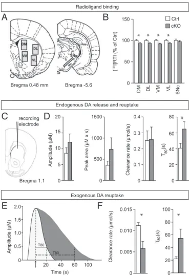
Normal Dopamine Receptor Distribution, but Reduced Striatal Dopamine Clearance in Vivo.To further explore the putative effect of the
con-ditional STN targeting ofVglut2on dopamine neurotransmission in
the basal ganglia circuitry, we addressed various components of the dopamine system. In addition to the described expression analysis of
TH, which encodes the rate-limiting enzyme for dopamine synthesis
and showed normal distribution in the cKO brain (Fig. S2), a series
of binding assays were performed and quantified in the SNc and
four striatal target areas (illustrated in Fig. 4A). Ligand binding
to D1R was normal in all four striatal areas examined as well as in
the SNr, which was also quantified (Fig. S5). The same observation
was made for the D2R, both in all striatal target areas analyzed as
well as presynaptically in the dopamine cell bodies of the SNc (Fig.
S5). In contrast, the binding capacity of DAT was significantly lower
in the cKO in all striatal target areas examined but preserved in the cell bodies of the SNc, indicating a selective reduction of striatal
150
100
50
0
DM DL VM VL
[ I]R
TI (% of Ctrl)
125
SNc
C
* * * *
E
0 20 40 60 80 100
T
(s)
Clearance rate (μmol/s)
20 40 60 100
Time (s) 0
0.5 1.0 1.5 2.0
T80 T80
Amplitude (μM)
F
*
B
Bregma 1.1 recording electrode
80
0 0.005 0.010 0.015
*
Amplitude (μM)
0 5 15 20
10
Peak area (μM x s)
0 500 1000 1500
T
(s)
0 20 40 60 80 Ctrl cKO
0 0.1 0.2 0.3 0.4
Clearance rate (μmol/s)
80
*
Endogenous DA release and reuptake Radioligand binding
Exogenous DA reuptake
D
A
Bregma -5.6 Bregma 0.48 mm
DAT expression in the cKO mice (Fig. 4B). To address whether dopamine kinetics is altered in the cKO mice, KCl-evoked dopa-mine release was measured and quantified in the dorsal striatum of anesthetized mice by high-speed in vivo chronoamperometry (Fig.
4C). No significant difference was found between control and cKO
mice in dopamine amplitude, but a trend was observed in the cKO mice toward increased peak area and decreased clearance rate (Fig.
4D). A significant decrease was observed for dopamine clearance
(T80,P=0.0474) (Fig. 4D). Local striatal application of exogenous
dopamine also revealed significantly reduced clearance in the cKO
mice (Fig. 4E; representative peaks derived from control and cKO
mice). Significantly lower clearance rate (P=0.0317) (Fig. 4F) as
well as prolonged time to clear 80% of dopamine from the
extra-cellular space (T80,P=0.0451) (Fig. 4F) were observed. Targeting
Vglut2in the STN thus leads to an effect on DAT activity that is likely to contribute to the hyperlocomotion phenotype observed.
Preserved Spatial Memory, Level of Impulsivity, Cognitive Flexibility, and Social Dominance.To analyze cognitive behavior in the cKO mice, spatial working and reference memory was measured in the radial eight-arm maze. cKO mice did not make more working memory or reference memory errors than controls; however, they had a significantly increased number of trials that were not
completed (Fig. S6). We also used the delay discounting task,
a paradigm that allows the animal to freely choose between a small but immediate reward and a larger, but delayed, one. The cKO mice did not differ significantly from controls in their ability to wait for a larger reward, but they refrained from choosing in response to the light stimulus more often. All animals registered similar omissions to collect the earned reward, average latencies to choose or collect rewards, and inappropriate head entries at
the small reward side and inactive holes (Fig. S7).
Discussion
Contrary to what was previously reported (15, 16), we found that the STN has not one, but at least three main populations of glutamatergic cells, as characterized by their expression of either Vglut1orVglut2or coexpression of both.Vglut2is expressed at
higher, and thus more readily detectable, levels thanVglut1and
is strongly correlated withPitx2expression. Despite the restricted
reduction of Vglut2 expression in the STN of our cKO mice,
leaving about two-thirds of the originalVglut2expression levels,
it gave rise to severely disrupted communication with the main target areas, the EP and the SNr, and the mice displayed a strong movement-specific phenotype while preserving, with minor excep-tions, normal cognitive and affective behavior. In addition to the reduced glutamatergic tone in the SNr and EP, decreased DAT levels and slower dopamine reuptake support an indication of increased extracellular dopamine in the striatum, likely related to the observed hyperlocomotion. Altogether, our results
demon-strate that shifting theVglut2expression profile in the STN from
high- to low-level expression is sufficient to cause behavioral consequences comparable with much larger and indiscriminate lesions in this area.
Since the discovery of the beneficial clinical effects of STN high-frequency stimulation, this nucleus has been the subject of hundreds of studies attempting to explain the phenomenon (28). Valuable efforts have been made in characterizing the anatom-ical (29) and electrophysiologanatom-ical (30) properties of neurons within this structure. Recently, Favier et al. (31) identified altered protein levels of VGLUT1 and VGLUT2 in multiple brain areas upon STN high-frequency stimulation. The identification of promoter-specific subgroups of neurons within the STN itself, however, has been somewhat neglected, likely owing to the homogeneity of the STN (having only one type of neurotransmitter, glutamate), and the relatively recent discovery of different glutamatergic trans-porters. Compared with previous attempts that performed a broad Vglutcharacterization of several brain areas at once (15, 16, 32),
we focused our efforts specifically on the STN and quantified the
expression ofVglut1andVglut2through both in situ hybridization
and single-cell RT-PCR techniques. Whereas Vglut2 expression
was easily detected by both techniques, Vglut1 expression—not
detected by in situ hybridization analysis—could be quantified by multiplex single-cell RT-PCR. We found a partial overlap between the expression of both transporters, a pattern that is also found in other areas of the central nervous system (33, 34). The advantage of characterizing the expression pattern of promoters is the ability to subsequently manipulate them, an approach we took here by
blunting glutamatergic release inVglut2-expressing cells that were
also positive forPitx2. This is the first description, to our
knowl-edge, of a behavioral role for a subpopulation of neurons within the STN. Our results are comparable to those obtained by tradi-tional methods of lesioning this nucleus, in which animals display a consistent improvement in locomotion (35), and also clearly observed in experiments with high-frequency stimulation of the STN (36, 37). Although we cannot determine whether disabling Vglut2-expressing neurons is necessary for this behavioral effect, we now have evidence that it is a sufficient condition. Moreover, the effect we observe here does seem specific to the role of Vglut2itself in the STN, and not to a general decrease in
glu-tamatergic signaling:Vglut2-heterozygous mice, in which all
glu-tamatergic transmission mediated by this transporter is reduced by up to 50%, present no abnormalities in locomotion (20), and the
same is true for mice heterozygous forVglut1(38). Therefore, it is
likely that the effects we achieved are due to a characteristic property of the cKO cells, such as their local connectivity or projection targets. The experiments we performed regarding syn-aptic activity in the two major targets of the STN corroborate this interpretation. The cKO mice showed a strong reduction in the excitatory activity the STN could produce on the EP and the SNr, as evidenced by both in vitro and in vivo electrophysiology. The
latter experiments, using ChR2 expression selectively inPitx2-Cre–
expressing cells of the STN, could confirm that these neurons are normally able to induce increased activity in the STN targets, but
that this function was lost afterVglut2conditional deletion.
Considering the classic model describing a direct and an in-direct pathway through the basal ganglia, we suggest that the locomotor effects we observed were likely mediated by this dis-ruption in the indirect pathway, weakening the STN chronic excitation on the SNr and EP and, thus, decreasing the total inhibitory output these structures have on the thalamus. Addi-tionally, consistent with high-frequency stimulation studies (39), no difference was found in the levels of dopamine receptor binding in the dorsal striatum; the altered DAT levels, however, in combination with slower dopamine clearance, are suggestive of overall elevated extracellular striatal dopamine levels. Besides lo-comotor activity, the behavioral tasks we performed generally did not show any differences between cKOs and controls, showing that
the role ofVglut2-expressing cells in the STN seems to be closely
related to locomotion. One notable exception was the forced swim test, in which the knockout mice spent more time active, a finding that suggests that, unlike interventions such as high-frequency stimulation of the STN (39), our cKOs do not show increased depression-like behavioral symptoms, but rather the opposite. In humans, lesions of the STN have also been reported to improve depression symptoms (8). The effect we saw could be due to some
particular pattern of projections of theVglut2population of cells
compared with other neurons in the STN, or simply due to the fact that the cKO mice are more active in general, possibly even during the water-based test. Aside from this, it seems our intervention was largely limited to locomotor consequences, presenting none of the affective or cognitive impairments observed in other experiments. It is possible to speculate that this promoter-based approach to decreasing STN function could be a way of avoiding some of the side effects commonly seen in treatments that require lesions or high-frequency stimulation of this nucleus.
NEUROSCI
Perhaps the most severe limitation in our study is the use of knockout animals, a method that does not discriminate between developmental and acute effects. However, it can be argued that, given the progressive nature of our knockout, this limitation was not as worrisome as most knockout techniques. Another limi-tation is the fact that we also targeted some neurons in other parts of the brain, in particular the PH and the MN, even though
neither area showed significantly reduced Vglut2 expression
levels as detected by quantitative in situ hybridization. These structures are not traditionally associated with locomotion, and lesion studies also do not support a contributing role as to the phenotype of our cKO mice (39, 40), but every manipulation can have unforeseeable consequences, and we cannot rule out that they are also involved in some of the results we reported. It is conceivable that our results could translate to other animals, and to humans as well. In monkeys, the STN seems to be
com-posed almost exclusively ofVglut2-expressing neurons (40),
al-though this report relies solely on in situ hybridization, which
we showed to be unreliable to detectVglut1in mice. Silencing
a particular subset of cells through genetic or pharmacological
means could be feasible in the future and might be clinically relevant for Parkinson disease.
Materials and Methods
All mice used in the study were housed in accordance with the Swedish regulation guidelines (Animal Welfare Act SFS 1998:56) and European Union legislation (Convention ETS123 and Directive 2010/63/EU). The Vglut2f/f;Pitx2mouse line was produced by breeding the Pitx2-Cre mice (21) to
Vglut2f/f(19) mice, thereby generating cKO (Vglut2f/f;Pitx2-Cre+
) and controls (Vglut2f/f;Pitx2-Cre-andVglut2wt/wt;Pitx2-Cre+
) as littermates with the identical ge-netic background. For a detailed outline of the experimental setups, seeSI
Materials and Methods.
ACKNOWLEDGMENTS.We thank Profs. James Martin and Sylvia Arber for generously sharingPitx2-Creandtau-mGFPmouse lines, respectively; and Emelie Perland, Thomas Viereckel, and Dr. Carolina Birgner for technical assistance. This work was supported by Swedish Medical Research Council Grants SMRC 2007-5742, 2011-4747, and 2011-5171, Uppsala University, the Swedish Brain Foundation, Parkinsonfonden, and the foundations of Bertil Hållsten, Kjell och Märta Bejers, Major Gösta Lind, Åhlén, Åke Wiberg, and Coordenação de Aperfeiçoamento de Pessoal de Nível Superior (CAPES) Stipend 9915-11-7.
1. Benabid AL, Chabardes S, Mitrofanis J, Pollak P (2009) Deep brain stimulation of the subthalamic nucleus for the treatment of Parkinson’s disease.Lancet Neurol8(1): 67–81.
2. Windels F, et al. (2000) Effects of high frequency stimulation of subthalamic nucleus on extracellular glutamate and GABA in substantia nigra and globus pallidus in the normal rat.Eur J Neurosci12(11):4141–4146.
3. Benazzouz A, et al. (2000) Effect of high-frequency stimulation of the subthalamic nucleus on the neuronal activities of the substantia nigra pars reticulata and ven-trolateral nucleus of the thalamus in the rat.Neuroscience99(2):289–295. 4. Magariños-Ascone C, Pazo JH, Macadar O, Buño W (2002) High-frequency stimulation
of the subthalamic nucleus silences subthalamic neurons: A possible cellular mecha-nism in Parkinson’s disease.Neuroscience115(4):1109–1117.
5. Tai C-H, et al. (2003) Electrophysiological and metabolic evidence that high-frequency stimulation of the subthalamic nucleus bridles neuronal activity in the subthalamic nucleus and the substantia nigra reticulata.FASEB J17(13):1820–1830.
6. Plaha P, Ben-Shlomo Y, Patel NK, Gill SS (2006) Stimulation of the caudal zona incerta is superior to stimulation of the subthalamic nucleus in improving contralateral par-kinsonism.Brain129(Pt 7):1732–1747.
7. Gradinaru V, Mogri M, Thompson KR, Henderson JM, Deisseroth K (2009) Optical deconstruction of parkinsonian neural circuitry.Science324(5925):354–359. 8. Alvarez L, et al. (2005) Bilateral subthalamotomy in Parkinson’s disease: Initial and
long-term response.Brain128(Pt 3):570–583.
9. Parsons TD, Rogers SA, Braaten AJ, Woods SP, Tröster AI (2006) Cognitive sequelae of subthalamic nucleus deep brain stimulation in Parkinson’s disease: A meta-analysis. Lancet Neurol5(7):578–588.
10. Winstanley CA, Baunez C, Theobald DEH, Robbins TW (2005) Lesions to the sub-thalamic nucleus decrease impulsive choice but impair autoshaping in rats: The im-portance of the basal ganglia in Pavlovian conditioning and impulse control.Eur J Neurosci21(11):3107–3116.
11. Baunez C, Robbins TW (1997) Bilateral lesions of the subthalamic nucleus induce multiple deficits in an attentional task in rats.Eur J Neurosci9(10):2086–2099. 12. Van Der Kooy D, Hattori T (1980) Single subthalamic nucleus neurons project to both
the globus pallidus and substantia nigra in rat.J Comp Neurol192(4):751–768.
13. Kita H, Kitai ST (1987) Efferent projections of the subthalamic nucleus in the rat: light and electron microscopic analysis with the PHA-L method.J Comp Neurol260(3): 435–452.
14. Maurice N, Deniau JM, Menetrey A, Glowinski J, Thierry AM (1998) Prefrontal cortex-basal ganglia circuits in the rat: Involvement of ventral pallidum and subthalamic nucleus.Synapse29(4):363–370.
15. Hisano S (2003) Vesicular glutamate transporters in the brain.Anat Sci Int78(4): 191–204.
16. Barroso-Chinea P, et al. (2007) Expression of the mRNAs encoding for the vesicular glutamate transporters 1 and 2 in the rat thalamus.J Comp Neurol501(5):703–715. 17. Köhler C, Schwarcz R (1983) Comparison of ibotenate and kainate neurotoxicity in rat
brain: A histological study.Neuroscience8(4):819–835.
18. Lein ES, et al. (2007) Genome-wide atlas of gene expression in the adult mouse brain. Nature445(7124):168–176.
19. Wallén-Mackenzie Å, et al. (2006) Vesicular glutamate transporter 2 is required for central respiratory rhythm generation but not for locomotor central pattern gener-ation.J Neurosci26(47):12294–12307.
20. Moechars D, et al. (2006) Vesicular glutamate transporter VGLUT2 expression levels control quantal size and neuropathic pain.J Neurosci26(46):12055–12066. 21. Martin DM, et al. (2004) PITX2 is required for normal development of neurons in the
mouse subthalamic nucleus and midbrain.Dev Biol267(1):93–108.
22. Sclafani AM, et al. (2006) Nestin-Cre mediated deletion of Pitx2 in the mouse.Genesis 44(7):336–344.
23. Birgner C, et al. (2010) VGLUT2 in dopamine neurons is required for psychostimulant-induced behavioral activation.Proc Natl Acad Sci USA107(1):389–394.
24. Hippenmeyer S, et al. (2005) A developmental switch in the response of DRG neurons to ETS transcription factor signaling.PLoS Biol3(5):e159.
25. Gerfen CR (1992) The neostriatal mosaic: Multiple levels of compartmental organi-zation.Trends Neurosci15(4):133–139.
26. Ammari R, Lopez C, Bioulac B, Garcia L, Hammond C (2010) Subthalamic nucleus evokes similar long lasting glutamatergic excitations in pallidal, entopeduncular and nigral neurons in the basal ganglia slice.Neuroscience166(3):808–818.
27. Sulzer D, Sonders MS, Poulsen NW, Galli A (2005) Mechanisms of neurotransmitter release by amphetamines: A review.Prog Neurobiol75(6):406–433.
28. McIntyre CC, Savasta M, Kerkerian-Le Goff L, Vitek JL (2004) Uncovering the mech-anism(s) of action of deep brain stimulation: Activation, inhibition, or both.Clin Neurophysiol115(6):1239–1248.
29. Nauta HJW, Cole M (1978) Efferent projections of the subthalamic nucleus: An au-toradiographic study in monkey and cat.J Comp Neurol180(1):1–16.
30. Beurrier C, Congar P, Bioulac B, Hammond C (1999) Subthalamic nucleus neurons switch from single-spike activity to burst-firing mode.J Neurosci19(2):599–609. 31. Favier M, et al. (2013) High-frequency stimulation of the subthalamic nucleus
modi-fies the expression of vesicular glutamate transporters in basal ganglia in a rat model of Parkinson’s disease.BMC Neurosci14(1):152.
32. Hur EE, Zaborszky L (2005) Vglut2 afferents to the medial prefrontal and primary somatosensory cortices: A combined retrograde tracing in situ hybridization study [corrected].J Comp Neurol483(3):351–373.
33. Kaneko T, Fujiyama F, Hioki H (2002) Immunohistochemical localization of candidates for vesicular glutamate transporters in the rat brain.J Comp Neurol444(1):39–62.
34. Todd AJ, et al. (2003) The expression of vesicular glutamate transporters VGLUT1 and VGLUT2 in neurochemically defined axonal populations in the rat spinal cord with emphasis on the dorsal horn.Eur J Neurosci17(1):13–27.
35. Bergman H, Wichmann T, DeLong MR (1990) Reversal of experimental parkinsonism by lesions of the subthalamic nucleus.Science249(4975):1436–1438.
36. Shi L-H, et al. (2004) High-frequency stimulation of the subthalamic nucleus reverses limb-use asymmetry in rats with unilateral 6-hydroxydopamine lesions.Brain Res 1013(1):98–106.
37. Vlamings R, et al. (2007) High frequency stimulation of the subthalamic nucleus im-proves speed of locomotion but impairs forelimb movement in Parkinsonian rats. Neuroscience148(3):815–823.
38. Tordera RM, et al. (2007) Enhanced anxiety, depressive-like behaviour and impaired recognition memory in mice with reduced expression of the vesicular glutamate transporter 1 (VGLUT1).Eur J Neurosci25(1):281–290.
39. Temel Y, et al. (2007) Inhibition of 5-HT neuron activity and induction of depressive-like behavior by high-frequency stimulation of the subthalamic nucleus.Proc Natl Acad Sci USA104(43):17087–17092.