Cytotoxicity Analysis of Ti-7.5Mo Alloy After Biomimetic Surface Treatment to Use as
Dental Materials
Ana Lúcia do Amaral Escadaa*, Samira Esteves Afonso Camargob, Luana Marotta Reis de
Vasconcellosb, Noala Vicensoto Moreira Milhanc, Ana Paula Rosiini Alves Claroa
Received: March 31, 2016; Revised: January 17, 2017; Accepted: August 01, 2017
Titanium (Ti) and its alloys are widely used for medical and dental ields due to their excellent biocompatibility, high corrosion resistance, high speciic strength and excellent mechanical properties. Diferent methods have been developed to improve the surface properties of titanium-based implant materials, and consequently the bone-bonding ability. The Ti-7.5Mo alloy was activated by an alkaline treatment with 5M NaOH, heat treatment and subsequent immersion in SBFx5 to investigate the in vitro response of osteoblastic-like cells (MG-63) on altered biomimetic surfaces. Sample surfaces were characterized by scanning electron microscopy. Cytotoxicity was assessed by the MTT assay, total protein content, alkaline phosphatase activity (ALP) and mineralized bone-like nodule formation. It was shown the que alkali treatment led to the formation of sodium titanate and immersion in SBFx5 formed a ilm of calcium phosphate. The alkaline treatment and heat treatment of 7.5 Ti-Mo alloys followed by soaking them in SBFx5 for 24 hours is a suitable technique once the inal samples were biocompatible, allowed the attachment of the osteoblastic-like cells (MG-G3), and increased the mineralized like-bone nodules formation by these cells.
Keywords: titanium alloys, biomimetic treatment, cytotoxicity.
*e-mail: [email protected]
1. Introduction
Titanium (Ti) and its alloys are widely used in the medical and dental ields due to their excellent biocompatibility, high corrosion resistance, high speciic strength and excellent mechanical properties1. The Ti-7.5Mo alloy is a biocompatible alloy, which has a low elastic modulus (55 GPa) and a high strength/modulus ratio. Thus, it is a potential candidate for orthopedic applications2,3. Despite these appropriate properties, titanium and its alloys are considered bioinert4.
Diferent methods have been developed to improve the surface properties of titanium-based implant materials, and consequently the bone-bonding ability. Through the surface treatment, it is possible to change the chemical composition, morphology, topography and roughness. Among the surface treatments, titanium plasma-spraying, acid-etching, grit-blasting, anodization and calcium phosphate coatings5 are the most common.
Metal implants have been coated with layers of calcium phosphates mostly composed of hydroxyapatite to enhance hard tissue integration. Diferent methods have been used to coat metal implants, such as the sol-gel coating, sputter-deposition, plasma-spraying, electrophoretic deposition and biomimetic precipitation. The plasma-spraying coating method
is the only one that has been used in clinical practice; however, it has disadvantages 5 including changes in crystal structure, phase composition, speciic surface area and morphology 6.
In order to avoid the limitations of plasma-sprayed HA coatings, a new coating method which is based on the natural process of biomineralization has been developed. This method, also called biomimetic method, comprises the precipitation of calcium phosphate apatite crystals onto the titanium surface, after contact with simulated body luid (SBF), forming a coating 7,8.
In this work, the inluence of alkaline treatment and heat treatment on the formation of calcium phosphate layer on the surface of a Ti-7.5Mo alloy, after soaking in simulated body luid (SBFx5) for 24 hours, was evaluated concerning the biological behavior of osteoblastic-like cells (MG-63) after contact with these samples.
2. Materials and Methods
2.1 Samples
The Ti-7.5Mo alloy was produced from sheets of commercially pure titanium (99.9%) and molybdenum (99.9%). Samples were melted in an arc furnace under an aDepartamento de Materiais e Tecnologia, Universidade Estadual Paulista - UNESP, Av. Dr. Ariberto
Pereira da Cunha, 333, Pedregulho, CEP 12.516-410, Guaratinguetá, SP, Brazil.
bDepartamento de Biociências e Diagnóstico Bucal, Universidade Estadual Paulista - UNESP, São José dos Campos, SP, Brazil.
argon atmosphere. They were cold worked by swaging and bars with 10 mm of diameter were produced. Then discs with 4 mm of thickness were cut, sanded and samples were divided into three groups: Group 1 (G1): Ti-7.5Mo alloy, control; Group 2 (G2): alkaline treatment and heat treatment; and Group 3 (G3): Ti-7,5Mo alloy with alkaline treatment and heat treatment + immersion in SBFx5.
For alkaline surface treatment, samples were immersed in a 5.0M NaOH aqueous solution at 80ºC for 72 hours, washed with distilled water, and dried at 40ºC for 24 h using a methodology proposed by Wei et al. 20029. After alkaline treatment, samples were heat-treated at 450°C for 1 h in air in an electric furnace.
For SBF treatment Ti-7.5Mo discs were immersed into a polypropylene tube with SBF (Simulated Body Fluid) proposed by Barrère et al. 200210 for 1 day at 36.5 °C to form an calcium phosphate layer on the sample surface.
This SBF solution was prepared by dissolving reagent NaCl, MgCl2.6H2O, CaCl2.2H2O, Na2HPO4, and NaHCO3 in distilled water under constant bubbling of carbon dioxide gas at 36.5 °C.
The sample surfaces were examined using a scanning electron microscope (SEM, LEO 1450 VP, Zeiss, Germany).
Contact angle study and surface energy were performed using an automated Goniometer (Ramé-Hard Instrument Co. - Advanced Goniometer model n ° 300-F1, Serial No. 709262). The equipment has a camera, which captures the image of the drop being deposited on the sample, through a device similar to an eyedropper. The proile of this drop is determined by a computer program that calculates the contact angle of the surfaces. For the supericial energy, the ethyleneglycol was used as the second liquid. For these analyzes an arithmetic mean of 3 trials was done.
2.2 Cell culture experiments and analysis
In this study, an established lineage of osteoblast-like cells (MG 63) was obtained from the Rio de Janeiro Cell Bank (Rio de Janeiro, RJ, Brazil). Cells were cultured in Modiied Eagle’s Medium (Gibco-Life Technologies, NY, USA) containing 10 % fetal bovine serum (Gibco), 100 mg/ mL streptomycin (Gibco) and 100 U/mL penicillin (Gibco) at 37ºC in a humidiied atmosphere of 5 % (v/v) CO2. The cell culture medium was changed every two days and cell growth was assessed using a reverse phase microscope (Model Axiovert 40C, Carl Zeiss Microscopy GmbH, Jena, Germany).
Initially, all the samples were placed into well plates and were sterilized in absolute ethanol (100%) and then UV-exposed for 3 hours. Next, 2 x10⁴ cells were placed
in contact with the samples (G1, G2 and G3), for in vitro analysis. Seven wells were used for each material in three independent experiments. All in vitro procedures were performed according to ISO 10993 guidelines.
2.3 SEM evaluation
The cell attachment and the morphology of the cells were investigated using SEM (Inspect S50, FEI Company, Brno, Tcheca Republik) after 3 days of MG-63 cells growth on the samples. The supernatant was removed and the samples were rinsed twice with PBS. The adhered cells in samples were ixed with 4% glutaraldehyde in PBS, dehydrated using a series of ascending ethanol concentrations (70%, 80%, 90% and 100% at 20 mins each), air-dried at room temperature for 24 h.
For SEM analysis, the samples were sputter-coated with palladium-gold alloy (Polaron SC 7620 Sputter Coater, Quorum Technologies, Newhaven, UK) at a thickness of 7-10 nm (10-15 mA, under a vacuum of 130 mTorr). The SEM was operated between 15 and 30 kV, spot 3 to 6.
2.4 Citotoxicity assay
The cells were maintained in DMEM supplemented with 10% PBS, penicillin (100 U/ ml) and streptomycin (100 mg/ mL) at 37°C for 24 hours in a humidiied atmosphere with 5 % CO2. After this period, the old medium was removed and the cell cultures were exposed to the samples (G1, G2 and G3) and maintained in the incubator for 3, 7 and 14 days. Next, the number of living cells was determined by MTT assay (3-(4,5 dimethylthiazol-2-yl)-2,5-diphenyltetrazolium bromide, Sigma, St Louis, MO, USA). The activity was quantiied by dissolution of MTT in 0.1N NaOH (6.25 v/v%) in DMSO (dimethyl sulfoxide) and then the reading of optical density of the resulting solution was measured by spectrophotometer (Biotek - EL808IU) at 570 nm. The cytotoxicity was expressed in percentage relative to the control group (only cells) (100%).
2.5 Total protein content
To assess the total protein content, the cells were plated on the samples into 24-well plates at a density of 20,000 cells/ well for 3, 7 and 14 days. After these periods, the old medium was removed, the wells were washed three times with PBS at 37°C and illed with 2 mL of 0.1% sodium lauryl sulfate (Sigma) according to the modiied Lowry method (Lowry, et al. 1951). After 30 min, 1 mL of this solution from each well was mixed with 1 mL of Lowry solution (Sigma) and left for 20 min at room temperature. Next, 0.5 mL of the solution of Folin-Ciocalteau phenol reagent (Sigma) was added for 30 min at room temperature. Absorbance was measured at 680 nm using a UV 1203 spectrophotometer (Shimadzu Europa GmbH, Duisburg, Germany). The total protein content was calculated based on an albumin standard curve and expressed in µg/mL.
2.6 Alkaline phosphatase activity (ALP)
monophosphate, using a commercial Kit (Labtest Diagnóstica, Belo Horizonte, BR) according to the manufacturer’s instructions. Initially, the cells were lysated with 2 mL of 0.1% sodium lauryl sulfate (Sigma). Then, 50 µL of thymolphthalein monophosphate were mixed with 0.5 mL of 0.3 M diethanolamine bufer for 2 min at 37 °C. After that, 50 µL of the lysates obtained from each well was added for 10 min at 37 °C. Also, 2 mL of 0.09M Na2CO3 and 0.25M NaOH were added for color development. The absorbance was measured by spectrophotometer (Shimadzu Europa GmbH UV 1203) at 590 nm and the obtained data were shown as alkaline phosphatase activity normalized by total protein content. The values were presented in µmol of thymolphthalein/h /mg protein/mL.
2.7 Mineralized bone-like nodule formation
For quantitative analysis of mineralized matrix nodules, the cells were plated on the samples into 24-well plates at a density of 20,000 cells/well and kept in an incubator for 21 days. Then, the samples were washed three times with Hank's solution (Sigma) at 37°C, and the cultures were ixed with 70% ethanol for 1 hour at 4ºC. Following ixation, the samples were washed twice with PBS and stained with 2% Alizarin red (Sigma), pH 4.2, for 15 minutes at 37°C. This solution is known for staining nodules of mineralized matrix. The samples were washed three times with deionized H2O and left to dry for 24 h at 37°C. The quantiication of mineralized formation was performed according to the method previously described by Gregory et al. (Gregory, et al. 2004). Next, 560 µL of 10% acetic acid was added to each well, followed by 40 µL of 10% ammonia hydroxide in order to neutralize the acid. The absorbance was measured by spectrophotometer (Biotek - EL808IU) at 405 nm. The values were expressed in optical density.
2.8 Statistical analysis
ALP activity, total protein content, citotoxicity assay and nodules formation are expressed as mean ± standard deviations. Statistical analyses were performed using GraphPad Prism software (GraphPad Software version 5.0, San Diego, CA, USA). Statistical comparisons were carried out via ANOVA one-way, followed by a multiple comparison Tukey test where applicable. Diferences were considered statistically signiicant at p<0.05. The results are representative of experiments performed with three distinct primary cultures.
3. Results
Figure 1a shows the image obtained in the scanning electron microscope of sample sanded control where it is possible to view the TiO2 passive layer formed on the of Ti-7.5Mo alloy surface. During immersion in 5M NaOH for 72 hours, the metal O passive layer dissolves
Figure 1. SEM micrographs of the Ti-7.5Mo alloy: Group 1 (G1)
sample control (1a); Group 2 (G2): NaOH alkaline treatment and heat treatment (1b); and Group 3 (G3): Ti-7.5Mo alloy with alkaline treatment and heat treatment + immersion in SBFx5 (1c)
to form metal-OH and a porous ilm of sodium titanate is formed (Figure 1b). When this layer is exposed to SBF, the titanium coating formed by chemical treatment releases Na+ ions of the sodium titanate layer in exchange for H3O
+ ions, wich increases the pH of the solution and promotes the nucleation process of some phosphates. The Ti-OH groups negatively charged react with Ca2
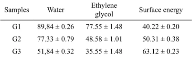
The hydrophilic potential is a very important parameter for biomaterials. Contact angle studies were performed to evaluate this hydrophilic potential provided by various surface coatings (Table 1). The results show that the contact angle of the samples decreased from 89.84 ° (G1) to 77.33 ° (G2) and 51.84 ° (G3) when this study was carried out with deionized water. In the study made with ethyleneglycol, the contact angle decreased from 77.55 ° (G1) to 48.58 ° (G2) and 35.55 ° (G3). These results were used to calculate the surface energy that increased from 40.22 ° (G1) to 50.31 ° (G2) and 63.12 ° (G3).
Table 1. Contac angle values and surface energy
Samples Water Ethylene
glycol Surface energy
G1 89,84 ± 0.26 77.55 ± 1.48 40.22 ± 0.20
G2 77.33 ± 0.79 48.58 ± 1.01 50.31 ± 0.38
G3 51,84 ± 0.32 35.55 ± 1.48 63.12 ± 0.23
3.1 SEM evalution
The cell attachment was observed in all groups. There was an intimate contact between the cells and all the surfaces. The cells were extended and spread on the samples. The SEM images showed that the groups G1 and G2 presented higher number of cells than G3 group (Figure 1).
3.2 Citotoxicity assay
Statistical analysis of the data obtained in the MTT test showed that none of the tested materials was cytotoxic after 3, 7 or 14 days of contact with the cells. The groups G1, G2 and G3 showed no values of absorbance statistically diferent from the control group (p>0.05). Moreover, the groups G1 and G3 showed absorbance values higher than 100% on the 3th and 14th day (Figure 2).
3.3 Total protein content
The values of total protein content were similar among the groups in all experimental periods (p>0.05) (Figure 3). In all groups, the production of total protein on the 3th day was lower than this production on the 7th day, which was lower than this production on the 14th day.
3.4 Alkaline phosphatase activity (ALP)
The values of ALP on the 3th and 14th days showed no statistically signiicant diference among the groups. On the other hand, on the 7th day a diference (p<0.05) was observed between G1 and G3 groups, with G3 presenting a higher
value of ALP than G1 group (Figure 4). Figure 2. Cell attachment on the samples at 3 days, as observed
Figure 3. Graph showing the mean percentage of absorbance,
obtained with MTT assay, of G1, G2 and G3 and control group (100 %) after 3, 7 and 14 days
Figure 4. Total protein content after 3, 7 and 14 days of cell culture on the samples. The values are reported as mean ± SD (n = 3)
Figure 5. ALP activity after 3, 7 and 14 days of cell culture on the samples. The values are reported as mean ± SD. a,b represent Tukey
test results where the groups presenting diferent superscript letters difer statistically
3.5 Mineralized bone-like nodule formation
The data obtained in this test showed that all groups allowed the mineralization of extracellular matrix after 21 days of osteoblast-like culture in contact with the samples.
Statistical analysis of these data showed that G3 group presented the highest formations mineralized bone-like nodules. A statistical diference was observed between this group and the other groups (p<0.05) (Figure 5). In addition, there was not a signiicant diference between G1 and G2 groups (p>0.05).
4. Discussion
The biomimetic method uses supersaturated aqueous solutions with ionic composition resembling the human plasma. This process allows coating of metal implants with apatite crystals 4,7,8. It is believed that biomimetic calcium phosphate (Ca-P) coatings enhances the bone integration as compared to the noncoated implants7.
Moreover, biomimetic Ca-P coatings on Ti6Al4V alloy using simulated body luids ive-fold concentrated (SBFx5) were performed with good results10. In this study, samples were submitted to the alkaline treatment and heat treatment to provide the formation of calcium phosphate layer on the surface of a Ti-7.5Mo alloy, after soaking in simulated body luid (SBFx5) for 24 hours. Thus, osteoblastic-like cells (MG-63) were evaluated regarding the biological responses after contact with theses samples.
The biomimetic methods have been proposed as they have some advantages when compared to the others10. Chen et al. (2003)13 reported that a microporous surface beneits the formation of calcium phosphate nuclei, and improves the adhesion between the porous coating and the substrate, and the authors found the presence of pores with a diameter between 1-2 µm. According to Citeau et al. (2005)14 surface
roughness is one of the essential factors for osseointegration of dental and orthopedic implants. It increases the mechanical anchoring of calcium phosphate which acts as a support for colonization and adherence of osteogenic cells.
The hydrophilic potential is a very important parameter for biomaterials. Contact angle studies were performed to evaluate this hydrophilic potential provided by various surface coatings. After alkaline treatment (G2) and immersion in SBFX5 (G3), a signiicant decrease in the contact angle was observed; however, an increase in surface energy was obtained, which improved the hydrophilic potential of this condition. The biomaterials surface energy is one of the most important surface properties (such as morphology and chemical composition), determining interactions between the biomaterials and the surrounding biological environment 15. As the relationship between the contact angle and the surface energy occurs inversely on the same surface, a decrease of this angle increases the wettability of the surface3, showing that the study presented coherence in its results.
The wettability of the implant surface (hydrophobic or hydrophilic) has a profound inluence on the cells behavior during the osseointegration process, which begins when the implant is in contact with the blood. According to Elias et al., (2008)16 the adsorption and adhesion behavior of proteins on the surface of an implant depends on its surface properties of the implant. On hydrophobic surfaces, traces of antibodies reduce cell adsorption. In hydrophilic surfaces, traces of thrombin and prothrombin predominate and increase the cellular adsorption. Therefore, in order to promote the proliferation of human osteoblasts, it is necessary to increase the surface area of the implant, which consequently increases the wettability of the surface. This increased wettability results in increased cell proliferation, indicating the importance of hydrophilicity for applications such as dental implants. In addition, the increase in surface energy can be attributed to the increase of the surface area caused by the calcium phosphate coating 17.
The biocompatibility of materials is related to cell behavior in contact with the materials18. Cell attachment is presumably the most important stage of the cell interaction with a biomaterial because the cell behavior depends on signaling cascades initiated via adhesion19 needed for other cellular activities, such as spreading, proliferation and biosynthesis. After initial attachment, the cells become lattened and inally fully spread 20.
The surface of the porous Ti-7.5Mo allow the attachment of cells, and the rough surfaces seems to be more favorable for cell growth 21. A previous study indicated that cell adhesion inluences the process of cell proliferation22. In this study, the qualitative analysis of SEM images showed that the treatment did not impair the attachment of the cells, which were extended and spread on all the samples. Although fewer cells have been observed on the samples of G3 group, the number of viable cells in contact with these samples was similar to the control group. The explanation for this inding may be related to the type of analysis performed in both tests. The cell attachment by MEV was evaluated just in some areas of the samples, in order to prove the cell attachment and spreading, which was observed in all groups. On the other hand, the number of viable cells was determined by a quantitative test. In addition, the tests related to osteogenesis indicated that osteoblast diferentiation was enhanced in G3 group, which suggests that the attachment was appropriate and allowed the diferentiation of the cells, especially in this group.
The cell viability was measured by citotoxicity assay, after contact with the samples, through MTT assay. This assay shows not only the number of cells, but also the level of its metabolic activity because it is based on the activity of enzymes, such as succicil dehydrogenase, which is present in viable cells23. A previous study which evaluated Ti-7.5Mo alloys showed that these alloys did not induce cell death, when the cells were plated on the samples or incubated for 24-72 h3. In our study, G3 group showed absorbance values higher than 100% on the 3th and 14th days, as occurred in G1. In this way, we noticed that independently of the treatment, the alloys were not cytotoxic for the cells, after 3, 7 and 14 days of contact with them.
Tests evaluating the total protein content, ALP and mineralized bone-like nodule (Figure 6) formation were performed in this study. These tests have been used in many in vitro studies in order to analyze the behavior of bone cells in contact with biomaterials, concerning the indicator of osteogenesis24,25,26,27.
Figure 6. Mineralized like-bone nodules formation after 21 days of
cell cell culture on the samples. Values are reported as mean ± SD. a,b represent tukey test results where the groups presenting diferent
superscript letters difer statistically
inluenced by the period of osteoblast-like culture in contact with the samples, with the highest values of total protein content occurring on the 14th day. Similarly, a previous study also demonstrated that the total protein content increases from 3th to 14th day25, result that corroborates with ours and indicates that in a suitable environment the ability of cells to synthesize proteins increases in the course of time.
Among all the proteins synthesized by the cells, alkaline phosphatase is an important one. Its production characterizes a functional parameter that relects the ability of cell diferentiation in vitro, representing an important indicator of osteoblast diferentiation27. It was previously demonstrated that the surface topography of hydroxyapatite did not cause alteration in the total protein content while the ALP is afected by it, as observed in our study, in which just the ALP was enhanced in G3 group28.
Correlation between ALP and mineralized matrix production by osteoblasts has been observed 24,26,29. A previous study demonstrated that enzimatic activity of alkaline phosphatase is necessary to form mineralized tissue29, indings that are in accordance with ours, since there was higher ALP on the 7th day in G3 group, which also demonstrated higher formation of mineralized like-bone nodules. In this way, the level of ALP activity necessary to produce mineralized like-bone nodules formation was reached in all the groups, especially in the group with biomimetic treatment.
In studies using osteogenic cultures, the mineralization is considered a inal stage of the cell diferentiation (Hoemann et al., 2009). Previous reports have demonstrated, through alizarin test, that bone-like formations enhances after diferent treatments24,26,30.
In addition, the formation of mineralized nodules is inluenced not only by the material composition but also by the surface topography 26. In this work, G3 group interestingly provided a more suitable environment for bone-like tissue formation. Therefore, our results indicate that biomimetic method on the Ti-7.5Mo alloy may enlarge the formation of mineralized like-bone nodules. It
probably occurs due to calcium phosphate coatings, which in a previous in vivo study demonstrated an early bone apposition on the BCA-coated dense Ti6Al4V and porous Ta cylinders7.
Previously, an in vivo study showed that Ti-7.5Mo alloy
improve the bone formation in rabbit femur compared to Ti-6Al-4V alloy. According to these authors, this facilitation of bone formation could be associated with the positive properties of this alloy, such as the low modulus and good biocompatibility3. Our in vitro results suggest that these good properties of Ti-7.5Mo alloy can be further improved with biomimetic calcium phosphate coatings.
5. Conclusion
The alkali treatment enables to obtain a sodium titanate porous ilm. The immersion in SBFx5 enables the formation of a calcium phosphate layer that has a bioactive surface. The alkaline treatment and heat treatment of Ti-7.5Mo alloys followed by soaking them in SBFx5 for 24 hours is a suitable technique once the inal samples were biocompatible, allowed the attachment of the osteoblastic-like cells (MG-63), and increased the mineralized like-bone nodules formation by these cells.
6. Acknowledgments
The authors acknowledge inancial support received from FAPESP (Project 2013/08200-9) and CNPq 486352/2013-7.
Laboratório de Imagens de Materiais - LAIMat, UNESP - Guaratinguetá. Laboratório de Estudos Interdisciplinar de Células - LEIC - ICT/UNESP. São José dos Campos.
7. References
1. Niinomi M. Recent metallic materials for biomedical applications.
Metallurgical and Materials Transactions A. 2002;33:477-486.
2. Lin CW, Ju CP, Lin JHC. A comparison of the fatigue behavior of cast Ti-7.5Mo with c.p. titanium, Ti-6Al-4V and Ti-13Nb-13Zr alloys. Biomaterials. 2005;26(16):2899-2907. 3. Lin DJ, Chuang CC, Lin JHC, Lee JW, Ju CP, Yin HS. Bone
formation at the surface of low modulus Ti-7.5Mo implants in rabbit femur. Biomaterials. 2007;28(16):2582-2589. 4. Nebe JB, Müller L, Lüthen F, Ewald A, Bergemann C, Conforto
E, et al. Osteoblast response to biomimetically altered titanium surfaces. Acta Biomaterialia. 2008;4(6):1985-1995. 5. Le Guéhennec L, Soueidan A, Layrolle P, Amouriq Y. Surface
treatments of titanium dental implants for rapid osseointegration.
Dental Materials. 2007;23(7):844-854.
6. Radin SR, Ducheyne P. Plasma spraying induced changes of calcium phosphate ceramic characteristics and the efect on
in vitro stability. Journal of Materials Science: Materials in
Medicine. 1992;3(1):33-42.
7. Barrère F, van der Valk CM, Meijer G, Dalmeijer RAJ, de Groot K, Layrolle P. Osteointegration of biomimetic apatite
coating applied onto dense and porous metal implants in femurs
of goats. Journal of Biomedical Materials Research Part B:
8. Escada AL, Machado JP, Schneider SG, Rezende MC, Claro AP. Biomimetic calcium phosphate coating on Ti-7.5Mo alloy for dental application. Journal of Materials Sciences: Materials
in Medicine. 2011;22(11):2457-2465.
9. Wei M, Kim HM, Kokubo T, Evans JH. Optimising the bioactivity of alkaline-treated titanium alloy. Materials Science
and Engineering: C. 2002;20(1-2):125-134.
10. Barrère F, van Blitterswijk CA, de Groot K, Layrolle P. Inluence of ionic strength and carbonate on the Ca-P coating formation from SBFx5 solution. Biomaterials. 2002;23(9):1921-1930. 11. Kokubo T, Miyaji F, Kim HM, Nakamura T. Spontaneous
Formation of Bonelike Apatite Layer on Chemically Treated Titanium Metals. Journal of the American Ceramic Society. 1996;79(4):1127-1129.
12. Nishiguchi S, Nakamura T, Kobayashi M, Kim HM, Miyaji F, Kokubo T. The efect of heat treatment on bone-bonding ability of alkali-treated titanium. Biomaterials. 1999;20(5):491-500. 13. Chen MF, Yang XJ, Liu Y, Zhu SL, Cui ZD, Man HC. Study
on the formation of an apatite layer on NiTi shape memory alloy using a chemical treatment method. Surface and Coatings
Technology. 2003;173(2-3):229-234.
14. Citeau A, Guicheux J, Vinatier C, Layrolle P, Nguyen TP, Pilet P, et al. In vitro biological efects of titanium rough surface obtained by calcium phosphate grid blasting. Biomaterials. 2005;26(2):157-165.
15. Wang Z, Ou J, Wang Y, Xue M, Wang F, Pan B, et al.
Anti-bacterial superhydrofobic silver on dicerses substrates based
on the mussel-inspired polydopamine. Surface and Coantings
Technology. 2015;280:378-383.
16. Elias CN, Lima JHC, Valiev R, Meyers MA. Biomedical applications of titanium and its alloys. Journal of The Minerals, Metals & Materials Society. 2008;60(3):46-49.
17. Lynge ME, Schattling P, Städler B. Recent developments in poly(dopamine)-based coatings for biomedical applications.
Nanomedicine (Lond). 2015;10(17):2725-2742.
18. Wilke A, Orth J, Lomb M, Fuhrmann R, Kienapfel H, Griss P, et al. Biocompatibility analysis of diferent biomaterials in human bone marrow cell cultures. Journal of Biomedical
Materials Research Part A. 1998;40(2):301-306.
19. Schlie S, Gruene M, Dittmar H, Chichkov BN. Dynamics of Cell Attachment: Adhesion Time and Force. Tissue Engineering
Part C: Methods. 2012;18(9):688-696.
20. Khalili AA, Ahmad MR. A Review of Cell Adhesion Studies for Biomedical and Biological Applications. International
Journal of Molecular Sciences. 2015;16(8):18149-18184.
21. Hsu HC, Hsu SK, Tsou HK, Wu SC, Lai TH, Ho WF. Fabrication and characterization of porous Ti-7.5Mo alloy scafolds for biomedical applications. Journal of Materials Science: Materials
in Medicine. 2013;24(3):645-657.
22. Sista S, Wen C, Hodgson PD, Pande G. The inluence of surface energy of titanium-zirconium alloy on osteoblast cell functions in vitro. Journal of Biomedical Materials Research. Part A. 2011;97(1):27-36.
23. Mosmann T. Rapid colorimetric assay for cellular growth and survival: Application to proliferation and cytotoxicity assays.
Journal of Immunological Methods. 1983;65(1-2):55-63.
24. Beloti MM, Martins W Jr, Xavier SP, Rosa AL. In vitro
osteogenesis induced by cells derived from sites submitted
to sinus grafting with anorganic bovine bone. Clinical Oral
Implants Research. 2008;19(1):48-54.
25. Beloti MM, Rosa AL. Osteoblast diferentiation of human bone
marrow cells under continuous and discontinuous treatment with
dexamethasone. Brazilian Dental Journal. 2005;16(2):156-161. 26. de Andrade DP, de Vasconcellos LM, Carvalho IC, Forte LF, de
Souza Santos EL, Prado RF, et al. Titanium-35niobium alloy as a potential material for biomedical implants: In vitro study. Materials Science & Engineering: C, Materials for Biological
Applications. 2015;56:538-544.
27. Hoemann CD, El-Gabalawy H, McKee MD. In vitro osteogenesis assays: Inluence of the primary cell source on alkaline phosphatase activity and mineralization. Pathologie Biologie. 2009;57(4):318-323.
28. Rosa AL, Beloti MM, Noort RV, Hatton PV, Devlin AJ. Surface topography of hydroxyapatite afects ROS17/2.8 cells response.
Pesquisa Odontológica Brasileira. 2002;16(3):209-215.
29. Sugawara Y, Suzuki K, Koshikawa M, Ando M, Iida J. Necessity of enzymatic activity of alkaline phosphatase for mineralization of osteoblastic cells. Japanese Journal of Pharmacology. 2002;88(3):262-269.