Influence that oscillating positive expiratory pressure using
predetermined expiratory pressures has on the viscosity and
transportability of sputum in patients with bronchiectasis*
Influência da técnica de pressão expiratória positiva oscilante utilizando pressões expiratórias pré-determinadas na viscosidade e na
transportabilidade do escarro em pacientes com bronquiectasia Ercy Mara Cipulo Ramos, Dionei Ramos, Daniela Mizusaki Iyomasa,
Graciane Laender Moreira, Kátia Cristina Teixeira Melegati,
Luiz Carlos Marques Vanderlei, José Roberto Jardim, Adriana Siqueira de Oliveira
Abstract
Objective: To determine the effectiveness of oscillating positive expiratory pressure (OPEP) using predetermined expiratory pressures on the viscosity and transportability of sputum in patients with bronchiectasis. Methods: The study involved 15 stable patients with bronchiectasis (7 males; mean age = 53 ± 16 years), submitted to two consecutive OPEP interventions, with a 24-h interval between the two, using positive expiratory pressures set at 15 cmH2O (P15) and 25 cmH2O (P25). The protocol consisted of a voluntary cough; another voluntary cough
20 min later, designated time zero (T0); a 10-min rest period; and two 10-min series (S1 and S2, using OPEP at P15 and P25 in both), with a 10-min interval between the two. The viscosity and transportability of sputum were evaluated by viscometry, relative transport velocity on frog palate, transport in a simulated cough machine and contact angle. Sputum samples were collected at T0, after S1 and after S2. Specific statistical tests were performed depending on the type of data distribution. Results: In comparison with the values obtained at T0, sputum viscosity decreased significantly after S1 at P15 and after S2 at P25. There were no significant differences among all of the samples in terms of transportability. Conclusions: The fact that sputum viscosity decreased whether OPEP was performed at P15 or at P25 suggests that there is no need to generate high expiratory pressure to achieve the desired result.
Keywords: Viscosity; Bronchiectasis; Physical therapy (specialty); Sputum.
Resumo
Objetivo: Verificar a efetividade da técnica de pressão expiratória positiva oscilante (PEPO) utilizando pressões expiratórias pré-determinadas sobre a viscosidade e a transportabilidade do escarro em pacientes com bron-quiectasia. Métodos: Foram incluídos no estudo 15 pacientes estáveis com bronquiectasia (7 homens; média de idade = 53 ± 16 anos), submetidos a duas intervenções PEPO consecutivas, com 24 h de intervalo entre si, utili-zando pressões expiratórias de 15 cmH2O (P15) e 25 cmH2O (P25). O protocolo consistiu de tosse voluntária; nova
expectoração voluntária após 20 min, denominado tempo zero (T0); repouso de 10 min; e utilização da técnica em duas séries de 10 min (S1 e S2) de PEPO em P15 e P25, com intervalo de 10 min entre si. A viscosidade e transportabilidade do escarro foram avaliadas pela viscosimetria, velocidade relativa de transporte no palato de rã, deslocamento em máquina simuladora de tosse e ângulo de adesão. As amostras de escarro foram coletadas em T0, após S1 e após S2. Testes estatísticos específicos foram aplicados de acordo com a distribuição dos dados. Resultados: Houve diminuição significante da viscosidade do escarro após S1 em P15 e após S2 em P25. Não houve diferenças significantes entre todas as amostras para a transportabilidade. Conclusões: Houve diminuição da viscosidade do escarro quando a PEPO foi realizada em P15 e P25, o que sugere que não seja necessário gerar alta pressão expiratória para obter o resultado desejado.
Descritores: Viscosidade; Bronquiectasia; Fisioterapia (especialidade); Escarro.
* Study carried out in the Department of Physical Therapy, School of Science and Technology, Universidade Estadual Paulista – UNESP, São Paulo State University – Presidente Prudente, Brazil and at the Universidade Federal de São Paulo/Escola Paulista de Medicina – UNIFESP/EPM, Federal University of São Paulo/Paulista School of Medicine – São Paulo, Brazil.
Correspondence to: Ercy Mara Cipulo Ramos. Departamento de Fisioterapia, Faculdade de Ciências e Tecnologia, UNESP, Rua Roberto Simonsen, 305, Centro Educacional, CEP 19060-900, Presidente Prudente, SP, Brasil.
Tel 55 18 3229-5388. E-mail: ercy@fct.unesp.br Financial support: None.
Studies evaluating the clinical use of the Flutter VRP1® have demonstrated improvements
in sputum expectoration,(8) pulmonary function
and clinical score,(12) as well as in oxygen
satura-tion and blood gas analysis.(13)
Although those clinical studies confirm the effectiveness of the use of OPEP, none of the physical variables of the Flutter VRP1®,
such as flow, expiratory pressure and oscilla-tion frequency, were monitored, and in vitro studies(6,14) have demonstrated that these
vari-ables influence the effects generated by the equipment. In addition, there have been no studies evaluating the influence that the use of this device, at different expiratory pressures, has on the rheological properties of sputum.
Therefore, the objective of the present study was to evaluate the influence that OPEP, using the Flutter VRP1® at controlled expiratory
pres-sures (15 and 25 cmH2O), has on the viscosity
and transportability of sputum.
Methods
The study involved 15 volunteers (7 were male) with cylindrical or varicose bronchiectasis and treated at the Bronchiectasis Outpatient Clinic of the Federal University of São Paulo/ Paulista School of Medicine. None of the 15 patients had vesicular bronchiectasis.
The participating patients presented the following characteristics (in mean ± SD): age = 53 ± 16 years; weight = 59 ± 14 kg; height = 1.58 ± 0.08 m; body mass index = 23 ± 5 kg/m2;
FEV1/FVC = 63 ± 17%; and FVC = 71.7 ± 13.8%. The study participants did not engage in any physical activity, nor did they participate in any pulmonary rehabilitation program, and only one used oxygen therapy.
The experimental procedures were approved by the Research Ethics Committee of the Faculdade de Ciências e Tecnologia/Universidade Estadual Paulista (FCT/UNESP, School of Science and Technology/São Paulo State University) in the city of Presidente Prudente, located in the State of São Paulo, Brazil (Process no. 093/2006). All participants were advised of the procedures and objectives of the study, and all gave written informed consent.
The criteria for inclusion in the study were as follows: having no cognitive changes, as deter-mined by the Mini-Mental State Examination(15);
being self-stabilized (with constant oral
hydra-Introduction
In chronic suppurative lung diseases, such as bronchiectasis, the irreversible bronchial changes and the persistent production of purulent sputum results, in the long term, in impaired defense mechanisms and impaired mucociliary clearance, with consequent accumulation of secretion in the respiratory tract.(1) Therefore,
mechanisms that promote the displacement and removal of secretions are essential to maintain the respiratory tract defenses against infections and the proliferation of bacteria.(2)
One of the most important mechanisms to promote the removal of secretions consists is changing their viscoelastic properties.(3) One
of the bronchial hygiene techniques employed in physical therapy uses the Flutter VRP1®
(Scandipharm, Birmingham, AL, USA), which combines high-frequency oscillation and posi-tive expiratory pressure, and its proposed effects are preventing airway obstruction and removing the accumulation of secretion.(4)
The Flutter VRP1® is a device containing a
cone, within which there is a metallic sphere. When the patient exhales through the device, the expired airflow raises the metallic sphere, which falls again under its own weight. The rapid succession of these events makes the air vibrate within the device, and this vibra-tion is transmitted to the patient airways,(5,6)
displacing the secretion and facilitating expec-toration.(5) However, it remains unclear whether
these effects result in changes in the rheological properties of sputum.(4)
Studies have confirmed some of the mecha-nisms of action of this device by analyzing the volume of sputum and the spirometric parame-ters.(7-10) One group of authors,(4) in a pilot study
of patients with bronchiectasis, evaluated the effect that two techniques, one of which being oscillating positive expiratory pressure (OPEP), have on sputum transportability and found that there was no change in transport after the use of either technique. Another group of authors,(11)
in a case study of a patient with bronchiectasis, compared the effects of the use of the Flutter VRP1® and the use of “tapotement” (percussive
interval between the two, in accordance with the protocol used in the CEAFIR Department of Respiratory Physical Therapy, totaling 20 min of exercise, which is in agreement with the tech-niques used in previous studies.(4,18)
In order to perform the technique, the patients were instructed to remain seated on a chair, with their torso inclined forward (coxofemoral joint flexion at 65°), their head in a neutral position, their elbows supported on a table and their hands holding the device. When exhaling through the device, the patients could change their posi-tion in terms of torso inclinaposi-tion if they did not feel an intrathoracic vibration effect, which was confirmed by the therapist. Similarly, in terms of the inclination of the device, the patients
reported the inclination at which (range, −30° to
+30°)the intrathoracic vibration effect was more evident. The posture adopted to perform the technique was that in which there was a greater intrathoracic air vibration effect, reported by the patient and confirmed by the therapist’s hands, which were placed on the chest of the patient. This practice was repeated in each session.
During or at the end of each series, the least aqueous portion of the sputum collected at T0 was stored in a plastic Eppendorf tube, containing mineral oil to prevent dryness, and
frozen at −20°C for subsequent analysis, which
was performed in the FCT/UNESP Laboratory for Mucus Secretion System Studies.
For the analysis of the transportability and viscosity of sputum, the samples were defrosted and submerged in petroleum ether to remove the mineral oil. Mucociliary transportability was tion); being clinically stable, as determined by
the absence of fever, the absence of evidence of a recent respiratory infection and the constant volume/aspect of the secretion; having had no changes in drug dosage; and being able to perform the technique using the Flutter VRP1®
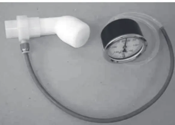
at the predetermined expiratory pressures. In order to measure and record expira-tory pressures, a mouthpiece was developed to connect a vacuum manometer (± 300 cmH2O; Gerar, São Paulo, Brazil) to the Flutter VRP1®
(Figure 1). Before starting the experimental protocol, to ensure the reliability of the values seen in the vacuum manometer, the manometer was connected to a pressure generator (water column) in order to determine whether the pres-sure generated and, consequently, the prespres-sure displayed in this equipment were equivalent. In addition, the calibration of the vacuum manom-eter was checked at each collection.
Before performing the technique using pres-sures set at 15 cmH2O (P15) and 25 cmH2O (P25), the participants underwent training, which consisted in using the technique until being able to perform three consecutive exhalations at the predetermined expiratory pressure. By watching the vacuum manometer display, the participants could maintain the established pressure, which was also checked by the therapist.
The choice of P15 was based on the obser-vation of the results obtained in clinical practice at the FCT/UNESP Centro de Estudos e Atendimentos em Fisioterapia e Reabilitação (CEAFIR, Center for Physical Therapy and Rehabilitation Studies and Treatment), in which we identified that the mean expiratory pressure generated by the patients, when instructed to perform the technique using the Flutter VRP1® for
10 consecutive min, was 15 cmH2O. In contrast,
P25 was considered since it is described in the literature as the maximum expiratory pressure obtained using the equipment.(16,17) The
selec-tion of the expiratory pressure to be used on the first day of the protocol was made randomly.
The standard technique used on the two days of the experiment was as follows: a) a voluntary cough to expectorate sputum, so that all partici-pants would start the experiment under the same baseline conditions; b) another voluntary cough, after a 20-min rest period, designated time zero (T0); c) 10 min after T0, two 10-min series (S1 and S2) using the Flutter VRP1®, with a 10-min
Figure 1 - Adaptation to allow the vacuum manometer and the Flutter VRP1® to be connected in
cm and Q = volumetric flow ratio). The mean viscosity value, which was obtained by meas-uring the viscosity of five samples of the same sputum that were collected at each time point, was used for the analysis.
For statistical analysis, first the Shapiro-Wilk test was used to assess the normality of the data. For data with normal distribution, the comparison between P15 and P25 in terms of the different time points (T0, S1 and S2) was performed using the Student’s t-test for paired data (simulated cough machine, contact angle, relative velocity of sputum on frog palate after S1 and viscosity after S1). For data with non-normal distribution, the Wilcoxon test was used (relative velocity of sputum on frog palate at T0 and after S2 and analyzed based on relative transport velocity on
frog palate, sputum transport in a simulated cough machine and contact angle.(19)
Relative transport velocity of sputum on frog palate (velocity of sputum/velocity of endogenous mucus) was measured using a stereoscopic magnifying glass equipped with a reticulated eyepiece (Wild M8; Wild Heerbrugg, Gais, Switzerland) and using a stopwatch (Track. Pro; Mondaine, Zurich, Switzerland) to measure transport time (in seconds) in a cephalocaudal direction along 10 mm of frog palate.(20,21) After
every four samples tested, the frog mucus was measured to control the integrity of the palate.
The in vitro representation of cough was performed using a simulated cough machine,(22)
consisting of a simple airway model (a PVC tube), measuring 4 mm in internal diameter and 30 cm in length, within which the sputum samples were placed. This model was connected to a latex tube that linked it to a solenoid valve, which was connected to a timing system. The propellant pressure was 4.2 kgf/cm2, and release
time was 1 s. In order to perform the test, a small quantity of sputum was placed in the proximal part of the model and the timing system was activated.
In order to measure the contact angle, the sputum sample was placed on a deionized slide, which was treated with sulfochromic acid, and was observed using an eyepiece (×20)equipped with a goniometer (angle microscope/042002; Holtermann, São Paulo, Brazil). The contact angle was measured (in degrees) immediately after the sample was placed on the slide and 5 min later. The angle formed between the sputum and the solid surface reflects the surface tension and strength of adhesion of sputum.(23)
Viscosity was analyzed using a dual capillary viscometer,(24,25) consisting of a steel capillary
within a glass capillary. The capillary system was connected to a water pressure column. A quantity of sputum filling 5 mm of the glass capillary was placed at its proximal end, which was connected to the water pressure column. Sputum detachment, pressure, time elapsed and recoil were determined by imposing pres-sure within the capillary. The viscosity value was obtained using Poiseuille’s equation (η = ϖ × r4 × ΔP/8 × l × Q; where η = viscosity,
r = tube radius in cm, ΔP = sample pressure within the tube in cmH2O, l = tube length in
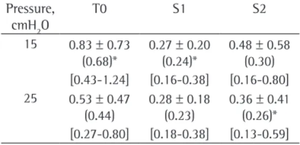
Table 1 - Sputum viscosity (×10³ P) for patients with bronchiectasis prior to and after the use of the Flutter VRP1® with pressures set at 15 and 25 cm H
2O. a
Pressure, cmH2O
T0 S1 S2
15 0.83 ± 0.73 (0.68)*
0.27 ± 0.20 (0.24)*
0.48 ± 0.58 (0.30) [0.43-1.24] [0.16-0.38] [0.16-0.80] 25 0.53 ± 0.47
(0.44)
0.28 ± 0.18 (0.23)
0.36 ± 0.41 (0.26)* [0.27-0.80] [0.18-0.38] [0.13-0.59]
T0: sputum samples collected after a 20-min rest period; S1: sputum samples collected after the 1st series using the Flutter VRP1®; and S2: sputum samples collected after the
2nd series using the Flutter VRP1®. aValues expressed as
mean ± SD (median) [95% CI]. *Values with a statistically significant difference in relation to T0 (Friedman test, followed by Dunn’s test; p < 0.05).
Table 2 - Relative transport velocity of sputum on frog palate (velocity of sample/normal velocity of frog mucus) for patients with bronchiectasis prior to and after the use of the Flutter VRP1® with pressures set
at 15 and 25 cm H2O.a
Pressure, cmH2O
T0 S1 S2
15 0.97 ± 0.43 (0.87)
0.87 ± 0.26 (0.82)
0.95 ± 0.37 (0.83) [0.74-1.21] [0.72-1.01] [0.75-1.16] 25 0.76 ± 0.20
(0.75)
0.94 ± 0.40 (0.84)
0.87 ± 0.30 (0.72) [0.65-0.89] [0.72-1.16] [0.71-1.03]
T0: sputum samples collected after a 20-min rest period; S1: sputum samples collected after the 1st series using the Flutter VRP1®; and S2: sputum samples collected after the
2nd series using the Flutter VRP1®. aValues expressed as
P25 were significantly lower than were those obtained at T0 at each pressure, respectively.
Table 2 presents the mean values for relative transport velocity of sputum at each time point and at each pressure applied using the device. There were no significant differences among the time points at each pressure or between the pressures studied.
Table 3 shows the mean values for sputum transport in a simulated cough machine at the different time points and expiratory pressures. There were no significant differences among the time points analyzed at each pressure or between the pressures studied.
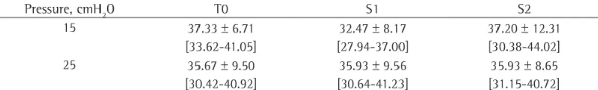
Table 4 presents the mean values for sputum contact angle. No significant differences were found among the time points analyzed or between the pressures studied.
Discussion
The results demonstrate that there were no significant differences in sputum viscosity between the values obtained at T0 (Table 1). The absence of differences ensured the similarity of sputum viscosity prior to the initiation of each intervention. In addition, there were significant differences between S1 and T0 at P15, as well as between S2 and T0 at P25, and there were no differences between the expiratory pressures viscosity at the same time points). The
compar-ison of the different time points (T0, S1 and S2) in terms of the same expiratory pressure was performed using ANOVA with repeated measures (simulated cough machine and contact angle at P15 and P25) or the Friedman test (relative velocity of sputum on frog palate and viscosity at P15 and P25), followed by Dunn’s test. The level of significance was set at 5%.(26)
The power of the study was calculated using the GraphPad StatMate software, version 2.00 for Windows (GraphPad Software, San Diego, CA, USA). The number of volunteers analyzed and the level of significance of 5% (two-tailed test) ensured a power greater than 80% to detect differences among the variables.
Results
None of the patients were excluded from the study, and all of them performed the technique with the Flutter VRP1® placed in a neutral
posi-tion, that is, parallel to the table. Table 1 shows the mean values for sputum viscosity prior to (T0) and after (S1 and S2) the sessions performed at P15 and P25. It is possible to observe that there was no significant difference between the values obtained at T0 for the two sessions performed (P15 and P25) and that the viscosity values obtained after S1 at P15 and after S2 at
Table 3 - Sputum transport in a simulated cough machine (in mm) for patients with bronchiectasis prior to and after the use of the Flutter VRP1® with pressures set at 15 and 25 cm H
2O. a
Pressure, cmH2O T0 S1 S2
15 141.93 ± 50.35
[114.05-169.82]
153.83 ± 52.30 [124.87-182.79]
153.03 ± 53.69 [123.29-182.76]
25 127.21 ± 53.59
[97.54-153.89]
140.93 ± 56.84 [109.46-172.41]
137.33 ± 53.59 [107.65-167.01]
T0: sputum samples collected after a 20-min rest period; S1: sputum samples collected after the 1st series using the Flutter VRP1®; and S2: sputum samples collected after the 2nd series using the Flutter VRP1®. aValues expressed as mean ± SD
[95% CI].
Table 4 - Sputum contact angle (in degrees) for patients with bronchiectasis prior to and after the use of the Flutter VRP1® with pressures set at 15 and 25 cm H
2O. a
Pressure, cmH2O T0 S1 S2
15 37.33 ± 6.71 32.47 ± 8.17 37.20 ± 12.31
[33.62-41.05] [27.94-37.00] [30.38-44.02]
25 35.67 ± 9.50 35.93 ± 9.56 35.93 ± 8.65
[30.42-40.92] [30.64-41.23] [31.15-40.72]
T0: sputum samples collected after a 20-min rest period; S1: sputum samples collected after the 1st series using the Flutter VRP1®; and S2: sputum samples collected after the 2nd series using the Flutter VRP1®. aValues expressed as mean ± SD
studied or the expiratory pressures used in terms of transportability as evaluated using the simu-lated cough machine. Since expiratory time was not controlled, it is not possible to ensure that the oscillation frequency was constant enough to alter elasticity, a property that was not evalu-ated in the present study, given that sputum clearance depends on a high viscosity/elasticity ratio.(28)
In terms of sputum contact angle, there were no significant differences among the time points analyzed at each pressure or between the pressures (Table 4). Assuming that the practices performed did not contribute to the worsening of the physicochemical properties of sputum, that the initial time points of the practices did not present statistical differences, that the ideal contact angle value was approximately 20°(21)
and that the sputum samples during those sessions presented an angle that differed signifi-cantly from the reference values, with a mean higher than 32°, we can infer that, at all time points analyzed, sputum transportability was decreased.
The high means for contact angle (Table 4) and the low means for relative transport velocity on frog palate (Table 2), in comparison with the reference values, show that sputum trans-portability was decreased and, regardless of the expiratory pressure employed while using the Flutter VRP1®, sputum clearance by the
muco-ciliary system would be more difficult.
The absence of significant differences in terms of sputum transportability after the tech-nique was performed at P15 and at P25 is in agreement with the results of one study,(4)
despite methodological differences related to expiratory pressure and time required to perform the technique.
In summary, as has been shown in a study of patients with cystic fibrosis,(12) the use of
the Flutter VRP1® is safe, efficient and easy in
patients with bronchiectasis. Under the condi-tions of our experiment, the use of the device at a controlled expiratory pressure is a viable technique that can be implemented in clinical practice. It is only necessary to adapt a mouth-piece so that a vacuum manometer can be connected.
In addition, our results showed that there were no significant differences in terms of sputum transportability at the different pres-at each time point studied, which suggests thpres-at
the use of the Flutter VRP1® reduced sputum
viscosity, facilitating sputum clearance, and its immediate effect was more evident at P15. This finding is in agreement with those of previous studies,(3,11) which found decreased sputum
viscosity with the use of the device.
The occurrence of significant differences in sputum viscosity at distinct time points, at the expiratory pressures studied, can be related to the difference in expiratory time in the two sessions. Although expiratory time was not controlled, we observed that, at a lower pressure (P15), the patients maintained longer expiratory time and, consequently, the amount of time to which the mucus was exposed to a constant oscillation frequency increased, making viscosity decrease after only one series. In order to perform the technique at P25, a greater effort is needed and shorter expiratory time was observed. Therefore, the amount of time to which the mucus was exposed to oscillation decreased, and the effect on sputum viscosity was achieved only after S2.
In the present study, three methods were used to evaluate sputum transportability: rela-tive transport velocity of sputum on frog palate, sputum transport in a simulated cough machine and contact angle.(21)
In terms of transport velocity on frog palate, no significant differences were found after the session performed at the two expiratory pres-sures or among each time point analyzed (Table 2). However, the means for relative muco-ciliary transport velocity were found to be lower than 0.97. According to one group of authors,(27)
a sputum sample with good transportability is that presenting relative velocity values between 1.0 and 1.1. The values found in the present study suggest that cough alone, as well as the use of OPEP at P15 or at P25, removes sputum with decreased transportability, there being no influence of the device.
In vitro mucociliary transport was also analyzed based on sputum transport in a simulated cough machine.(22) Physiologically,
secretion clearance by cough can be facilitated by a high viscosity/elasticity ratio.(28) However,
sputum samples with extremely high viscoe-lastic components can present decreased in vitro transport,(28) as evaluated using the simulated
chronic bronchitis: effectiveness of three methods. Arch Phys Med Rehabil. 2000;81(5):558-60.
11. Pires Neto RC, Ramos EMC, Ramos D. Transportabilidade e viscoelasticidade do muco brônquico, de um paciente com bronquiectasia, expectorado após a tapotagem e o aparelho flutter-VRP1 - estudo de caso. Rev Bras Fisioter. 2004;8(2):165-68.
12. Homnick DN, Anderson K, Marks JH. Comparison of the flutter device to standard chest physiotherapy in hospitalized patients with cystic fibrosis: a pilot study. Chest. 1998;114(4):993-7.
13. Chatham K, Marshall C, Campbell IA, Prescott RJ. The use of the Flutter VRP1 device in post thoracotomy patients. Physiotherapy. 1993;79(2):95-8.
14. Brooks D, Newbold E, Kozar LF, Rivera M. The flutter device and expiratory pressures. J Cardiopulm Rehabil. 2002;22(1):53-7.
15. Folstein MF, Folstein SE, McHugh PR. “Mini-mental state”. A practical method for grading the cognitive state of patients for the clinician. J Psychiatr Res. 1975;12(3):189-98.
16. de Lima LC, Duarte JB, Lépore Neto FP, Abe PT, Gastaldi AC. Mechanical evaluation of a respiratory device. Med Eng Phys. 2005;27(2):181-7.
17. Volsko TA, DiFiore J, Chatburn RL. Performance comparison of two oscillating positive expiratory pressure devices: Acapella versus Flutter. Respir Care. 2003;48(2):124-30.
18. Gondor M, Nixon PA, Mutich R, Rebovich P, Orenstein DM. Comparison of Flutter device and chest physical therapy in the treatment of cystic fibrosis pulmonary exacerbation. Pediatr Pulmonol. 1999;28(4):255-60. 19. Gastaldi AC, Jardim JR, King M. The influence of
temperature and length of time of storage of frog mucus samples. Biorheology. 2000;37(3):203-11. 20. Rubin BK, Ramirez O, King M. Mucus-depleted frog
palate as a model for the study of mucociliary clearance. J Appl Physiol. 1990;69(2):424-9.
21. Macchione M, Guimarães ET, Saldiva PH, Lorenzi-Filho G. Methods for studying respiratory mucus and mucus clearance. Braz J Med Biol Res. 1995;28(11-12):1347-55.
22. King M, Brock G, Lundell C. Clearance of mucus by simulated cough. J Appl Physiol. 1985;58(6):1776-82. 23. Girod S, Zahm JM, Plotkowski C, Beck G, Puchelle E.
Role of the physiochemical properties of mucus in the protection of the respiratory epithelium. Eur Respir J. 1992;5(4):477-87.
24. Barnett B, Dulfano MJ, Philippoff W, Han CD. A method for determining the viscoelastic properties of bioogical fluids. Biorheology. 1970;7(1):55-67.
25. Kim CS, Berkley BB, Abraham WM, Wanner A. A micro double capillary method for rheologic measurements of lower airway secretions. Bull Eur Physiopathol Respir. 1982;18(6):915-27.
26. Norman GR, Streiner DL. Biostatistics – The Bare Essentials. St. Louis: Mosby Year Book; 1994.
27. Ramos D, Ramos EMC, Jardim JRB, Saldiva PHN, Faresin SM. Efeito da aerossolterapia nas propriedades físico-químicas do muco brônquico. Rev Bras Fisioter. 2004;8(1):61-4.
28. Zayas JG, Man GC, King M. Tracheal mucus rheology in patients undergoing diagnostic bronchoscopy. Interrelations with smoking and cancer. Am Rev Respir Dis. 1990;141(5 Pt 1):1107-13.
sures during the sessions using the device. However, the decreased sputum viscosity after the sessions at P15 and P25 suggests a better rheological profile and greater sputum thinning after the use of the device. Due to the similarity of the results obtained at the two pressures studied, we can conclude that the patient does not have to generate high pressure to achieve the desired result.
In the present study, expiratory pressure measurement (P15 and P 25) was the only vari-able that was pre-established and controlled during the series using the device. Therefore, the absence of data regarding expiratory time, flow and oscillation frequency can be considered a limitation of the present study. For this reason, future studies should include the standardiza-tion of these variables, during the use of the device, so that their effect on the rheological properties of sputum can be understood more fully.
References
1. Zanchet RC, Magalhães AC, Correia AF, Feijó G. A influência de bactérias patogênicas na transportabilidade do escarro e na qualidade de vida de portadores de bronquiectasia. Rev Bras Fisioter. 2006;10(4):457-63. 2. Angrill J, Agustí C, Torres A. Bronchiectasis. Curr Opin
Infect Dis. 2001;14(2):193-7.
3. App EM, Kieselmann R, Reinhardt D, Lindemann H, Dasgupta B, King M, et al. Sputum rheology changes in cystic fibrosis lung disease following two different types of physiotherapy: flutter vs autogenic drainage. Chest. 1998;114(1):171-7.
4. Valente AM, Gastaldi AC, Cravo SL, Afonso JL, Sologuren MJ, Guimarães RC. The effect of two techniques on the characteristics and transport of sputum in patients with bronchiectasis: a pilot study. Physiotherapy. 2004;90(3):158-64.
5. Morsch AL, Amorim MM, Barbieri A, Santoro LL, Fernandes AL. Influence of oscillating positive expiratory pressure and the forced expiratory technique on sputum cell counts and quantity of induced sputum in patients with asthma or chronic obstructive pulmonary disease. J Bras Pneumol. 2008;34(12):1026-32.
6. Alves LA, Pitta F, Brunetto AF. Performance analysis of the Flutter VRP1 under different flows and angles. Respir Care. 2008;53(3):316-23.
7. Hardy K. A review of airway clearance: new techniques, indications and recommendations. Respir Care.1994;39(5):440-55.
8. Konstan MW, Stern RC, Doershuk CF. Efficacy of the Flutter device for airway mucus clearance in patients with cystic fibrosis. J Pediatr. 1994;124(5 Pt 1):689-93. 9. Pryor JA. Physiotherapy for airway clearance in adults.
Eur Respir J. 1999;14(6):1418-24.
About the authors
Ercy Mara Cipulo Ramos
Professor. Department of Physical Therapy, School of Science and Technology, Universidade Estadual Paulista – UNESP, São Paulo State University – Presidente Prudente, Brazil.
Dionei Ramos
Professor. Department of Physical Therapy, School of Science and Technology, Universidade Estadual Paulista – UNESP, São Paulo State University – Presidente Prudente, Brazil.
Daniela Mizusaki Iyomasa
Physical Therapist. School of Science and Technology, Universidade Estadual Paulista – UNESP, São Paulo State University – Presidente Prudente, Brazil.
Graciane Laender Moreira
Masters Student in Physical Therapy. School of Science and Technology, Universidade Estadual Paulista – UNESP, São Paulo State University – Presidente Prudente, Brazil.
Kátia Cristina Teixeira Melegati
Physical Therapist. School of Science and Technology, Universidade Estadual Paulista – UNESP, São Paulo State University – Presidente Prudente, Brazil.
Luiz Carlos Marques Vanderlei
Professor. Department of Physical Therapy, School of Science and Technology, Universidade Estadual Paulista – UNESP, São Paulo State University – Presidente Prudente, Brazil.
José Roberto Jardim
Professor. Universidade Federal de São Paulo/Escola Paulista de Medicina – UNIFESP/EPM, Federal University of São Paulo/Paulista School of Medicine – São Paulo, Brazil.
Adriana Siqueira de Oliveira