3
Universidade Estadual Paulista “Júlio de Mesquita Filho”
Faculdade de Medicina de Botucatu
AVALIAÇÃO DO POTENCIAL CANCERÍGENO DO
DIURON [3-(3,4-DICLOROFENIL)-1,1-DIMETILURÉIA]
NO MODELO DE INICIAÇÃO-PROMOÇÃO CUTÂNEA
EM CAMUNDONGOS SWISS
Bianca Ferrucio
Dissertação apresentada ao Programa de Pós-Graduação em Patologia da Faculdade de Medicina de Botucatu, Universidade Estadual Paulista –
UNESP, para obtenção do título de Mestre em Patologia.
4
Universidade Estadual Paulista “Júlio
de Mesquita Filho”
Faculdade de Medicina de Botucatu
AVALIAÇÃO DO POTENCIAL CANCERÍGENO DO
DIURON [3-(3,4-DICLOROFENIL)-1,1-DIMETILURÉIA]
NO MODELO DE INICIAÇÃO-PROMOÇÃO CUTÂNEA
EM CAMUNDONGOS SWISS
Mestranda: Bianca Ferrucio
Orientador: João Lauro Viana de Camargo
Co-orientador: Deilson Elgui de Oliveira
Dissertação apresentada ao Programa de Pós-Graduação em Patologia da Faculdade de Medicina de Botucatu, Universidade Estadual Paulista –
UNESP, para obtenção do título de Mestre em Patologia.
5
Dedico este trabalho aos meus pais e maiores
fãs, Paulo e Tereza. Com o apoio e a
dedicação de vocês, difícil é não progredir!
Obrigada por acreditarem em mim.
6
AGRADECIMENTO ESPECIAL
Ao meu orientador, João Lauro. Serei para sempre grata pela oportunidade
e honra de ser sua orientada. Com certeza sentirei falta de um orientador
“Father Figure” no doutorado!
7
AGRADECIMENTOS
Ao meu irmão Bruno Cochoco: irmão mais velho, mais careca, e mais
companheiro do mundo!
À minha vó Helena, exemplo de força e vitalidade. Obrigada pelo carinho e
por todas as comidas congeladas que eu almocei durante estes 2 anos.
Ao meu vô Alcides, por sua excentricidade e por ainda me surpreender, e
minha vó Consuelo (
in memorian
) por fazer parte das minhas melhores
lembranças.
Ao Henrique, meu melhor amigo e melhor namorado, por tudo o que
construímos e a tudo que sobrevivemos juntos. Te amo!
Às amigas Paulinha e Lu, porque vocês são meu porto seguro e porque
vocês são divertidas.
À Ana Rachel, por tantos momentos bons e especiais que passamos juntas,
e à Alice, pela segurança de que posso contar com você, mesmo tão
distante.
Ao meu co-orientador Deilson, pela ajuda na elaboração deste projeto.
Aos amigos do TOXICAM: Iza, Mitscheli, Shadia, Marize, Gabrielli, Meri
(e Edson), Meire, Tony, João, Alexandre, Paulo, Mara, Ivana e PC, e
especialmente à Carla, por tantos conselhos e presentinhos, e à querida
“equipe” Ana Paula e Vivi.
À Cris, por todas as impressões, telefonemas, scanners, etc...e pelo seu alto
astral constante.
Aos funcionários do Laboratório de Patologia Molecular: Marquinhos,
Selene, Fernando e Cris por todas as vezes que me socorreram.
8 ÍNDICE
REVISÃO DE LITERATURA
Carcinogênese cutânea 9
Bioensaios para detecção de cancerígenos químicos 13
O herbicida diuron 15
REFERÊNCIAS BIBLIOGRÁFICAS 18
MANUSCRITO 24
Abstract 26
Introduction 27
Material and methods 30
Results 33
Discussion 35
Aknowledgements 39
References 39
Tables 44
Figures 50
APÊNDICE
9 REVISÃO DE LITERATURA
Carcinogênese cutânea
De acordo com a Organização Mundial da Saúde, um terço de todos os casos de câncer diagnosticados são de pele, e sua incidência tem aumentado nas últimas décadas. Esta observação evidencia a importância da prevenção do câncer de pele, pela identificação dos agentes causadores e minimizando a exposição aos mesmos. Por outro lado, por serem de fácil acesso, neoplasias cutâneas podem ser detectadas precocemente e curadas com sucesso, permitindo a redução das taxas de morbidade e mortalidade.
Visando entender os mecanismos pelos quais agem os agentes cancerígenos, há décadas que a pele de animais de experimentação tem sido estudada em modelos de carcinogênese química, tendo sido o órgão investigado pioneiramente por Yamagiwa & Ichikawa em 1915 (Williams, 1999). Em 1947, Berenblum & Shubik descreveram o atual paradigma da carcinogênese de duas etapas, iniciação e promoção, baseados em estudos sobre a atividade cancerígena da resina do óleo de cróton na pele de camundongos. Atualmente, sabe-se que a carcinogênese cutânea é um processo que evolui em múltiplas etapas, caracterizadas experimentalmente como as de iniciação, promoção e progressão (Slaga et al., 1996). A iniciação requer a lesão do DNA e
posterior “fixação” desta alteração por pelo menos um ciclo de proliferação; tem por
10 e mutabilidade, como o TP53 (Frame et al., 1998). O oncogene H-RAS é crítico na
carcinogênese cutânea de camundongos, já que sua ativação é suficiente para iniciar células epidérmicas, encontrando-se comumente mutado em papilomas e em carcinomas (Hennings et al., 1993). A promoção, por sua vez, é a etapa em que ocorre a expansão clonal das células iniciadas e, consequentemente, a expressão da alteração do genoma ocorrida na iniciação (DiGiovanni, 1992). O promotor, sendo um agente químico ou físico, geralmente age por estímulo à proliferação celular, e tem por característica a existência de um limiar-de-dose abaixo do qual não exerce efeito tumorigênico. Diferente dos agentes iniciadores, os promotores possuem ação epigenética, i.e., não agridem o DNA. Se a exposição aos promotores for interrompida antes do aparecimento de tumores, as alterações estabelecidas por eles, e.g., hiperplasias, regridem, ou seja, são reversíveis (Yuspa et al., 1976).
Finalmente, na etapa da progressão, as células tumorais adquirem as
características básicas da malignidade, como aneuploidia e capacidade de invasão tecidual e de metástases, levando à manifestação clínica da doença (Yuspa et al., 1981). A conversão de papilomas a carcinomas cutâneos (progressão) é um evento que raramente ocorre na ausência de novos estímulos externos. É necessária ao menos uma nova alteração genética no clone das células promovidas, além daquelas ocorridas na iniciação (Boukamp, 2005). A progressão se desenvolve por várias mudanças moleculares. Exemplo disso, a expressão das citoceratinas 1 e 10, presente em pele normal e em papilomas, deixa de ser observada em carcinomas, que apresentam expressão da citoceratina 8, característica que se associa com a aquisição do fenótipo maligno (Abel et al., 2009). Da mesma maneira, enzimas como a telomerase e γ
11 importantes marcadores da progressão, como o TGF-β, pois sua ausência em papilomas
representa alto risco para conversão à malignidade (Hennings et al., 1993).
No processo de carcinogênese, inclusive na pele, são observadas alterações nas vias de sinalização celular, decorrentes de modificação nas funções de enzimas com participação em diferentes pontos da transdução de sinais. Dentre as vias mais estudadas nesse contexto estão as cascatas de MAP (mitogen-activated protein) cinases, a via
fosfatidilinositol-3-cinase (PI3K) – Akt (ou proteína cinase B, PKB) e a via de proteínas
da família NF-kappaB (El-Abaseri & Hansen, 2007; Kobielak & Fuchs, 2006; Wattenberg, 2007).
A via das MAP cinases (MAPK) é basicamente constituída por enzimas que fosforilam e ativam uma enzima cinase subseqüente na cascata. A ativação dessa via pode ser deflagrada por fatores de crescimento, agentes mitógenos, receptores acoplados a proteínas G, estresse e citocinas pró-inflamatórias. No topo da cascata estão as MAP3Ks, enzimas que fosforilam e ativam as MAP2Ks, e estas as MAPKs, que por sua vez ativam fatores de transcrição que regulam a expressão de conjunto heterogêneo de genes, em última análise proporcionando aumento da proliferação celular. Entre as MAP3Ks, destacam-se as MEKK1-4 (MAP and ERK kinase kinase), que atuam junto à
porção citoplasmática da membrana celular, e as proteínas das famílias Src e Raf (Avruch, 2007). As cinases da família Src desempenham papel crítico na adesão, invasão, proliferação e sobrevivência celular e angiogênese, e assim podem estar envolvidas em todas as etapas da carcinogênese cutânea (Kim et al., 2009).
12 demonstrado que o desenvolvimento e a manutenção de tumores induzidos pela proteína Ras são altamente dependentes da proteína Raf-1, que por sua vez é dispensável para a homeostase epidérmica (Ehrenreiter et al., 2009). Neste mesmo estudo, relatou-se que a presença da cinase Raf-1 foi também necessária para a expressão da proteína Myc. Esta proteína, presente nas camadas basais da epiderme, induz a proliferação e inibe a diferenciação de queratinócitos, e está associada à regulação de células-tronco (Watt, 1998).
A via PI3K-Akt, iniciada pela ativação de receptores com atividade tirosina cinase intrínseca (RTK) e receptores acoplados a proteínas G, requer a ativação do mensageiro secundário fosfatidilinositol-3,4,5-trifosfato (PIP3). Uma das principais conseqüências da ativação desta cascata de sinalização é a supressão da apoptose e aumento da síntese protéica, o que pode contribuir para a tumorigênese (Osaki et al., 2004). A via de sinalização dos fatores de transcrição NF-κB é ativada por citocinas
pró-inflamatórias (e.g., TNF, IL1) e por padrões moleculares associados a patógenos, e é dependente da ativação de diversas proteínas cinases, notadamente TAK1 e IKKs. A ativação desta via leva à translocação de dímeros de NF-κB do citoplasma para o
núcleo, sendo essencial na regulação da imunidade inata e da imunidade adaptativa (Bonizzi & Karin, 2004). Adicionalmente, a ativação da via NF-κB proporciona
estímulos para aumento da sobrevivência celular (pela diminuição da susceptibilidade à apoptose) e aumento da proliferação. Ativação constitutiva de NF-κB é documentada
13 Bioensaios para detecção de cancerígenos químicos
Existem numerosos protocolos para testar o potencial cancerígeno de substâncias químicas. O ensaio convencional, que tem duração de cerca de dois anos e envolve a administração de altas doses do composto em teste, é muitas vezes inviável devido ao elevado consumo de tempo e de recursos financeiros. Além disso, não considera as múltiplas etapas da carcinogênese química e é pouco eficiente para detectar agentes promotores fracos (Williams, 1999).
Devido a essas limitações, foi realizado considerável esforço para o estabelecimento de testes alternativos para a detecção do potencial cancerígeno de agentes químicos. Os ensaios de carcinogênese de duas etapas apresentam duração mais reduzida (semanas) e consideram também o aparecimento de lesões pré-neoplásicas como desfechos (end-points). Os ensaios de iniciação-promoção podem ser delineados
tanto para detectar o potencial promotor de agentes químicos, como o potencial iniciador. No primeiro caso, estes ensaios consistem na indução de células iniciadas pela aplicação de um agente iniciador (mutagênico) e, após certo intervalo, aplicações consecutivas da substância-teste (possível agente promotor), por período pré-determinado; no segundo caso, a substância-teste é administrada previamente ao tratamento com um promotor de tumores conhecido (Tsuda et al., 1999). Assim, este modelo é particularmente importante na investigação do mecanismo de ação de cancerígenos químicos, já que permite a distinção operacional entre as várias etapas do desenvolvimento de tumores (DiGiovanni, 1992).
14 atividade cancerígena do composto testado, por serem consideradas precursoras de papilomas e carcinomas de células escamosas (Williams, 1999).
Na mesma linha, foram desenvolvidos bioensaios alternativos de carcinogênese cutânea com animais produzidos por biotecnologia. Tais avanços metodológicos permitem reduzir a duração dos experimentos e o número de animais necessários, além de gerar resultados mais precisos e informações acerca do modo /mecanismo de ação das substâncias testadas (Brown & Balmain, 1995). Entre os modelos mais utilizados, estão as linhagens Tg.AC e RasH2, que carregam o transgene H-RAS ativado. Assim,
estes animais são geneticamente iniciados e indicados para a identificação de cancerígenos genotóxicos e não-genotóxicos (Lynch et al., 2007). Na mesma linha, camundongos nocaute Trp53+/- apresentam apenas um alelo funcional do gene supressor tumoral p53 e são altamente suscetíveis ao desenvolvimento de tumores por agentes mutagênicos (French et al., 2001). A linhagem de camundongos SENCAR tem sido utilizada na identificação de agentes iniciadores, promotores e progressores. Embora não seja geneticamente modificada, esta linhagem foi selecionada durante algumas gerações para expressar suscetibilidade elevada à carcinogênese cutânea de duas etapas, apresentando menor período de latência dos tumores (Slaga, 1986).
Em 2003, Pritchard et al. avaliaram a precisão das linhagens transgênicas
Trp53+/-, Tg/AC e RasH2 na identificação de cancerígenos humanos, em comparação
com os ensaios tradicionais de dois anos. De modo geral, estas linhagens geneticamente modificadas apresentaram boa preditividade, e a combinação de dois modelos diferentes foi considerada a melhor estratégia para avaliar o potencial cancerígeno de substâncias químicas.
15 prevalente em humanos (Enzmann, 1998). Estes ensaios procuram ainda mimetizar a exposição a baixas doses, como tipicamente ocorre na população humana. Além disso, a carcinogênese cutânea em camundongos desenvolve-se de maneira bastante similar àquela em humanos, o que contribui para a justificativa da utilização deste modelo animal. Exemplo disso, a mutação em células tronco epidérmicas é, aparentemente, fator desencadeante da carcinogênese em ambas as espécies (Kangsamaksin et al., 2007). Adicionalmente, muitas das mutações em genes considerados críticos na carcinogênese, como o H-Ras e o Tp53, são comuns tanto em camundongos como em humanos (French et al., 2001; Janowski, 1991). Por outro lado, algumas disparidades dificultam a extrapolação interespécies. Entre elas, está o fato de que camundongos desenvolvem predominantemente papilomas, tumor que não apresenta equivalente humano e que raramente progride a carcinoma de células escamosas, que por sua vez, é o tumor mais frequente na espécie humana (Glick et al., 2007).
O herbicida Diuron
16 Quando foi submetido ao teste convencional de longa duração em roedores, o Diuron mostrou atividade cancerígena para o urotélio de ratos Wistar e para a mama de camundongos NMRI (Iyer, 2002). Por esta razão, ele é classificado como “provável cancerígeno para a espécie humana” pela Agência de Proteção Ambiental norte
-americana (USEPA, 2004). No momento, este herbicida está liberado para uso agrícola em diversos países, inclusive no Brasil, o que não aconteceria se sua atividade mutagênica/cancerígena fosse bem definida (Lei Federal de Agrotóxicos, 1991). É necessário esclarecer qual o modo de ação (MOA) pelo qual o Diuron exerce a atividade cancerígena verificada em roedores e qual seu significado para a espécie humana. Estudos prévios em nosso laboratório demonstraram que o diuron e/ou seus metabólitos induzem tumores na bexiga urinária de ratos Wistar através de citotoxicidade química direta, seguida de proliferação celular regenerativa; no entanto, o mecanismo de ação específico ainda não foi esclarecido (Rocha et al., 2009).
Apesar de ter sido avaliado em vários ensaios in vitro e em ensaios in vivo em
mamíferos, a genotoxicidade do Diuron é controversa, dependendo do modelo experimental, dose e parâmetros finais estudados (Grutman et al., 1984; Iyer, 2002; Liu, 2001) (Tabela VII - Apêndice). No ensaio de Ames com Salmonella thyphimurium e
ativação metabólica, por exemplo, o Diuron apresentou fraca atividade mutagênica, e no teste do micronúcleo em medula óssea os resultados foram negativos (Seiler, 1978). Em testes in vivo com camundongos Swiss, este herbicida mostrou atividade mutagênica
pelo teste de micronúcleo em medula óssea (Agrawal et al., 1996) e pelo teste do dominante letal em células germinativas (Agrawal & Melhrota, 1997). No entanto, pelo teste Mutatox com uma variante escura (M169) da Photobacterium phosphoreum, os
17 questionada, por apresentarem baixa preditividade da carcinogenicidade em roedores (Witte et al., 2007). Além disso, o teste do cometa (ensaio de eletroforese em gel de célula alcalina única) foi sugerido como o método preferível para avaliar a genotoxicidade, uma vez que é relativamente insensível a citotoxicidade; assim, resultados falso-positivos são menos frequentes, mesmo quando altas concentrações do composto teste claramente afetam a viabilidade celular (Witte et al., 2007). Estudos prévios em nosso laboratório indicaram ausência de genotoxicidade do Diuron pelo teste do cometa em células uroteliais e do sangue periférico de ratos Wistar machos (Nascimento et al., 2006). Além disso, uma versão modificada do ensaio desenvolvida
in vitro em células CHO sugeriu que o produto também não é capaz de induzir ligações
cruzadas (crosslinks) no DNA (Rocha et al., 2009).
18 REFERÊNCIAS BIBLIOGRÁFICAS
Abel, E.L., Angel, J.M., Kiguchi, K., DiGiovanni, J. (2009). Multi-stage chemical carcinogenesis in mouse skin: Fundamentals and applications. Nat protoc 4(9),
1350-62.
Agrawal, R.C., Kumar, S., Mehrotra, N.K. (1996). Micronucleus induction by diuron in mouse bone marrow. Toxicol Lett 89, 1-4.
Agrawal, R.C., Mehrotra, N.K. (1997). Effect of diuron on germ cells of mice. Indian J
Exp Biol 35(11), 1256-7.
Antony, M., Shukla, Y., Mehrotra, N.K. (1989). Tumour initiatory activity of a herbicide diuron on mouse skin. Cancer Lett 48, 125-8.
Avruch J. (2007) MAP kinase pathways: The first twenty years. Biochim Biophys Acta
1773(8), 1150-60.
Berenblum, I., Shubik, P. (1947). A new, quantitative approach to the study of the stages of chemical carcinogenesis in the mouse`s skin. Br J Cancer 1,383-91.
Bonizzi, G., Karin, M. (2004). The two NF-κB activation pathways and their role in
innate and adaptative immunity. Trends Immunol 25(6), 280-8.
Boukamp, P. (2005). Non-melanoma skin cancer: what drives tumor development and progression? Carcinogenesis 26(10), 1657-67.
Brow, K., Balmain, A. (1995). Transgenic mice and squamous multistage skin carcinogenesis. Cancer Metastasis Rev 14(2), 113-24.
19 Canna-Michaelidou, S., Nicolaou, A.S. (1996). Evaluation of the genotoxicity potential
(by MutatoxTM test) of ten pesticides found as water pollutants in Cyprus. Sci
Total Environ 193, 27-35.
Carpenter, W.S., Lee, B.C., Gunderson, P.D., Stueland, D.T. (2002). Assessment of personal protective equipment use among midwestern farmers. Am J Ind Med
42, 236-47.
Chan, K.S., Sano, S., Kiguchi, K., Anders, J., Komazawa, N., Takeda, J., DiGiovanni, J. (2004) Disruption of Stat3 reveals a critical role in both the initiation and the promotion stages of epithelial carcinogenesis. J Clin Invest 114, 720-8.
Chaudhary, S.C., Alam, M.S., Siddiqui, M.S., Athar, M. (2009). Chemopreventive effect of farnesol on DMBA/TPA-induced skin tumorigenesis: Involvement of inflammation, Ras-ERK pathway and apoptosis. Life Sci 85(5-6), 196-205.
DiGiovanni, J. (1992). Multistage carcinogenesis in mouse skin. Pharmacol Ther 54(1),
63-128.
Ehrenreiter, K., Kern, F., Velamoor, V., Meissl, K., Galabova-Kovacs, G., Sibilia, M., Baccarini, M. (2009). Raf-1 Addiction in Ras-Induced Skin Carcinogenesis.
Cancer Cell 16, 149-60.
Enzmann, H., Bomhard, E., Iatropoulos, M., Ahr, H.J., Schlueter, G., Williams, G.M. (1998). Short- and intermediate-term carcinogenicity testing – a review. Part 1:
the prototypes mouse skin tumour assay and rat liver focus assay. Food Chem
Toxicol 36, 979-95.
El-Abaseri, T.B., Hansen, L.A. (2007). EGFR Activation and Ultraviolet Light-Induced Skin Carcinogenesis. J Biomed Biotechnol 3,1-4.
20 carcinogenesis in the mouse: correlating the genetics and the biology. Phil Trans
R Soc Lond B 353, 839-45.
French, J., Storer, R.D., Donehower, L.A. (2001). The nature of the heterozygous Trp53
knockout model for identication of mutagenic carcinogens. Toxicol Pathol 29,
24-9.
Glick, A.B., Ryscavage, A., Perez-Lorenzo, R., Hennings, H., Yuspa, S., Darwiche, N. (2007). The high-risk benign tumor: evidence from the two-stage skin cancer model and relevance for human cancer. Mol Carcinog 46(8), 605-10.
Grutman, G., Schoofs, L., Lontie, J.F., van Larebeke, N. (1984). The mutagenicity in prokaryotes of herbicides. Residue Rev 91, 1–46.
Hennings, H., Glick, A.B., Greenhalgh, D.A., Morgan, D.L., Strickland, J.E., Tennenbaum, T., Yuspa, S.H. (1993). Critical aspects of initiation, promotion and progression in multistage epidermal carcinogenesis. Soc Exp Biol Med,
202,1-18.
Iyer, P. (2002). Evidence on the development and reproductive toxicity of diuron. Draft. Reproductive and Cancer Hazard Assessment Section. Office of Environmental Health Hazard Assessment, California Environmental Protection Agency, 43. Janowski, M. (1991) ras proteins and the ras-related signal transduction pathway.
Radiat Environ Biophys 30(3), 185-9.
Kangsamaksin, T., Park, H.J., Trempus, C.S., Morris, R.J. (2007). A perspective on murine keratinocyte stem cells as targets of chemically induced skin cancer. Mol
Carcinog. 46, 579–584.
Karin, M. (2006). Nuclear factor-κB in cancer development and progression. Nature
21 Kim, L.C., Song, L., Haura, E.B. (2009). Src kinases as therapeutic targets for cancer.
Nat Rev Clin Oncol 6(10), 587-95.
Kobielak, A., Fuchs, E. (2005). Links between α-catenin, NF-κB, and squamous cell
carcinoma in skin. PNAS 103(7), 2322–2327.
Liu, J. (2001). Phenylurea herbicides. In Handbook of Pesticide Toxicology – Agents
(K.E. Krieger, ed.), Vol. 2, pp. 1521-3. Academic Press, San Diego.
Lynch, D., Svoboda, J., Putta, S., Hofland, H.E.J., Chern, W.H., Hansen, L. (2007) Mouse skin models for carcinogenic hazard identification: utilities and challenges. Toxicol Pathol 35(7), 853-64.
Nascimento, M.G., de Oliveira, M.L.C.S., Lima, A.S., de Camargo, J.L.V. (2006). Effects of diuron [3-(3,4-dichlorophenyl)-1,1-dimethylurea] on the urinary bladder mucosa of male Wistar rats. Toxicology 224, 66-73.
Organização Mundial da Saúde (OMS) – World Health Organization, Health Topics,
Skin cancer. http://www.who.int/uv/faq/skincancer/en/index1.html.
Osaki, M., Oshimura, M., Ito, H. (2004) PI3K-Akt pathway: its functions and alterations in human cancer. Apoptosis 9, 667-76.
Perry, M.J., Marbella, A., Layde, P.M. (2002). Compliance with required pesticide-specific protective equipment use. Am J Ind Med 41, 70-3.
Pitot III, H.C., Dragan, Y.P. (2001). Chemical carcinogenesis. In Casarett & Doull’s
Toxicology the Basic Science of Poisons (C.D. Klaassen, ed), pp. 241-319. McGraw-Hill, New York.
Pritchard, J.B., French, J.E., Davis, B.J., Haseman, J.K. (2003). The Role of Transgenic Mouse Models in Carcinogen Identification. Environ Health Perspect 111(4),
22 Rocha, M.S., Nascimento, M.G., Cardoso, A.P.F., Lima, P.L.A., Zelandi, E.A., de
Camargo, J.L.V., de Oliveira, M.L.C.S. (2009). Cytotoxicity and regenerative proliferation as the mode of action for diuron-induced urothelial carcinogenesis in the rat. Toxicol Sci [Epub ahead of print], doi:10.1093/toxsci/kfp241.
Sancheti, G., Goyal, P.K. (2006) Effect of rosmarinus officinalis in modulating 7,12-dimethylbenz(a)anthracene induced skin tumorigenesis in mice. Phytother Res
20(11), 981-6.
Seiler, J.P. (1978) Herbicidal phenylalkylureas as possible mutagens I. Mutagenicity tests with some urea herbicides. Mutat Res 58(2-3), 353-9.
Slaga, T.J. (1986). SENCAR mouse skin tumorigenesis model versus other strains and stocks of mice. Environ Health Perspect 68, 27-32.
Tsuda, H., Park, C.B., Moore, M.A. (1999). Short- and medium-term carcinogenicity tests: Simple initiation-promotion assay systems. In The use of short- and medium-term tests for carcinogens and data on genetic effects in carcinogenic hazard evaluation (D.B. McGregor, J.M. Rice, S Venitt, eds). Vol. 146, pp 203-49. IARC Scientific Publications, Lyon, France.
USEPA (2004). Chemicals evaluated for carcinogenic potential. Office of Pesticide Programs, Health Effects Division. Science Information Management Branch, 106.
Watt, F.M. (1998). Epidermal stem cells: markers, patterning and the control of stem cell fate. Phil Trans R Soc Lond B 353, 831-7.
23 Williams, G.M. (1999) Chemically induced preneoplastic lesions in rodents as
indicators of carcinogenic activity. In The use of short- and medium-term tests for carcinogens and data on genetic effects in carcinogenic hazard evaluation (D.B. McGregor, J.M. Rice, S. Venitt, eds), Vol. 146, pp. 185-202. IARC Scientific Publications, Lyon, France.
Witte, I., Plappert, U., de Wall, H., Hartmann, A. (2007). Genetic toxicity assessment: employing the best science for human safety evaluation - Part III: The Comet Assay as an alternative to in vitro clastogenicity tests for early drug candidate selection. Toxicol Sci 97(1), 21-26.
Yuspa, S.H., Hennings, H., Saffiotti, U. (1976). Cutaneous chemical carcinogenesis: past, present and future. J Invest Dermatol 67(1), 199-208.
Yuspa, S.H., Hennings, H., Lichti, U. (1981). Initiator and promoter induced specific changes in epidermal function and biological potential. J Supramol Struct Cell
24 MANUSCRITO
O manuscrito apresentado a seguir, já submetido para publicação em Toxicologic
Pathology (ISSN 0192-6233, Fator de Impacto 2008 = 1.642), compreende os trabalhos
de Iniciação Científica e de Mestrado da biomédica Bianca Ferrucio, realizados no Departamento de Patologia, Faculdade de Medicina de Botucatu, UNESP, no período 2006-2010.
Auxílios financeiros
FAPESP Processos # 06/60506-1, 06/04630-5 e 08/01809-0;
25 Evaluation of diuron [3-(3,4-dichlorophenyl)-1,1-dimethyl urea] in a two-stage
mouse skin carcinogenesis assay
Bianca Ferrucio* - biferrucio@hotmail.com
Carla Adriene da Silva Franchi* - carlafranchi@fmb.unesp.br Natália Ferreira Boldrin* - nataliaboldrin@gmail.com
Maria Luiza Cotrim Sartor de Oliveira* - mdeolive@fmb.unesp.br João Lauro Viana de Camargo* - decam@fmb.unesp.br
*Center for the Evaluation of the Environmental Impact on Human Health (TOXICAM), Department of Pathology, Botucatu Medical School, UNESP - São Paulo State University, Botucatu, São Paulo, Brazil. 18618-000
Address correspondence to:
João Lauro Viana de Camargo, M.D. Ph.D.
Departamento de Patologia, Faculdade de Medicina, UNESP 18618-000, Botucatu, São Paulo, Brasil
E-mail: decam@fmb.unesp.br
Phone number: +55 14 38116238; Fax: +55 14 38152348
26 ABSTRACT
Diuron [3-(3,4-dichlorophenyl)-1,1-dimethyl urea] is a herbicide with carcinogenic activity in rats and mice which have developed respectively urothelial and mammary gland tumors in long-term studies. Accordingly, diuron has been categorized as a
“likely human carcinogen” by the USEPA. Although the carcinogenesis initiating
27 INTRODUCTION
Diuron [3-(3,4-dichlorophenyl)-1,1-dimethyl-urea] is a widely used herbicide for weed control in soy, cotton, sugarcane, citrus fruits, wheat and coffee crops, as well as in non-crop areas such as airport runways, railroads and oil-pipes. Occupational exposure is the main concern about this herbicide. Agricultural workers frequently neglect safety practices, including the use of personal protective equipments that aim to avoid dermal exposure during pesticides mixing or application (Carpenter et al., 2002; Perry et al., 2002). Moreover, skin protective equipment (SPE) may fail to cover the skin surface entirely and transfer may occur when touching the contaminated exterior of the SPE; additionally, SPE may enable permeation depending on the manipulated substance and/or the SPE material (Brouwer et al., 2005).
Diuron was recognized as carcinogenic to rodents orally exposed to 2,500 ppm through the feed in two-year long bioassays (Iyer, 2002). In those studies, Wistar rats developed urothelial papillomas and carcinomas, and NMRI female mice developed mammary gland adenocarcinomas. According to these evidences, the U.S. Environmental Protection Agency has categorized this herbicide as “likely human carcinogen” (US Environmental Protection Agency, 2004). Since diuron is released for
agricultural use, it is necessary to clarify the mode of action (MOA) through which this product has caused rodent neoplasia and verify whether it is relevant to humans. In a recent study from this laboratory it was demonstrated that diuron and/or its metabolites induce urinary bladder carcinogenesis in Wistar rats through direct chemical cytotoxicity followed by regenerative cell proliferation; the specific mechanism of action, however, has not been clarified yet (Rocha et al., 2009).
Despite having been evaluated in several in vitro and mammalian in vivo genetic
28 experimental protocol, dose levels and endpoints evaluated (Grutman et al., 1984; Iyer, 2002; Liu, 2001). A weak mutagenic activity of diuron was reported in a metabolically
activated Ames’ test with Salmonella thyphimurium, but results were negative in a
mouse bone marrow micronucleus assay (Seiler, 1978). When tested in Swiss mice, diuron was positive in the bone marrow micronucleus assay (Agrawal et al., 1996) and in the germ cells dominant lethal assay (Agrawal & Mehrotra, 1997). However, in the Mutatox test with a dark variant (M169) of the Photobacterium phosphoreum the
genotoxicity of diuron resulted only suspected (Canna-Michaelidou & Nicolaou, 1996). The biological significance of positive results obtained in genotoxicity cytogenetic assays has been questioned based on their low predictivity of rodent carcinogenicity (Witte et al., 2007). Additionally, it was suggested that the comet assay (single-cell gel assay) is the most preferable method to assess genotoxicity once it is relatively insensitive to cytotoxicity; hence, false positive results are less frequent, even when high concentrations of the test compounds clearly affect cell viability (Witte et al., 2007). Previous studies from this laboratory reported absence of diuron genotoxicity in the standard alkaline version of the comet assay with urothelial and peripheral blood cells (Nascimento et al., 2006). Besides, a modified version of the comet assay developed in vitro with CHO cells indicated that diuron also does not induce DNA
cross-links (Rocha et al., 2009).
29 genotoxic (Pitot III & Dragan, 2001). However, no initiating potential was verified by Antony et al. (1989) when diuron was applied at a single 250 mg/kg b.w. dose, which could indicate a possible threshold, inconsistent with its supposed initiating potential. In this case, a promoting influence on the mouse skin carcinogenesis process would be more likely, since in this condition a threshold level and a non-genotoxic MOA would be more suitable (Williams, 1999). Antony et al. (1989) did not justify the diuron dose levels they evaluated, adding to the unsubstantial aspect of their study.
30 The present studies aimed to evaluate the carcinogenic potential of diuron in a female Swiss mouse skin carcinogenesis assay. Besides, the potential role of DMSO or acetone – the herbicide solvents – was also investigated in this process. Regardless of
the solvent used, diuron did not exert any carcinogenesis initiating or promoting activities in the mouse skin.
MATERIAL AND METHODS
Animals and chemicals
These experiments were approved by the Committee for Ethics in Animal Experimentation of the UNESP Medical School, SP, Brazil [protocols 2006-567 and 2008-672]. Four-week-old Swiss female mice obtained from the Multidisciplinary Center for Biological Investigation (CEMIB, UNICAMP, Campinas, SP, Brazil) were kept in polypropylene cages (five animals/cage) with metallic grill covers, and maintained in a room at 22 ± 2◦C, 55 ± 10% humidity and a 12h light/dark cycle.
Animals were provided with commercial pelletized diet (Nuvital, Nuvilab, Curitiba, PR, Brazil) and tap water ad libitum.
After a two-week acclimatization period, mice were randomized to the experimental groups according to body weights. The dorsal skin was shaved using electric clippers one day before the first treatment and weekly thereafter, except for the untreated control.
31 areas. Skin patches with lesions (ulcers and tumors) were cut randomly for histological analyses. Liver, spleen, kidneys and lymph nodes were also collected and weighted. All tissues were fixed in 10% buffered formalin, followed by routine histological processing.
Treatments
Positive control groups were initiated with 9,10-dimethyl-1,2-benzantracene (DMBA, Sigma–Aldrich Co., St Louis, MO), and promoted with
12-O-tetradecanoylphorbol 13-acetate (TPA, Sigma–Aldrich Co, St Louis, MO). Diuron
(CAS No. 30-54-1) was purchased from Sigma Chemical Co., St. Louis, MO.
The chemical substances were alternatively or successively applied with micropipette on a shaved dorsal area of approximately 9 cm2, at the following concentrations: DMBA, 52µg/100µL acetone; TPA, 3.5µg/100µL acetone; diuron, 250mg/kg b.w. dissolved in 100µL DMSO or in 300µL acetone; acetone, 100µL and 300µL (vehicle controls). DMBA was applied once and TPA was applied three times/week until the end of each experiment. Diuron was applied three times/week during three weeks when tested as an initiator, and three times/week until the end of each experiment when tested as a promoter. Acetone was applied once when tested as an initiator and three times/week until the end of the experiment when tested as a promoter.
32 diuron/DMSO. A three-day interval was given between initiation and promotion treatments in groups G4 and G5, and one-week interval in groups G2, G3 and G6.
Experiment-2 consisted of nine groups with 15 animals each (Figure 2): Group 1 (G1), untreated control; Group 2 (G2), DMBA followed by acetone 300µL; Group 3 (G3), DMBA followed by diuron/acetone; Group 4 (G4), DMBA followed by diuron/DMSO; Group 5 (G5), acetone 100µL followed by diuron/acetone; Group 6 (G6), acetone 100µL followed by diuron/DMSO; Group 7 (G7), DMBA followed by TPA; Group 8 (G8), diuron/acetone followed by TPA; Group 9 (G9), diuron/DMSO followed by TPA. One-week interval was given between initiation and promotion treatment in all groups.
Experiments -1 and -2 were planned to last until week 25. However, a humane decision was taken to anticipate termination at weeks 23 and 21 respectively, owing to the high number of animals with severe skin ulcers.
Statistical analysis
The Fisher’s Exact Test was applied for comparison of skin lesions incidence
between experimental groups and for comparison between ulcer-bearing and “non -ulcer”-bearing animals in relation to the incidences of carcinomas per group. The
Mann-Whitney test for independent samples was used to compare the variation of the number of papillary tumors per mouse between ulcer-bearing and “non-ulcer”-bearing animals
33 RESULTS
From the first to the last week of experiment, mean body weights raised from 25.32 ± 0.20 g to 39.32 ± 1.82 g in the first study, and from 26.63 ± 0.25 g to 38.85 ± 1.60 g in the second study (data not shown). Taking into account the lowest and highest mean body weights, diuron doses ranged from 6.30 mg/application to 9.80 mg/application, representing 250 mg/kg b.w. Food and water consumptions varied between groups in both studies (data not shown), but apparently did not affect significantly mice body weight gain and viability since all animals grew progressively during the studies.
34 ulcerated lesions, their incidences being significantly higher in the groups treated with TPA.
Table II shows the incidences of cutaneous lesions in the Experiment-2 (Figure 3). Again, macroscopic papillary tumors were only registered in the TPA-treated groups (G7, G8 and G9) but only the positive control group (G7, DMBA+TPA) yielded a significantly increased incidence (100%) of tumors, which started to appear at week 7. Starting from week 13, ulcers occurred in animals exposed to TPA during the promotion step, irrespective of previous initiating treatment with DMBA (G7) or with diuron diluted in acetone (G8) or in DMSO (G9). Histologically, lesions were similar to those found in the Experiment-1. Accentuated acanthosis occurred in all animals treated with TPA. Squamous cell carcinomas occurred at the same incidences in the positive control group (G7) and in the groups initiated with diuron diluted either in acetone or DMSO and promoted with TPA (G8 and G9).
In Experiment-2, gross papillary tumor counts per mouse were recorded at weeks 9, 12 and 16. Since in diuron-initiated TPA-promoted animals (G8 and G9) these tumors occurred in few animals, which developed no more than one papillary tumor each, tumor countings were restricted to the positive control group (G7, Table III). In general, at week 16 ulcer-bearing animals presented less tumors when compared with
“non-ulcer”-bearing animals. The association between the number of gross papillary
tumors and ulcers development was statistically evaluated considering tumor multiplicity in ulcer-bearing and in “non-ulcer”-bearing animals (Table IV). The
number of papillary tumors in ulcer-bearing animals decreased by 34.6% between weeks 9 and 16 and by 43.7% between weeks 12 and 16; in “non-ulcer”-bearing
35 development compromised the persistence of papillary tumors while in “non-ulcer”
-bearing mice, the multiplicity of these tumors increased continuously throughout the weeks.
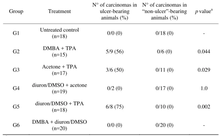
Tables V and VI present the comparison between ulcer-bearing and “non-ulcer”
-bearing animals in relation to the incidences of histologically-detected carcinomas in both experiments. In Experiment-1, carcinomas occurred at incidences of 56%, 50% and 75%, only in the animals that developed ulcers during TPA treatment (G2, G3 and G5 respectively). Association between ulcers and carcinomas was statistically significant in those groups, pointing to the critical role of TPA in establishing ulcerations and carcinomas. In the Experiment-2, carcinomas occurred in ulcer-bearing animals at incidences of 42%, 60% and 60% in the groups G7, G8 and G9, all of them treated with TPA during the promotion step. One “non-ulcer” animal in the positive control group
(G7, DMBA+TPA) developed a papillary carcinoma. The association between ulcers and carcinomas was significant for group G8 (p = 0,044), but not for G7 and G9.
DISCUSSION
36 extended period that characterizes the promotion step, once initiating agents are often complete carcinogens (Iversen, 1994).
Therefore, under the experimental conditions adopted, diuron did not exert carcinogenesis initiating or promoting activities in the Swiss female mouse skin, and varying the herbicide solvent did not influence the results. The present data strongly diverge from previous findings that indicated tumor initiatory activity of diuron in a mouse skin assay (Antony et al., 1989). Besides, they suggest that diuron is not a genotoxic agent, what is in line with information from this laboratory that reported non-genotoxicity of the herbicide under two different comet assay protocols (Nascimento et al., 2006; Rocha et al., 2009).
A striking observation in these studies was the development of severe skin ulcers and histologically-detected carcinomas in the groups exposed to TPA. This non-genotoxic compound has been reported to be a weak but complete carcinogen when applied repeatedly in the hairless Oslo mouse skin, in which even a positive dose-response has been demonstrated (Iversen, 1985; 1994). In the Iversen’s studies,
6.17µg/100µL of TPA diluted in acetone applied twice a week to non-initiated mouse skin produced 9% incidence of tumor-bearing animals after 55 weeks. The NTP has designed a protocol in which 5µg of TPA applied once a week for 51 weeks was sufficient to induce tumors in 13% of non-initiated Swiss female mouse (National Toxicology Program, 1996). In a recent review on the multistage skin chemical carcinogenesis protocol, it was suggested that TPA should be applied only once or twice a week, depending on the dose and mouse strain used (Abel et al., 2009).
37 levels adopted by Antony et al. (1989) were sufficient to induce tumors even in the absence of any initiating procedure, leading to the 73% tumor incidence they verified in that group.This possibility, however, does not explain the absence of tumors verified by those authors in the group initiated with a single dose of diuron and promoted with the same TPA schedule (Antony et al., 1989), and this controversy remains to be elucidated. The TPA dose level used in our studies was slightly lower (3.5 µg/100µL) than those suggested by others (NTP, 1996; Abel et al., 2009) but was applied three times a week, the same as done by Antony et al. (1989) and in dissonance with Abel et al. (2009) recommendations. It is intriguing that Antony et al. (1989) did not account on histological lesions and did not refer to ulcer or carcinoma developments in their study. The occurrence of these lesions in the current study is discussed below.
The present experiments were planned to last 25 weeks, as indicated previously. This duration was based on the verification of conspicuous development of papillary tumors at this moment in the Antony et al. (1989) study, and also on other reports using the mouse skin carcinogenesis protocol, which did have the same or shorter lengths (Chan et al., 2004; Ridd et al., 2006; Chaudhary et al., 2009). However, due to the severe ulcers developed in the TPA-treated animals, experiments were terminated at weeks 23 and 21 for humane reasons. Highly reproducible papilloma burden is expected after 20 weeks of promotion, and progression to squamous cell carcinoma usually occurs within 20 to 50 weeks of treatment with tumor-promoting agent (Abel et al.,
2009). Therefore, it can be assumed that the experiments’ duration adopted was
sufficient to detect potentially diuron-induced skin papillary tumors.
38 Herein, it was demonstrated that several papillary tumors were eradicated by the process of ulceration, as the number of tumors per mouse decreased concurrently with ulcers development.
Similarly to the ulcer incidences, the significantly higher incidences of histologically-detected carcinomas in the TPA-treated groups is probably a consequence of TPA cytotoxicity, regardless of the initiating treatment. Besides TPA being a weak skin carcinogen (Iversen, 1985; 1994), it is possible that the repetitive cell injury it provoked led to epithelial disruption and ulceration, inducing the surviving epithelium at the ulcer borders to regenerate and eventually progress to malignancy. This possibility is supported by the strong association registered between carcinomas and ulcers. Although a significant positive association was not verified in all groups from Experiment-2, there was a clear biological relationship between ulcers and carcinomas, and it is very likely that the absence of statistical significance of this association may be accounted on the size of these groups. Additionally, there are no significant differences between the incidences of carcinomas in the positive control group and the other TPA-treated animals in both Experiment-1 and -2, suggesting that TPA treatment itself, and not the initiating treatment, was critical for the development of epithelial malignancies. These observations reinforce the recommendations that experimental skin treatment by TPA should follow careful selection of doses and frequency of applications (Abel et al., 2009) in order to avoid incidental events that may hinder the interpretation of results due to the citotoxicity of this compound.
39 incidences of epidermal hyperplasia – an indirect indication of enhanced cell
proliferation – only when associated to DMBA or TPA; otherwise, when applied with
either DMSO or acetone, the incidences of epithelial hyperplasia were not relevant. Therefore, contrary to what is observed in the rat urinary mucosa, diuron seems not to induce epidermal cell proliferation. Overall, the present data indicate that diuron acts neither as an initiator nor as a promoter of carcinogenesis in the female Swiss mouse skin assay. Taking into account the limits of extrapolation between rodent and human skin biological events (Enzmann et al., 1998), it could be assumed that potential cutaneous occupational exposure to this herbicide probably does not represent a risk for human skin cancer development.
ACKNOWLEDGMENTS
This work was supported by the Fundação de Amparo à Pesquisa do Estado de São Paulo (FAPESP) [Grants # 06/60506-1, 06/04630-5 and 08/01809-0] and Núcleo de Avaliação do Impacto Ambiental sobre a Saúde Humana (TOXICAM). Paulo Roberto Cardoso, Maria Luiza Falaguera, Paulo Cesar Georgete and Cristina Aparecida Alquati Dorico provided helpful technical assistance.
REFERENCES
Abel, E.L., Angel, J.M., Kiguchi, K., and DiGiovanni, J. (2009). Multi-stage chemical carcinogenesis in mouse skin: Fundamentals and applications. Nat protoc 4(9),
1350-62.
40 Agrawal, R.C., and Mehrotra, N.K. (1997). Effect of diuron on germ cells of mice.
Indian J Exp Biol 35(11), 1256-7.
Antony, M., Shukla, Y., and Mehrotra, N.K. (1989). Tumour initiatory activity of a herbicide diuron on mouse skin. Cancer Lett 48, 125-8.
Brouwer, D.H., Aitken, R.J., Oppl, R., and Cherrie, J.W. (2005). Concepts of skin protection: considerations for the evaluation and terminology of the performance of skin protective equipment. J Occup Environ Hyg 2(9), 425-34.
Canna-Michaelidou, S., and Nicolaou, A.S. (1996). Evaluation of the genotoxicity potential (by MutatoxTM test) of ten pesticides found as water pollutants in Cyprus. Sci Total Environ 193, 27-35.
Carpenter, W.S., Lee, B.C., Gunderson, P.D., and Stueland, D.T. (2002). Assessment of personal protective equipment use among midwestern farmers. Am J Ind Med
42, 236-47.
Chan, K.S., Sano, S., Kiguchi, K., Anders, J., Komazawa, N., Takeda, J., and DiGiovanni, J. (2004). Disruption of Stat3 reveals a critical role in both the initiation and the promotion stages of epithelial carcinogenesis. J Clin Invest
114(5), 720-8.
Chaudhary, S.C., Alam, M.S., Siddiqui, M.S., and Athar, M. (2009). Chemopreventive effect of farnesol on DMBA/TPA-induced skin tumorigenesis: Involvement of inflammation, Ras-ERK pathway and apoptosis. Life Sci 85(5-6), 196-205.
Enzmann, H., Bomhard, E., Iatropoulos, M., Ahr, H.J., Schlueter, G., and Williams, G.M. (1998) Short- and intermediate-term carcinogenicity testing – a review.
Part 1: the prototypes mouse skin tumour assay and rat liver focus assay. Food
41 Fu, W., McCormick, T., Qi, X., Luo, L., Zhou, L., Li, X., Wang, B.C., Gibbons, H.E.,
Abdul-Karim, F.W., and Gorodeski, G.I. (2009). Activation of P2X7-mediated apoptosis inhibits DMBA/TPA-induced formation of skin papillomas and cancer in mice. BMC Cancer 9, 114.
Grutman, G., Schoofs, L., Lontie, J.F., and van Larebeke, N. (1984). The mutagenicity in prokaryotes of herbicides. Residue Rev 91, 1–46.
Iversen, O.H. (1985). TPA (12-O-tetradecanoyl-phorbol-13-acetate) as a carcinogen for mouse skin. A positive dose-response relationship. Virchows Arch B Cell Pathol
Incl Mol Pathol49(2), 129-35.
Iversen, O.H. (1994). A course of very small doses of DMBA, each of them allegedly with no promoting potency, acts with clear synergistic effect as a strong promoter of DMBA-initiated mouse skin carcinogenesis. A comparison of the tumorigenic and carcinogenic effects of DMBA (7,12-dimethylbenz-alpha-anthracene) and TPA (12-O-tetradecanoyl-phorbol-13-acetate) used as initiators and promoters in classical two-stage experimental protocols. APMIS Suppl 41,
1-38.
Iyer, P. (2002). Evidence on the development and reproductive toxicity of diuron. Draft. Reproductive and Cancer Hazard Assessment Section. Office of Environmental Health Hazard Assessment, California Environmental Protection Agency, 43. Klein-Szanto, A.J.P., Conti, C.J., Aldaz, C.M., Clapp, N., Nesnow, S., and Slaga, T.J.
(1986). Effects of chronic topical application of 1 2-O-tetradecanoylphorbol-1 3-acetate on the skin and internal organs of SENCAR mice. Environ Health
Perspect 68, 75-80.
Liu, J. (2001). Phenylurea herbicides. In Handbook of Pesticide Toxicology – Agents
42 Nascimento, M.G., de Oliveira, M.L.C.S., Lima, A.S., and de Camargo, J.L.V. (2006).
Effects of diuron [3-(3,4-dichlorophenyl)-1,1-dimethylurea] on the urinary bladder mucosa of male Wistar rats. Toxicology 224, 66-73.
National Toxicology Program (1996). Comparative initiation/promotion skin paint studies of B6C3F1 mice, Swiss (CD-1®) mice, and SENCAR mice. NTP TR 441, NIH Publication No. 96-3357. NIH, Research Triangle Park, NC.
Perry, M.J., Marbella, A., and Layde, P.M. (2002). Compliance with required pesticide-specific protective equipment use. Am J Ind Med 41, 70-3.
Pitot III, H.C., and Dragan, Y.P. (2001). Chemical carcinogenesis. In Casarett &
Doull’s Toxicology the Basic Science of Poisons (C.D. Klaassen, ed), pp. 241
-319. McGraw-Hill, New York.
Ridd, K., Zhang, S.D., Edwards, R.E., Davies, R., Greaves, P., Wolfreys, A., Smith, A.G., and Gant, T.W. (2006). Association of gene expression with sequential proliferation, differentiation and tumor formation in murine skin.
Carcinogenesis 27(8), 1556-66.
Rocha, M.S., Nascimento, M.G., Cardoso, A.P.F., Lima, P.L.A., Zelandi, E.A., de Camargo, J.L.V., and de Oliveira, M.L.C.S. (2009). Cytotoxicity and regenerative proliferation as the mode of action for diuron-induced urothelial carcinogenesis in the rat. Toxicol Sci [Epub ahead of print],
doi:10.1093/toxsci/kfp241.
43 Slaga, T.J., and Fischer, S.M. (1983). Strain differences and solvent effects in mouse
skin carcinogenesis experiments using carcinogens, tumor initiators and promoters. Prog Exp Tumor Res 26, 85-109.
Stoll, R.E., Furst, S.M., Stoltz, J.H., Lilly, P.D., and Mennear, J.H. (2001). Dermal carcinogenicity in transgenic mice: effect of vehicle on responsiveness of hemizygous Tg.AC mice to phorbol 12-myristate 13-acetate (TPA). Toxicol
Pathol 29(5), 535-40.
US Environmental Protection Agency (2004). Chemicals evaluated for carcinogenic potential. Office of Pesticide Programs, Health Effects Division. Science Information Management Branch, 106.
Volden, G., Thorud, E., and Iversen, O.H. (1983). Inhibition of methylcholanthrene-induced skin carcinogenesis in hairless mice by the membrane-labilizing agent DMSO. Br J Dermatol 109(25), 133-6.
Williams, G.M. (1999) Chemically induced preneoplastic lesions in rodents as indicators of carcinogenic activity. In The use of short- and medium-term tests for carcinogens and data on genetic effects in carcinogenic hazard evaluation (D.B. McGregor, J.M. Rice, S. Venitt, eds), Vol. 146, pp. 185-202. IARC Scientific Publications, Lyon, France.
44
Table I – Experiment 1: Incidences of cutaneous lesions at the end of week 23 in a female Swiss mouse two-step skin carcinogenesis assay using DMSO as the solvent for diuron.
Groups Treatment
Macroscopic lesions Histological lesions
Papillary tumors1,5 Ulcers2,5 Epidermal hyperplasia3 Carcinomas4
G1 Untreated control 0/18a 0/18a 0/18a 0/18a
G2 DMBA + TPA 15/15b 9/15b 15/15b,* 5/15b
G3 Acetone + TPA 3/17a 6/17b,c,d 17/17b,* 3/17a,b
G4 diuron/DMSO +
acetone 0/19
a 2/19a,c 2/19a 0/19a
G5 diuron/DMSO + TPA 3/18a 8/18b,d 18/18b,* 6/18b
G6 DMBA +
diuron/DMSO 0/20
a 0/20a 19/20b 0/20a
45
Table II – Experiment 2: Incidences of cutaneous lesions at the end of week 21 in a female Swiss mouse two-step skin carcinogenesis assay using acetone or DMSO as the solvent of diuron.
Groups Treatment
Macroscopic lesions Histological lesions
Papillary tumors1,5 Ulcers2,5 Epidermal hyperplasia3 Carcinomas4
G1 Untreated control 0/15a 0/15ª 0/15a 0/15a
G2 DMBA + acetone 0/15a 0/15ª 2/15a,b 0/15a
G3 DMBA + diuron/acetone 0/15a 0/15ª 6/15b,c 0/15a
G4 DMBA + diuron/DMSO 0/15a 0/15ª 11/15c 0/15a
G5 Acetone + diuron/acetone 0/15a 0/15ª 1/15a 0/15a
G6 Acetone + diuron/DMSO 0/15a 0/15ª 3/15a,b 0/15a
G7 DMBA + TPA 15/15b 12/15b 15/15d,* 6/15b
G8 diuron/acetone + TPA 2/15a 10/15b 15/15d,* 6/15b
G9 diuron/DMSO + TPA 1/14a 10/14b 14/14d,* 6/14b
46 Table III: Female Swiss mouse two-step skin carcinogenesis bioassay. Experiment 2 – Number of papillary tumors per mouse and ulcers
occurrence at week 16 in the positive control group (G7, DMBA+TPA).
Animal identification
Week Ulcers at
week 16
9 12 16
1 30 35 35
2 29 39 39
3 12 25 29
4 4 13 19
5 2 7 18
6 22 32 36
10 6 21 29
12 30 37 35
7 10 23 13 +
8 25 28 12 +
9 32 34 13 +
11 26 29 17 +
13 29 29 26 +
14 23 32 37 +
15 21 22 10 +
47 Table IV: Female Swiss mouse two-step skin carcinogenesis bioassay. Experiment 2, positive control group (G7, DMBA+TPA) – Association between ulcer development
and number of gross skin papillary tumors per mouse from week 9 to 16*. Ulcer-bearing
animals (n=8)a
“Non-ulcer”-bearing
animals (n=7) p value b
Variation (%) of the number of papillary tumors per mouse between weeks 9 and 16
- 34.6 (-52.3; 30.0) 102.6 (21.2; 381.2) 0.008
Variation (%) of the number of papillary tumors per mouse between weeks 12 and 16
- 43.7 (-57.1; -10.3) 14.2 (0.0; 44.3) 0.005
48
Table V: Female Swiss mouse two-step skin carcinogenesis bioassay. Experiment 1 - association between ulcer development and incidences (%) of carcinomas per group.
Group Treatment
N° of carcinomas in ulcer-bearing
animals (%)
N° of carcinomas in
“non-ulcer”-bearing
animals (%)
pvaluea
G1 Untreated control
(n=18) 0/0 (0) 0/18 (0) -
G2 DMBA + TPA (n=15) 5/9 (56) 0/6 (0) 0.044
G3 Acetone + TPA (n=17) 3/6 (50) 0/11 (0) 0.029
G4 diuron/DMSO + acetone
(n=19) 0/2 (0) 0/17 (0) 1.0
G5 diuron/DMSO + TPA (n=18) 6/8 (75) 0/10 (0) 0.002
G6 DMBA + diuron/DMSO (n=20) 0/0 (0) 0/20 (0) - aFish
49 Table VI: Female Swiss mouse two-step skin carcinogenesis bioassay. Experiment 2 - Association between ulcer development and incidences (%) of carcinomas per group.
Group Treatment
N° of carcinomas in ulcer-bearing
animals (%)
N° of carcinomas in
“non-ulcer”-bearing
animals (%)
pvaluea
G7 DMBA + TPA
(n=15) 5/12 (42) 1/3 (33) 1.0
G8 diuron/acetone + TPA (n=15) 6/10 (60) 0/5 (0) 0.044
G9 diuron/DMSO + TPA (n=14) 6/10 (60) 0/4 (0) 0.085 a
52
Tabela VII – Doses e tipos de exposição ao diuron entre diferentes testes de genotoxicidade.
Autor Teste Dose Exposição Genotoxicidade
Seiler, 1978 Ames1 1000 mg/kg Oral (ração) + (fraco)
Seiler, 1978 Micronúcleo2 1000 mg/kg Oral (ração) -
Agrawal et al., 1996 Micronúcleo2 170 e 340 mg/kg Dose única, i.p. +
Agrawal & Mehrotra, 1997 Dominante letal3 170 e 340 mg/kg, 3400 ppm aguda e crônica Oral (ração), +
Nascimento et al., 2006 Ensaio do Cometa4 125, 500 e 2500 ppm Oral (ração),
crônica -
Rocha et al., 2009 Ensaio do Cometa5 12,5, 25, e 50µg/mL In vitro -
1
Salmonella thyphimurium; 2Medula óssea de camundongos Swiss; 3Células germinativas em camundongos Swiss; 4Células
53
Tabela VIII – Doses, frequência de aplicações de TPA, e incidência de tumores entre diferentes estudos.
Autor Iniciação Dose
TPA
Frequência de aplicações
Duração (semanas)
Incidência de tumores
Linhagem de camundongos
Iversen, 1984 - 6,7µg 2x/semana 55 9% Oslo
NTP, 1996 - 5µg 1x/semana 51 13% Swiss
Abel et al., 2009 DMBA 5,23µg 2x/semana 20-52 90% CD-1
Abel et al., 2009 DMBA 2µg 1x/semana 20-52 53% CD-1
Antony et al., 1989 Diuron 5µg 3x/semana 52 73% Swiss
Ferrucio et al., 2009 Diuron ou acetona
50 Figure 1 – Experimental design 1
Figure 2 – Experimental design 2
Untreated control
DMBA 52μg/ 100μL acetone (single application)
TPA 3.5μg/ 100μL acetone (3 applications/week)
100μL acetone (3 applications/week)
Diuron 250mg/ Kg b.w. diluted in 100μL DMSO (3 applications/week)
51
Euthanasia
N = Number of animals at the end of the experiment
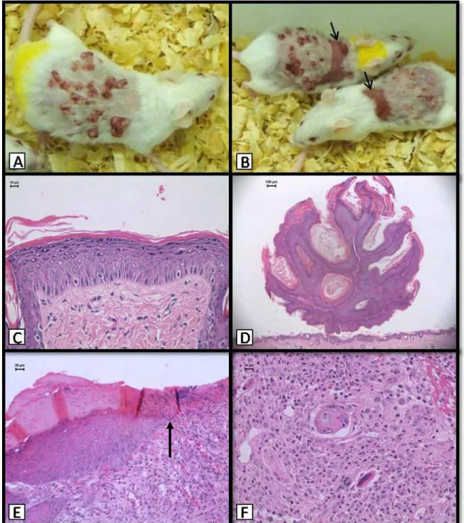
Figure 3 – TPA-treated animals. Macroscopic lesions: A, B) Papillomas and ulcers