Common and Low Frequency Variants in
MERTK
Are Independently Associated with
Multiple Sclerosis Susceptibility with
Discordant Association Dependent upon
HLA-DRB1
15
:
01
Status
Michele D. Binder1,2*, Andrew D. Fox1,3, Daniel Merlo2, Laura J. Johnson1,
Lauren Giuffrida2¤a, Sarah E. Calvert1, Rainer Akkermann2, Gerry Z. M. Ma1,2¤b, ANZgene¶,
Ashwyn A. Perera1, Melissa M. Gresle4, Louise Laverick4, Grace Foo4, Marzena J. Fabis-Pedrini5, Timothy Spelman4, Margaret A. Jordan6, Alan G. Baxter6, Simon Foote7, Helmut Butzkueven4, Trevor J. Kilpatrick1,2, Judith Field1,2
1Multiple Sclerosis Division, The Florey Institute of Neuroscience and Mental Health, Parkville, Victoria, Australia,2Department of Anatomy and Neuroscience, University of Melbourne, Parkville, Victoria, Australia,3Bioinformatics Core, The Florey Institute of Neuroscience and Mental Health, Parkville, Victoria, Australia,4Department of Medicine, University of Melbourne, Parkville, Victoria, Australia,5Western Australian Neuroscience Research Institute, Nedlands, Western Australia, Australia,6Comparative Genomics Centre, James Cook University, Townsville, Queensland, Australia,7John Curtin School of Medical Research, Australian National University, Acton, Australian Capital Territory, Australia
¤a Current address: Cancer Immunology Program, Peter MacCallum Cancer Centre, East Melbourne, Victoria, Australia
¤b Current address: MRC Centre for Regenerative Medicine, The University of Edinburgh, Edinburgh, United Kingdom
¶ Membership of ANZgene is provided in the Acknowledgments.
Abstract
Multiple Sclerosis (MS) is a chronic inflammatory demyelinating disease of the central ner-vous system. The risk of developing MS is strongly influenced by genetic predisposition, and over 100 loci have been established as associated with susceptibility. However, the bio-logically relevant variants underlying disease risk have not been defined for the vast major-ity of these loci, limiting the power of these genetic studies to define new avenues of research for the development of MS therapeutics. It is therefore crucial that candidate MS susceptibility loci are carefully investigated to identify the biological mechanism linking genetic polymorphism at a given gene to the increased chance of developing MS.MERTK has been established as an MS susceptibility gene and is part of a family of receptor tyro-sine kinases known to be involved in the pathogenesis of demyelinating disease. In this study we have refined the association ofMERTKwith MS risk to independent signals from both common and low frequency variants. One of the associated variants was also found to be linked with increased expression of MERTK in monocytes and higher expression of MERTK was associated with either increased or decreased risk of developing MS, depen-dent uponHLA-DRB1*15:01status. This discordant association potentially extended
OPEN ACCESS
Citation:Binder MD, Fox AD, Merlo D, Johnson LJ, Giuffrida L, Calvert SE, et al. (2016) Common and Low Frequency Variants inMERTKAre Independently Associated with Multiple Sclerosis Susceptibility with Discordant Association Dependent uponHLA-DRB115:01Status. PLoS Genet 12(3): e1005853. doi:10.1371/journal.pgen.1005853
Editor:Greg Gibson, Georgia Institute of Technology, UNITED STATES
Received:October 14, 2015
Accepted:January 18, 2016
Published:March 18, 2016
Copyright:© 2016 Binder et al. This is an open access article distributed under the terms of the
Creative Commons Attribution License, which permits unrestricted use, distribution, and reproduction in any medium, provided the original author and source are credited.
Data Availability Statement:The GEO accession number for the RNAseq data from which all samples can be accessed is GSE77598. The NCBI dbVar accession number for the structural variants identified is nstd124. All other relevant data are in the paper and its Supporting Information files.
beyond MS susceptibility to alterations in disease course in established MS. This study pro-vides clear evidence that distinct polymorphisms withinMERTKare associated with MS susceptibility, one of which has the potential to alterMERTKtranscription, which in turn can alter both susceptibility and disease course in MS patients.
Author Summary
Multiple sclerosis (MS) is the most common neurological disease of young Caucasian adults. Oligodendrocytes are the key cell type damaged in MS, a process that is accompa-nied by loss of the myelin sheath that these cells produce, resulting in demyelination and ultimately in secondary damage to nerve cells. Susceptibility to MS is strongly influenced by genes, and over 100 genes have now been linked with the risk of developing MS. How-ever, surprisingly little is known about the biological mechanism by which any one of these genes increases the probability of developing MS. In this study we have explored in detail the links between one known MS risk gene,MERTK, and MS susceptibility. We found that a number of different alterations in theMERTKgene are independently associ-ated with the risk of developing MS. One these changes was also linked with changes in the level of expression of MERTK in monocytes, an immune cell type known to be involved in the etiology of MS. In an unexpected result, we found this expression-linked alteration inMERTKwas either protective or risk-associated, depending on the genotype of the individual at another well known MS risk gene known asHLA-DRB1. In addition, we found that not only were alterations inMERTKassociated with MS susceptibility, but potentially with ongoing disease course, indicating thatMERTKmay be a good target for the development of novel MS therapeutics.
Introduction
Multiple Sclerosis (MS) is an inflammatory demyelinating disease of the central nervous system (CNS). Although the initiating insult in MS remains unknown, it is clear that the pathology of the disease involves a complex interaction between the immune system, neurons and glia, in which cells of the immune system target oligodendrocytes, ultimately resulting in central demyelination and secondary axonal damage.
A genetic basis for MS susceptibility has long been suggested by the observation of an increased familial risk in twins and in first-degree relatives [1], and there is substantial evidence that the increased risk seen in family members of MS patients is not simply the result of shared environment [1–4]. Although estimates of sibling relative risk (λs) vary, a recent meta-analysis
has calculatedλs as 16.8, with an overall heritability (h2) of 54% [5].
The association of MS susceptibility with specific genes began with studies in the 1970s describing an increase in the frequency of certain human leukocyte antigens (HLA) in MS pop-ulations[6–8]. The HLA genes are located on chromosome 6 and include the major histocom-patibility class (MHC) I and II loci. Improved methods of subtyping HLA loci, as well as studies with increased sample size, have allowed the identification of an extended HLA haplo-type,HLA DRB115:01,DQA10102,DQB10602, within the MHC class II region that is strongly associated with the risk of developing MS [9–11]. The association between HLA and MS susceptibility remained for many decades the only convincing association with MS risk. The advent of genome-wide association studies (GWAS) in the last decade has significantly and Medical Research Council Grant Number
APP1032486 to HB, JF and AGB, and by the Australian Research Council Linkage Grant Number LP110100473 to HB, JF abd AGB. The Florey Institute of Neuroscience and Mental Health acknowledges the strong support from the Victorian Government and in particular the funding from the Operational Infrastructure Support Grant. The funders had no role in study design, data collection and analysis, decision to publish, or preparation of the manuscript.
altered the landscape of MS genetics. In 2007, the first MS GWAS detected the first non-HLA loci [Interleukin-2 receptor alpha (IL2RA)] to be associated with MS at a genome-wide signifi-cance level [12]. A number of GWAS studies of increasing power have since extended the num-ber of loci established as associated with MS risk to 103 [13–15].
One clear finding from MS-GWAS has been that, outside the HLA genes, the majority of the associated variants exert small effects as measured by odds-ratios (ORs<1.10), compared with, for example, theHLA-DRB115:01(DR15) association which has an estimated OR of 3.1 [13]. The combined effect of the known MS-susceptibility loci is estimated to account for only around 28% of the heritability of MS [15]. A number of distinct, and not necessarily exclusive, hypotheses exist as to the nature of the "missing heritability" of MS and other common dis-eases, including the existence of rare variants of large effect, structural genomic alterations such as copy-number variants (CNVs), or epigenetic modifications, all of which are either poorly captured by GWAS or, in the case of epigenetic changes, not captured at all [16]. It is therefore crucial that candidate MS-susceptibility loci identified by GWAS are carefully investi-gated to identify the biological mechanism linking genetic polymorphism at a given locus to the increased chance of developing MS.
We and others have demonstrated that signalling via the TAM (TYRO3, AXL and MERTK) family of receptor tyrosine kinases (RTKs) profoundly influences the outcome of demyelin-ation. The TAM receptors were identified as a distinct RTK subfamily in 1991 [17], share a common domain structure [18,19], and are activated by two closely related ligands, GAS6 and PROTEIN S (PROS) [20–22]. The TAM receptors were first linked to demyelination in 2008 when we showed that loss of Gas6 leads to increased disease severity in cuprizone-induced demyelination in mice[23]. More recently, work from our group and others has shown that TAM receptor signalling is involved in both the etiology and the pathogenesis of MS. In 2011, we showed that polymorphisms within theMERTKgene are associated with MS susceptibility [24], a finding replicated by a large international GWAS [13], andMERTKremains on the cur-rent list of established MS-risk loci [15]. The MERTK and AXL receptors, as well as the soluble forms of these receptors, have also been found to be upregulated in MS lesions, and may be cor-related with extended lesion activity [25]. Alterations in the circulating levels of the TAM ligands, GAS6 and PROS, have been detected in MS patients, and the level of circulating PROS has been associated with severity measures in MS [26].
These data indicate thatMERTKis not only an important susceptibility gene for MS, but it could potentially have an ongoing role in determining disease severity. We therefore performed fine-mapping studies to refine the genetic association within this locus in order to identify bio-logically relevant variants withinMERTK. We identified both rare and common variants within theMERTKgene independently and significantly associated with the risk of developing MS. Two of the associated variants were found to operate intranswith theHLA-DRB1locus, with one SNP showing discordant association depending upon DR15 status. In exploring the functional basis of the associations, we found that associated variants were also expression quantitative trait loci (eQTLs) forMERTKin human monocytes, a cell type central to MS pathology. Finally, we provide evidence that theMERTKMS risk-associated variants may also alter disease course in established MS.
Results
Identification of a low-frequency SNP within
MERTK
associated with MS
susceptibility
large study by the IMSGC [13], confirming the association ofMERTKwith MS susceptibility. As is frequently found for GWAS-identified risk variants, all the risk-associated SNPs within theMERTKgene identified in the initial discovery phase were common, none were within exons, nor were they located within any obvious regulatory regions. In an attempt to identify functionally relevant variants underlying the association ofMERTKwith MS susceptibility, we conducted a follow-up fine-mapping study in which SNPs located within and adjacent to a number of known MS susceptibility genes, includingMERTK, were genotyped to better define the most associated genetic variations leading to increased risk of developing MS [27].
Included in this follow-up study were 239 SNPs on chromosome 2, including 124 within
MERTK, selected from dbSNP and the 1000 Genomes project. These SNPs were genotyped in 3268 MS cases and 3579 controls and analysed for association with MS. We identified a peak of association within theMERTKregion on chromosome 2, with 36 SNPs within theMERTK
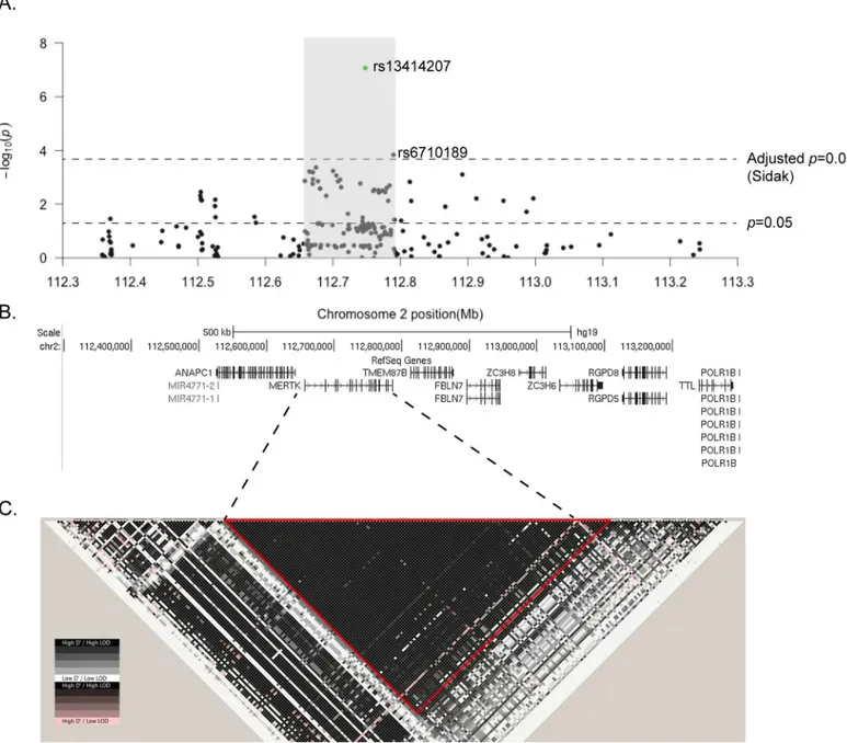
gene showing suggestive association (p<0.05), including 2 SNPs significantly associated with MS susceptibility following adjustment for multiple testing (Fig 1A,p<2.2 x 10⁻⁴). The SNPs within theMERTKgene that were associated with MS susceptibility (p<0.05), included 8 of the 10 most associated SNPs on chromosome 2 (Table 1). The SNPs withinMERTKwere con-tained within a region of high linkage disequilibrium (LD) that included the previously associ-ated rs17174870 (Table 1,Fig 1B). The lead SNP within this region, rs13414207, was strongly associated with the risk of developing MS (p= 8.5 x 10−08), with an OR of 1.6 (Table 1). In con-trast to the majority of variants previously associated with MS susceptibility[15], the risk asso-ciated allele at the lead SNP is low frequency [minor allele frequency (MAF)<0.05]. We also identified a striking over-representation of individuals homozygous for the risk allele at rs13414207 amongst cases (n = 14) compared with healthy controls (n = 3) amongst the 3268 people with MS and 3579 controls that we assessed, suggesting an allele dosage effect, which fits with a number of potential models (Table 2).
MERTK
SNPs define MS risk and protective haplotypes associated with
MS susceptibility
Given the substantial LD within the region, we examined the relationship of the lead SNP to other SNPs within theMERTKgene that showed suggestive association with MS susceptibility. We found that 28 SNPs form a single block of very high LD (D'>0.99; LOD2) containing 5 haplotypes with a frequency of>1% (Fig 2). The minor allele of the lead SNP (rs13414207) tags a low frequency haplotype that is significantly associated with the risk of developing MS (Fig 2,p= 8.53 x 10−07). This risk haplotype also contains variants previously shown to carry MS risk in other studies, including the risk allele of rs17174870 [13,24], as well as a newly iden-tified risk SNP (rs6710189). However, these SNPs do not appear to be independently associated with MS susceptibility outside of this haplotype (Fig 2).
In addition to the haplotype significantly associated with risk, a separate haplotype is signifi-cantly more frequent in controls compared with cases, forming an apparent protective haplo-type (p= 0.0012). As this apparently protective haplotype is tagged by the opposite alleles of the SNPs significantly associated with risk, it may be the effect is not truly protective but repre-sents the absence of risk.
Haplotype-based variant identification using whole-genome and
resequencing strategies
structural or regulatory alteration resulting from variation at rs13414207 makes the latter more probable. We used the high LD and the strong genotype effect as the basis for two complemen-tary strategies to identify potential causal variants within theMERTKgene, with a particular focus on low frequency and novel variants. Specifically, we identified individuals who were homozygous for the risk-associated haplotype (n = 17). We designed and validated a series of overlapping amplicons (n = 31) and employed long-range PCR to amplify the entireMERTK
gene plus ~10kb upstream and downstream of the gene (Chr2:112,645,298–112,789,127 on
Fig 1. A low-frequency SNP withinMERTKis significantly associated with MS susceptibility.124 SNPs on chromosome 2 were directly genotyped in 3268 MS cases and 3579 controls. (A) The negative log of the unadjustedp-value of each SNP is plotted against the relative position on chromosome 2, with a schematic of the gene structures shown underneath (B). Two SNPs, both withinMERTK, reach significance using the Sidak adjustment for multiple testing at a nominalα= 0.05. Associationp-values were determined using a Chi-square test. (C) Schematic of the pattern of LD across the whole region, with a large single block of high LD (D'>0.99), including the whole of theMERTKgene, highlighted with a red triangle.
GRch37/hg19 build). Amplicons for each individual were pooled at equimolar amounts and sequenced. As it was hypothesised that this strategy would identify a large number of variants, many of which would not be associated with MS susceptibility, we also included a number of individuals homozygous for other identified haplotypes to allow for prioritisation of variants for follow-up association testing (Table 3).
As targeted sequencing strategies based on amplification and using paired-end next-genera-tion sequencing technologies are not ideal for detecnext-genera-tion of large insernext-genera-tions/delenext-genera-tions (in-dels) and other large structural variants, we also undertook whole-genome sequencing (WGS) of a single individual homozygous for the risk haplotype.
Both strategies produced high quality data, with a sequencing depth of 35-40x (WGS) and 50x for>96% bases and 100x for>90% bases (targeted strategy), and the vast majority of vari-ants (>99%) were identified in both methods. The WGS strategy was successful in identifying two variants that failed to map using the targeted method. The first of these variants was a long repeat expansion within intron 1 (chr2:112,682,854; build hg19) from the reference TA8T21to TA21T>26<200. The second was a retrotransposon insertion polymorphism (RIP) involving an insertion of an AluYf4 retrotransposon in intron 4 (chr2:112,716,560; hg19). Overall we identi-fied 580 variants (SNPs, in-dels and larger variants) within or nearby theMERTKgene in indi-viduals homozygous for the risk haplotype (Table 4). As our resequencing strategy was focused upon the risk haplotype, it is not surprising that a comparison of the variation pattern com-pared with reference sequence showed a significant number of variants within this group (Fig 3). In contrast, the haplotype 3 group, despite sharing many alleles with haplotype 5 in our
Table 1. Top 10 SNPs withinMERTKassociated with Multiple Sclerosis susceptibility.
SNP ID Positiona Minor Allele Major Allele MAFbcases MAF controls Odds Ratioc Unadjustedp-value
rs13414207 112,748,053 A G 0.04893 0.03082 1.618 8.5x10-08
rs6710189 112,789,792 G A 0.2704 0.242 1.161 0.000145
rs1516639 112,750,085 G C 0.4753 0.4452 1.165 0.000427
rs884448 112,700,328 A T 0.4736 0.4442 1.126 0.000572
rs17174870 112,665,201 T Cd 0.2219 0.2469 0.87 0.000607
rs1400322 112,670,185 G A 0.4758 0.4466 1.125 0.000637
rs1400321 112,670,046 A C 0.2241 0.2491 0.871 0.000649
rs3761700 112,705,185 C A 0.473 0.4444 1.122 0.000854
rs1400323 112,670,461 C A 0.4749 0.4471 1.118 0.001179
rs4848901 112,710,828 A G 0.472 0.4444 1.118 0.001222
a. SNP positions are relative to the Human February 2009 (GRC37/hg19) assembly. b. MAF = minor allele frequency.
c. The odds ratio applies to the minor allele. d. These alleles are inferred from data obtained from sequencing the opposite strand but are presented as CT to maintain consistency with the remainder of the study.
doi:10.1371/journal.pgen.1005853.t001
Table 2. Gene model association of rs13414207 with Multiple Sclerosis.
Test Minor Allele Major Allele Affected Not affected p-value
Genotype A G 14/284/2890 3/207/3246 4.84x10-07
Trend A G 312/6064 213/6699 1.24x10-07
Allelic A G 312/6064 213/6699 8.50x10-08
Dominant A G 298/2890 210/3246 5.36x10-07
Recessive A G 14/3174 3/3453 0.0045
initial analysis of 28 SNPs (Fig 2) does not overall share many variants with the risk haplotype in the expanded resequencing analysis (Fig 3). Conversely, the haplotype 2 group also contains many non-reference alleles, many of which are shared with the risk haplotype (Fig 3).
We then used a number of methods to prioritise variants for downstream functional analy-sis and association testing. As we were,a priori, attempting to identify polymorphisms in
MERTKthat showed population level association with MS susceptibility, all private mutations were excluded from further analysis. Secondly, any SNPs previously tested in other large data sets, including our fine-mapping data set, and found not to be associated with MS susceptibil-ity, were excluded. We then used our control groups, particularly the haplotype 2 group, to exclude a further set of variants that showed a high degree of sharing between the risk and non-risk haplotypes. At the conclusion of this prioritisation strategy, we developed a final list of 74 variants for further analysis and association testing.
Low frequency and common variants within
MERTK
are independently
associated with MS susceptibility
Following the prioritisation of variants for association testing, we then designed genotyping strategies. A number of SNPs failed at the design or QC stage and were unable to be tested for association. Failed variants included the two novel variants identified via WGS. The TAnTn proved to be highly variable in length, with longer expansions correlating with the risk haplo-type and the presence of the lead SNP (S2 Table), however the repetitive nature of the
Fig 2. SNPs withinMERTKdefine both risk and protective haplotypes associated with MS susceptibility.28 SNPs within theMERTKgene form a single block of very high LD (D'>0.99, LOD2). The five most frequent haplotypes (population frequency>1%) are shown in this schematic, along with thep -value of association of each haplotype with MS susceptibility as determined using a Chi-square test. Arrowhead indicates the haplotype-tagging allele of rs13414207 in haplotype 5. The alleles presented for rs17174870 are inferred from data obtained from sequencing the opposite strand but are presented as CT to maintain consistency with the remainder of the study.
doi:10.1371/journal.pgen.1005853.g002
Table 3. Individuals used for resequencing ofMERTK.
Haplotype rs13414207 genotype Cases Healthy controls
2 GG 0 10
3 GG 20 12
4 GG 0 6
5 AA 14 3
expansion was refractory to both sequencing and cloning. Although we showed that the RIP AluYf4 insertion was in perfect LD with the lead SNP (S3 Table), the design of our association testing for newly identified variants (see below) did not allow independent association testing of this variant. At the conclusion of the design process a total of 52 variants (SNPs and in-dels) were taken to association testing.
A total of 1500 cases and an equivalent number of controls were randomly selected from the original fine-mapping data set, excluding the samples used for identification of variants, plus a small number of newly acquired samples. A completely independent sample was not available for association testing, as the original dataset comprised a substantial portion of all available MS cases in Australia and New Zealand. Given these limitations, and the need to specifically exclude all known homozygotes for our original lead SNP (and thus the AluYf4 RIP) it was not possible to independently confirm association of this lead SNP with MS susceptibility, although both rs13414207 and rs17174870 were included in the testing for examination of haplotype structure.
Table 4. Summary of variants identified inMERTK.
Variant Type Method of Identification All Intronic/ Exonic Novel (exonic) Private mutations
SNP WGSb/Targeted NGSc 337 332/5 88 (0) 44
In-Delsa WGS/Targeted NGS 241 241/0 201 (0) 29
Long Repeat expansion WGS 1 1/0 1(0) 0
Large In-Del WGS 1 1/0 1(0) 0
TOTAL 580 575/5 291 (0) 73
a. In-del = Insertion-Deletion.
b. WGS = Whole Genome Sequencing. c. NGS = Next-Generation Sequencing
doi:10.1371/journal.pgen.1005853.t004
Fig 3. Heat map of variants found inMERTKgrouped according to haplotype.The sequence of each group following resequencing was compared with the reference genome (GRCh37/hg19). Coloured lines indicate a base that is variant compared with the reference genome. Mapping of the groups shows that haplotype groups 3 and 4 were most closely related to the reference sequence, showing many invariant nucleotides. Conversely haplotype groups 2 and 5 showed the greatest differences to the reference sequence and an apparently close relationship, sharing many variants.
Given that our variant identification strategy was particularly focused on low frequency and novel variants, we first tested association under the genotypic model (2 d.f.). Under this model we observed a number of SNPs, both low frequency and common variants, which showed sig-nificant association with MS susceptibility (Table 5). The low frequency SNPs that show associ-ation are either contained within the same haplotype block or are connected across blocks (Fig 4), and all have a clear excess of minor allele homozygotes in the affected group (Table 5), sug-gestive of a recessive effect. Testing these SNPs under a recessive model showed a significant association of rs72825667 with MS susceptibility (p= 0.019, OR = 2.3) and this SNP also tags a significantly associated haplotype (Fig 4,p= 0.046).
Given that MERTK is expressed on a number of immune cells which also express MHC II molecules, and which have a well characterised association with MS susceptibility, we wished to determine if any of theMERTKvariants identified through resequencing showed an interac-tion with DR15, the predominant risk allele within the HLA locus. We therefore tested all 52 SNPs for interaction with DR15 and in addition to the low frequency variants associated with MS risk, we observed an apparently independent effect of other common SNPs that also showed a statistically significant interaction withDR15(Table 6). We therefore separately assessed the association of these SNPs in theDR15negative andDR15homozygote population and found that whilst these SNPs were both associated in theDR15negative population one of these SNPs, rs7422195, was associated in both populations, but the association was discordant, with the opposite allele associated with risk in theDR15negative versus DR15 positive group [Table 6, DR15 negativep= 0.0383 (A-allele); DR15 homozygous p = 0.0387 (G-allele)]. This SNP also tags a significantly associated haplotype (Fig 4,p= 0.049), but importantly tags a dif-ferent haplotype to that tagged by rs72825667, suggesting an independent effect. When we examined the disease frequency in populations stratified by genotype at rs7422195, it is clear that the minor allele is associated with risk in theDR15negative population, but in the smaller
DR15homozygote population, where the baseline disease rate is quite high, two copies of the minor allele has a clear protective effect (Fig 5).
MS susceptibility variants in
MERTK
are associated with altered
expression in human monocytes
The data above clearly link polymorphisms within theMERTKgene to MS susceptibility, but due to the multiple independent association signals and high LD within the region, resequen-cing has not allowed clear determination of a direct disease causing variant. As a complemen-tary approach to dissecting the association ofMERTKwith MS susceptibility, we used an expression quantitative trait loci (eQTL) mapping study to determine the effect of genetic vari-ants uponMERTKgene expression in immune cells. We collected peripheral blood mononu-clear cells from participants and purified CD14+vemonocytes, CD19+veB cells, CD4+veT cells, CD8+veT cells and CD56+veCD3-veNatural Killer cells and used Immunochip, which includes
Table 5. Results of the association testing for variants identified inMERTK.
SNP ID Position1 Minor Allele Major Allele MAF Affected Unaffected Chi-square p-value
rs56361454 112774105 T C 0.34 171/704/615 173/638/683 7.96 0.01868
rs13419523 112781917 C T 0.083 16/214/1261 7/239/1254 7.041 0.02959
rs7422195 112775064 A G 0.374 199/717/531 200/661/602 6.974 0.03059
rs6755828 112787215 C G 0.083 16/216/1257 7/236/1250 6.635 0.03625
rs72825667 112779732 T G 0.052 25/114/1353 11/124/1361 6.554 0.03775
rs10198880 112787805 T A 0.083 15/215/1259 7/237/1254 5.995 0.0499
theMERTKrisk-associated SNP rs17174870, to stratify expression based uponMERTK geno-type. We found that in purified monocytes, but not in the other immune cell types we studied, expression of theMERTKgene is strongly correlated with genotype (Table 7,p= 2.2 x 10⁻⁵, FDRpadj= 0.006). In CD4+veT-cells there is suggestive association of genotype withMERTK
Fig 4. The haplotype structure of variants identified inMERTK.The 52 variants inMERTKgenotyped for association with MS susceptibility fall into 4 separate blocks. Haplotype blocks are connected with thick lines if connections are observed in>10% samples and thin lines if connections are observed in >1% samples. A schematic of theMERTKgene is shown underneath indicating the relationship of the blocks to the physical structure ofMERTK. The haplotypes coloured in red are significantly associated with MS susceptibility (p<0.05) and the haplotype coloured in blue is associated with protection (p<0.05). Thep-value of association was determined using a Chi-square test. #This variant represents a tri-nucleotide in-del (T/- = TGG; -)
doi:10.1371/journal.pgen.1005853.g004
Table 6. Association ofMERTKSNPs in populations stratified byDR15status.
SNP ID Position1 DR15Interactionp-value Population MAF cases MAF controls p-value Odds Ratio
rs7422195 112775064 0.0296 All subjects 0.3853 0.3626 0.06516
DR15 negative 0.3935 0.3563 0.0383 (CHISQ) 1.172 0.00522 (Interaction Model) 1.228
DR15 heterozygous 0.3862 0.3698 0.4539 (CHISQ) 1.072 0.8547 (Interaction Model) 1.015
DR15 homozygous 0.3387 0.4722 0.03872 (CHISQ) 0.5725 0.1594 (Interaction Model) 0.7906
rs10864895 112764394 0.0286 All subjects 0.3922 0.3743 0.1841
DR15 negative 0.4 0.3665 0.05 (CHISQ) 1.152 0.01631 (Interaction Model) 1.187
DR15 heterozygous 0.3956 0.3919 0.8649 (CHISQ) 1.016 0.5595 (Interaction Model) 1.047
DR15 homozygous 0.3373 0.4583 0.05981 (CHISQ) 0.6015 0.1082 (Interaction Model) 0.7688
expression (Table 7,p= 7.6 x 10⁻⁴,padj= 0.091), although as no enrichment for T cells was per-formed beyond selection for CD4 positivity, the signal in this group could potentially be from CD4+vemonocytes. In contrast, expression was not correlated with phenotype in this popula-tion in any cell type (Table 7,p>0.05). The risk genotype was associated with higher expression in monocytes, with a clear allele dosage effect (Fig 6A).
In order to better define potential functional variants within theMERTKgene we then com-bined our expression data with genotype information obtained from our resequencing strategy. Given that each haplotype block contained multiple potential regulatory regions (Fig 4) we first stratified our samples by haplotype blocks to determine which region was most associated with alteredMERTKexpression. We then compared expression in individuals homozygous for the most frequent haplotypes in blocks 2 (from intron 1 to intron 15) and 3 (from intron 15 to
Fig 5. Discordant effect of rs7422195 in the presence or absence ofHLA-DRB1*15:01.All samples (n = 3000) were first stratified according to the number ofDR15alleles then by genotype at rs7422195. (A) The frequency of the A-allele of rs7422195 was calculated for cases and controls within each
DR15genotype group, showing a clear decrease in the frequency of the A-allele with increasing copies ofDR15within MS cases, and the opposite effect in healthy controls. The total number of samples within eachDR15genotype group is included below the group name on the x-axis, with the number of individuals used to calculate each point represented in brackets on the graph. (B) Disease frequency for each group was calculated as the number of MS cases divided by the total number of cases and healthy controls for each genotype. The minor allele at rs7422195 shows an increase in the disease risk in the absence ofDR15, but a clear decrease in the disease frequency amongst individuals carrying two copies ofDR15.
doi:10.1371/journal.pgen.1005853.g005
Table 7. rs17174870 genotype dependent expression ofMERTKin immune cell subtypes.
Cell type Number of samples Unadjustedp-value FDRq-value Genotype-Phenotype Interactionp-value
Monocytes 67 cases; 98 controls 2.215 x 10⁻⁵ 0.006 0.4551
B cells 38 cases; 89 controls 0.8052 0.99 0.4756
CD4+veT cells 43 cases; 88 controls 7.614 x 10⁻⁴ 0.091 0.9277
CD8+veT cells 56 cases; 74 controls 0.06279 0.69 0.5467
Natural Killer cells 56 cases; 93 controls 0.0385 0.69 0.7777
intron 18) (Fig 4). We were unable to compare other blocks or less frequent haplotypes due to insufficient sample numbers. The tag-SNPs within each block, irrespective of their marginal association with MS-risk, were identified and used to stratify haplotypes. When stratified by block 2, we observed significantly lower expression associated with the haplotype tagged by the minor allele (T) at rs17174870 (Fig 6B,p<0.0001), but no differences in expression between the two haplotypes tagged by the alleles at rs1084895 (Fig 6B,p>0.05). This data suggests firstly that rs1084895 is not likely to be the causal factor for expression differences; and secondly, that the non-risk associated allele at rs17174870 is associated with lower expression, either as a tag-SNP or potentially as the biologically relevant variant.
Conversely, in block 3 (Fig 4), the major haplotype tagged by the minor allele at rs7422195 is associated with significantly higher expression of theMERTKgene (Fig 6C,p<0.001), impli-cating this SNP as the functionally relevant variant associated with increased expression of
MERTK. When all samples were stratified by genotype at rs7422195, we observed the minor allele (A) was significantly associated with increasedMERTKgene expression (Fig 6D,
p<0.0001). However, rs7422195 is in very strong LD (r2= 0.99) with rs56361454, a SNP not contained within any block, and when samples are stratified by this SNP, the minor allele (T) is similarly associated with significantly increased expression ofMERTK(Fig 6E). These data therefore strongly implicate either rs7422195 or rs56361454 as the functionally relevant variant alteringMERTKexpression within monocytes. An examination of the genomic region contain-ing these variants shows that both are within the same 2kb region that contains known tran-scription factor binding sites for TCF7L2 and FOS, as well as other genomic features associated with transcriptional activity such as DNase hypersensitivity sites, (Fig 6F). However, we cannot formally exclude that rs7422195 may be acting as a tag-SNP for another variant directly affect-ingMERTKtranscription, including the TAnTnnovel repeat expansion identified in this study and for which we could not design a genotyping assay, as the A-allele at rs7422195 appeared to be associated with the presence of larger expansions (S1 Fig).
The data above clearly show that variants withinMERTKare significantly associated with changes inMERTKgene expression. We used flow cytometric analysis to explore whether changes in transcription of theMERTKgene were reflected in alterations in the expression of the MERTK receptor on the surface of monocytes. We found that the expression ofMERTK
gene was significantly correlated with the percentage of MERTK-positive cells in both the clas-sical (CD14Hi) monocyte population (Fig 6G, r2= 0.34,p= 0.0066), and the non-classical (CD14Lo) monocyte population (Fig 6H, r2= 0.36,p= 0.005). When we stratified the classical (CD14Hi) monocyte population by genotype at rs7422195 and determined the proportion of monocytes that were MERTKHi, we found that the minor allele was significantly associated
Fig 6. Expression of MERTK in monocytes is genotype dependent.Immune cell subsets were purified from peripheral blood mononuclear cells obtained from MS cases and healthy controls using magnetic cell sorting. (A) Gene expression was measured using the Affymetrix Human ST1.0 array. The genotype for each participant was determined using Immunochip (Illumina) and expression data stratified by genotype at rs17174870. Expression of theMERTKgene in monocytes was significantly associated with genotype (p= 2.215 x 10⁻⁵,p
adj= 0.006) but not with phenotype (p>0.05). We then stratified individuals
carrying various combinations of the haplotype blocks identified in association testing and stratified expression for individuals homozygous for those in block 2 (B) and block 3 (C), with the tag-SNP and relevant allele shown for each haplotype. (B) Individuals homozygous for the haplotype tagged by rs17174870(T) showed significantly lowerMERTKexpression compared with haplotypes A or B (p<0.0001). (C) Individuals homozygous for the haplotype tagged by rs7422195(A) showed significantly increased expression ofMERTKcompared with the haplotype tagged by the alternative (G) allele. (D) When samples are stratified by rs7422195 expression ofMERTKis increased with increased copies of the minor (A) allele [Mean expression±SD: 8.654±0.369 (GG), 9.012 ±0.5514 (AG)p<0.001 vs GG, 9.186±0.3434 (AA)p<0.0001 vs GG] (E) When samples are stratified by rs56361454 expression of MERTK is increased with increased copies of the minor (T) allele [Mean expression±SD: 8.655±0.4489 (CC), 9.041±0.5529 (CT)p<0.0001 vs CC, 9.139±0.3603 (TT)p<0.01 vs CC]. (F) Schematic of the genomic region surrounding rs7422195 and rs56361454 showing transcription factor binding sites and DNase sensitive regions. MERTK expression on the surface of monocytes was determined using flow cytometric analysis of whole blood and correlated with MERTK gene expression. MERTK surface expression was significantly correlated with gene expression in (G) CD14Hi(r2= 0.3434,p =0.0066) and (H) CD14Lo(r2= 0.3624,p= 0.005) monocytes. (I) The proportion of CD14HiMERTKHimonocytes is increased with increasing dose of the minor (A) allele of rs7422195 (p<0.01 GG vs AA). All grouped expression data are plotted as Tukey box and whiskers.
with a 4.5 fold increase in the proportion of CD14HiMERTKHimonocytes (Fig 6I,p<0.001), consistent with the increased transcription ofMERTKassociated with this allele.
The
MERTK
gene contains a novel alternative final exon, but exon
usage is not altered by MS susceptibility variants in
MERTK
As the initially identified low frequency risk associated SNP (rs13414207) was in perfect LD with an AluYf4 insertion, and such insertions have been shown in other diseases to be associ-ated with exon-skipping [28,29], we wished to test whether risk-associassoci-ated SNPs within
MERTKwere associated with differential exon usage in monocytes. We therefore purified monocytes from MS cases (n = 5) all of whom were homozygous for rs13414207(A), and which were also homozygous for the AluYf4 RIP. In addition, due to haplotype structure in the region, all patients were also homozygous for the rs7422195(A) allele associated with increased
MERTKgene expression but homozygous for the low expression associated C-allele at rs56361454. We also purified monocytes from healthy controls (n = 3) all of whom were homozygous for the opposite alleles for the aforementioned SNPs [ie. rs13414207(G)-rs7422195(G)-rs56361454(T)] and who were negative for the AluYf4 RIP. We then extracted mRNA from the purified monocytes and performed RNA sequencing (RNAseq). Whole tran-scriptome analysis for differential gene expression revealed thatMERTKtranscripts were increased 3.63 fold (p= 3.7 x 10⁻⁵, FDRpadj= 0.03) in the group homozygous for the
rs7422195 allele previously associated with high expression, confirming the association of this allele with increasedMERTKexpression. Conversely, as this group were all homozygous for the (T) allele at rs56361454, which we were previously unable to separate from rs7422195 due to high LD, this data suggests that the minor (A) allele at rs7422195 is the biologically relevant variant linked withMERTKgene expression.
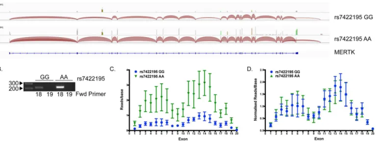
In addition to assessing overall expression effects, we also analysed the transcriptome for altered RNA splicing. We observed no differences in the location of splice junctions inMERTK
in either the high or lowMERTKexpressing group and the majority of junctions aligned with canonical 19 exons within theMERTKgene (Fig 7A). The only exception to this was that all samples contained a junction from the 3' end of exon 18 to a small putative alternative final exon that excluded the canonical final exon 19 (Fig 7A). Alignment of the transcript reads showed the putative final exon to be approximately 1273bp at position 112,796,928–
112,798,200 (genome build GRCh37/hg19). We confirmed the inclusion of this alternative exon in someMERTKtranscripts using reverse transcription PCR (Fig 7B). The inclusion of this exon, and the exclusion of the canonical final exon, would lead to an early stop signal within the tyrosine kinase domain of MERTK and potentially the production of a membrane-bound non-signalling MERTK receptor. However, the inclusion or exclusion of the alternative final exon does not appear to be altered by polymorphisms within theMERTKgene, as we observed no significant differences in exon usage between the two groups. Although overall expression of each exon was higher in the high expressing group (Fig 7C), once expression was normalised to account for the 3.63 fold overall expression increase in this group, the pattern of exon usage was the same in the two groups, independent of genotype (Fig 7D).
MERTK
risk variants may alter disease course in established MS
in the same centre (Victorian MS cohort), for the cumulative probability of MS progression over time. We stratified our data by genotype at rs7422195 and divided the cohort intoDR15
negative andDR15homozygous groups. We found that in both groups, there was a strong trend towards altered probability of progression dependent upon genotype at rs7422195 (Fig 8) but that, consistent with the association data, the opposite allele was associated with altered
Fig 7. TheMERTKgene contains a novel alternative final exon but usage is not genotype-dependent.(A) Exon junctions in RNA sequencing data were visualised using IGV_2.3.35. Representative junction maps for a low (rs7422195 GG) and high (rs7422195 AA) expressing sample are shown. Each shows the presence of a junction from exon 18 to a putative novel alternative final exon excluding exon 19. (B) RT-PCR analysis of a high and low expressing sample show the presence of this exon only in combination with exon 18 and not exon 19. (C) Exon usage analysis shows overall increased expression of each exon in samples homozygous for the rs7422195 minor (A) allele, but no difference in exon usage following normalisation for overall expression level (D).
doi:10.1371/journal.pgen.1005853.g007
Fig 8. Disease course is altered in the presence ofMERTKsusceptibility-associated variants.Individuals initially presenting with a relapsing-remitting course of MS were stratified by bothDR15status and genotype at rs7422195. (A) In the presence of the minor (A) allele of rs7422195,DR15negative individuals (n = 370) showed a strong trend towards increased probability of progression (p= 0.07) (B) In the presence of the major (G) allele of rs7422195
DR15homozygous individuals (n = 68) showed a strong trend towards increased probability of progression (p =0.081)
progression, such that in theDR15negative group, patients homozygous for the risk-associated (A) allele have an increased probability of progression, although the effect does not reach statis-tical significance (Fig 8A,p= 0.07, OR = 1.9, 95% CI 0.948 to 3.87). Conversely, in theDR15
homozygous group, where the (G) allele is associated with risk, patients homozygous for this allele have an increased probability of progression (Fig 8B,p =0.08, OR = 5.6, 95% CI 0.811 to 38.2).
Discussion
In this study we have refined the association ofMERTKwith MS susceptibility to independent signals from both common and low frequency variants. We have shown that one of the associ-ated variants, which is an eQTL for MERTK expression, is intranswith theHLA-DRB1locus, and shows discordant association dependent upon DR15 status. In addition, we have identified a number of low-frequency variants, all contained within the same haplotype, which are associ-ated with MS susceptibility and operate in a recessive manner. Finally, we have shown that polymorphisms withinMERTKaffect not only MS susceptibility, but may also affect severity in established MS.
The main focus of this study was to refine the association ofMERTKwith MS susceptibility and to identify potential causal variants. In the process of this refinement, we identified both common and low frequency variants withinMERTKindependently associated with the risk of developing MS. The variants identified in this study are all in LD with the common variant withinMERTK, rs17174870, previously identified by us, and replicated by the IMSGC, as asso-ciated with MS susceptibility [13,24]. The majority of variants captured in GWAS studies are common (MAF>0.05), and those found to be associated with disease are often intronic and outside any obvious regulatory regions, leading to the speculation that the signals detected in GWAS are in fact the result of synthetic associations between rare variants and common vari-ants [31]. However, a recent study has cast doubt upon this hypothesis, and found limited sup-port for the influence of rare coding sequence variants upon disease susceptibility in a
combined analysis of six common autoimmune diseases, including MS [32]. In contrast, it has recently been shown that a rare null mutation in the P2X7 receptor is strongly associated with protection against MS [33]. This study ofMERTKsuggests that, at least in some cases, associa-tions from low frequency variants may underlie the signal observed in GWAS analysis of com-mon variants, and that careful selection of individuals for resequencing is crucial for the detection of the true underlying signal, as has previously been suggested [34].
associated with overall burden of these low frequency alleles. The two larger variants in particu-lar warrant further investigation, as these types of variants have been linked with other human diseases. For example, a pathogenic retrotransposon insertion in the 3'UTR of the Fukutin gene leads to the development of Fukuyama-type congenital muscular dystrophy [29], whereas an ATndinucleotide repeat polymorphism within theCTLA-4gene has been shown to be pro-tective against development of Graves' Disease in childhood [35].
In addition to the recessive low frequency variants, we also identified other more common variants associated with MS susceptibility, one of which showed discordant association depending upon theDR15status of the individual. Discordant associations have been previ-ously reported across different autoimmune diseases (reviewed in [36]), but to our knowledge this is the first such association described within a single disease. In this case, the minor allele at rs7422195 is associated with MS susceptibility in the absence ofDR15, but this is converted to a protective effect on aDR15homozygous background.
What might mediate this discordant effect of rs7422195? We have shown that in monocytes, but not other immune cell types, this SNP is associated with altered expression of MERTK, at both the gene and protein level, and that the minor allele of rs7422195 is associated with increased expression of MERTK. Monocytes are the precursors of a number of cell types, including macrophages and dendritic cells, that have been shown to be central to the etiology of MS (reviewed in [37,38]). We further found that there was suggestive genotype dependent expression ofMERTKin CD4+vecells, the majority of which are T cells. Although this associa-tion would need to be validated in order to exclude the possibility that the detected altered expression was not the result of contaminating CD4+vemonocytes, it has recently been shown that Mertk is expressed in the Th17+vesubset of CD4+veT cells in mice following induction of experimental autoimmune encephalomyelitis, a mouse model of MS [39].
MERTK is an important regulator of immune activation, and the expression of MERTK is essential to the maintenance of immune homeostasis—maintaining the balance of immune activation when required and immune suppression following challenge. In animal models it has been shown that the relationship of MERTK to autoimmune disease is complex, and in some cases dependent upon genetic background. For example in the NOD (non-obese dia-betic) mouse model of spontaneous diabetes, knockdown ofMERTKresults in resistance to the development of diabetes, and this effect appears to be mediated by bone-marrow derived den-dritic cells [40]. In contrast in a different NOD genetic background, in this case in the presence of a transgenic T-cell receptor, deficiency of Mertk leads to an exacerbation of the rate of spon-taneous disease [41]. In humans, MERTK expressed on dendritic cells has been shown to be an important negative regulator of T cell activation, whereby MERTK expressed by tolerogenic dendritic cells suppresses T cell activation and proliferation [42]. The ability of MERTK to sup-press activated T cells may extend to previously activated memory T cells [42], providing a clear and plausible link between increased MERTK expression and protection against MS sus-ceptibility, as observed in ourDR15homozygous population: a population in which increased expression of the antigen presenting molecules encoded by theDR15haplotype is hypothesised to lead to more efficient presentation of encephalitogenic peptides and increased activation of autoreactive T cells [43]. What is not clear is why this link breaks down in the DR15 negative population, where increased MERTK expression is associated with MS risk. In this negative population, where antigen presentation and subsequent T cell activation may not be such a clear driving force for susceptibility, it is possible that other factors may be at work, perhaps involving similar biological processes to that observed in NOD mice, where deficiency of Mertk reduces susceptibility to spontaneous diabetes.
disease susceptibility, the pathways most amenable to intervention are those still active in con-trolling clinical course in established disease. In this study we have presented evidence that, in addition to disease susceptibility, MERTK expression associated variants may also be related to severity measures, specifically the rate of conversion from the relapsing-remitting phase of RRMS to progression. We found that, consistent with the discordant association in the pres-ence or abspres-ence ofDR15, the allele associated with high expression of MERTK was associated with a decreased prevalence of MS inDR15positive homozygotes, and a higher prevalence of MS in the absence ofDR15. These data highlight the potential of MERTK as a therapeutic tar-get, although theDR15status of the patient would likely determine whether activation or repression of MERTK signalling would be the appropriate intervention.
Materials and Methods
Ethics statement
Recruitment of subjects and collection of tissue and DNA was approved by the Melbourne Health Human Research Ethics Committee (Project number: 2013.111), the Eastern Health Human Research and Ethics committee (Reference number: SERP27/1314) and the Australian Bone Marrow Donor Registry Ethics Committee (Project number: 2006/02). All Human Research Ethics Committees which provided approval for this research are guided by national standards as outlined in the National Statement on Ethical Conduct in Human Research (https://www.nhmrc.gov.au/guidelines-publications/e72) issued by the National Health and Medical Research Council (Australia). All cases and controls provided written consent for the use and storage of DNA and tissue samples.
Study subjects and DNA samples
The 3268 MS cases and 3579 healthy controls genotyped for the fine-mapping component of this study formed part of a larger ANZgene MS loci fine-mapping study, and were phenotyped according to established criteria [27]. To assess for potential population stratification, principal component analysis was performed and genomic inflation was determined to be 1.10, and out-lier samples removed as previously described [14]. We estimated the power of the sample used for fine mapping using the online genetic power calculator (http://pngu.mgh.harvard.edu/~ purcell/gpc/). The sample was estimated to have high power (>90%) for both variants of mod-erate frequency with modmod-erate risk (eg. MAF = 0.3, marker = 0.3, D' = 0.99, disease preva-lence = 0.001 and relative risk = 1.2; alpha = 0.05), as well as for variants of low frequency with a stronger risk (MAF = 0.05, marker = 0.05, D' = 0.99, disease prevalence = 0.001 and relative risk = 1.6; alpha = 0.05).
A subset of these samples (1500 cases and 1500 controls) were used for association testing of the variants identified in the resequencing component of the study. The subjects used for the resequencing component of the study were excluded from association analysis of identified variants. We used the online genetic power calculator (http://pngu.mgh.harvard.edu/~purcell/ gpc/) with the following assumptions and calculated a minimum sample size for 80% power (at alpha = 0.05) of 1465 cases: MAF 0.02 (and "marker" at the same frequency since we predicted that re-sequencing will identify biologically relevant variants that we directly test); D' = 1, dis-ease prevalence = 0.001 and relative risk = 1.618. A small subset of samples used in resequen-cing were re-recruited for RNA sequenresequen-cing analysis (n = 5 cases; n = 3 controls).
Whole genome sequencing
A sequence-ready DNA library of short sequences for whole genome sequencing was prepared using the TruSeq PCR-free library preparation kit (Illumina, San Diego, CA). The sample library was uniquely barcoded and sequenced on a single lane of an Illumina HiSeq 2000 sequencer (Illumina, San Diego CA).
Amplicon design and targeted resequencing
PCR amplicons were designed to tile completely the genomic region of the MERTK gene (GRCh37/hg19region chr2:112,645,298−112,789,127). Illumina short read sequencing libraries
were constructed for each individual DNA sample after targeted capture and amplification of the
MERTKlocus based on the amplicon tiling design. Libraries for sequencing were prepared by the Australian Genome Research Facility (AGRF) and uniquely barcoded. All libraries were then sequenced in a multiplexed fashion within a single lane of an Illumina MiSeq sequencer by AGRF.
Variant discovery
Sequencing output was demultiplexed and base calling quality scores were verified using FastQC software which confirmed that reads were of uniformly high quality (Q>30). We mapped all reads to the human reference genome build GRCh37/hg19 using the BWA aligner [45]. Across all samples a minimum of 90% of sequence reads mapped successfully to the refer-ence genome, and this resulted in an average read depth of at least 40X in the target (MERTK) locus, giving more than sufficient depth to make robust sequence variant calls. We used Picard (http://picard.sourceforge.net) to remove PCR and optical duplicate reads. To call sequence variants [SNPs and insertions-deletions (in-dels)] we used GATK v3.1 [46] and implemented the GATK DNA-sequence variant calling best practice guidelines [47,48].
Genotyping
Genotyping of the full sample set for the fine-mapping phase was performed using 1536 cus-tom GoldenGate assay according to manufacturer's instructions [27]. Target specific primers for KASP (Kompetitive Allele Specific PCR) genotyping were designed for short variants (SNPs and in-dels) identified during the variant discovery phase and chosen for association testing, with each assay validated using samples of known genotypes. Assays that failed at either the design phase or during validation were then redesigned for the Sequenom MassArray sys-tem using iPLEX Gold chemistry, and assays validated using samples of known genotypes. Assays that also failed redesign were discarded. KASP assays were conducted by LGC genomics (Teddington, UK) and Sequenom MassArray genotyping was performed by the Garvan Molec-ular Genetics Service (Darlinghurst, Australia). All assays were plotted using cluster analysis software [KRAKEN (LGC Genomics, Teddington, UK)] and scored visually to ensure geno-types were correctly assigned. Two large variants not suitable for KASP genotyping (AluYf4 retrotransposon insertion and TAnTnrepeat expansion) were assessed using PCR in a subset of samples and not included in association analysis. Sequences of primers were as follows: AluYf4 retrotransposon insertion forward (ATCACTGGGCCTGAAATCTG), reverse (CATGCCTT
GGCATCACTTTT); TAnTnrepeat expansion forward (GGGTCCTAGCACCTAACCTG),
reverse (CCACGAAACCTACCCTGAAA).
Association analysis
group were then carefully re-assessed for quality of genotyping by cluster plot analysis and comparison of called genotypes to known genotypes from sequencing data, or to HLA typing where available in the case ofHLA-DRB115:01. Markers that showed evidence of poor quality genotyping were excluded from further analysis.
Association of variants (SNPs and in-dels) was measured using the allelic association test [1 degree of freedom (d.f)] or the genotyping test (2 d.f.) as appropriate using PLINK (v1.07; http://pngu.mgh.harvard.edu/~purcell/plink/).
We used standard logistic regression (R v3.1.2;https://www.r-project.org/) to identify potential interactions withDR15in the context of association. To test for statistical interaction between rs7422195 genotype andHLA-DR15status in the context of MS disease association, we constructed a standard logistic regression model with a response (output) term for MS dis-ease status, and covariate (input) terms for rs7422195 genotype,HLA-DR15status and the cor-responding multiplicative interaction term between these two covariates (the rs7422195-DR15
interaction term). The interactionp-value is thep-value associated with the rs7422195-DR15
interaction term in this logistic regression model. A significant interactionp-value implies that the disease risk profile ofDR15homozygous individuals across the possible rs7422195 geno-types (GG, AG, AA) is significantly different to the disease risk profile ofDR15negative and heterozygous individuals across these rs7422195 genotypes.
Haplotype construction and association analysis
Haplotypes were constructed and analysed for association using Haploview (v4.2;https://www. broadinstitute.org/scientific-community/science/programs/medical-and-population-genetics/ haploview/haploview) using the confidence intervals algorithm. As default settings exclude MAF<0.05, all analyses were altered to include MAF>0.01. All blocks identified using this algorithm were tested for association within Haploview using chi-square tests.
Cell subset purification
Immune cell subsets for RNA isolation were purified from Peripheral Blood Mononuclear Cells (PBMCs) using whole blood collected between 9am and 12pm. PBMCs were isolated from MS patients and controls using histopaque (Sigma-Aldrich, St Louis, MO) density gradi-ent separation. CD4+vecells, CD8+vecells and B lymphocytes (CD19+ve) were purified using magnetic bead separation (positive selection) using Human Microbeads directed against the indicated cell surface markers as per the manufacturer’s instructions (Miltenyi Biotec, Mac-quarie Park, Australia). For CD14+vemonocyte purification, a monocyte enrichment kit (Stem-cell Technologies, Tullamarine, Australia) was used prior to selection of CD14+vecells with human CD14 Microbeads (Miltenyi Biotec, Macquarie Park, Australia) and magnetic bead purification. For Natural Killer (NK) cell (CD3-veCD56+ve) purification, an NK enrichment kit (Stemcell Technologies, Tullamarine, Australia) was used as per manufacturer’s instructions prior to positive selection using human CD56 Microbeads (Miltenyi Biotec, Macquarie Park, Australia). Purified subsets were stored in RLT buffer (Qiagen) for subsequent RNA extraction. Purity of the subsets was determined by flow cytometry.
Flow cytometric analysis
cytometry of labeled PBMC and purified cell subsets was performed using a CyAn ADP ana-lyzer (Beckman Coulter) and the data analysed using WEASEL (v3.0). Purified fractions for each of the cell subsets were only used in subsequent analyses if purity was 90% or greater.
In order to assess MERTK expression on the surface of monocytes, PBMCs were isolated from whole blood and monocytes identified using antibodies directed against the markers CD14-PE [TUK4 (Miltenyi Biotec, Macquarie Park, Australia)] and CD16-FITC [VEP13 (Mil-tenyi Biotec, Macquarie Park, Australia)] as previously described [44]. Cell surface MERTK protein was detected using human Mer APC-conjugated antibody [Clone #125518 (R&D Sys-tems, Minneapolis, MN)] and compared with the appropriate isotype control [APC-conjugated Mouse IgG1, IS5-21F5 (Miltenyi Biotec, Macquarie Park, Australia)].
Expression analysis of immune cell subsets
We extracted RNA from purified immune cell subsets and expression microarray hybridiza-tions were performed using the WT Expression kit (Life Technologies, CA, USA), WT Termi-nal Labelling and Controls Kit (Affymetrix, CA, USA) and Affymetrix Human Gene_1.0ST arrays. The probed arrays were washed and stained using the GeneChip Hybridization Wash and Stain Kit (Affymetrix, CA, USA) and scanned using the GeneChip Scanner 3000. Images (. dat files) were processed using GeneChip Command Console (Affymetrix, CA, USA) and the CEL files generated were used for further analysis. Expression data was linearised by transfor-mation to a log(2) scale and normalised using the removal of unwanted variation, 2-step (RUV-2) method[49]. Combined case and control datasets were used to identifyciseQTL ciations across each cell type using an additive linear model. Statistically significant eQTL asso-ciations were defined as having p<0.05 following correction for multiple testing (ie. P<0.05 and FDR<0.05).
MERTK
expression analysis in monocytes using RNA sequencing
(RNAseq)
Total RNA was isolated from purified monocytes using the Qiagen RNeasy minikit (Qiagen, Hilden, Germany) according to manufacturer's instructions. We then enriched the samples for mRNA using the Ambion polyA purist kit (ThermoFisher Scientific, Waltham, MA) according to manufacturer's instructions. Libraries for sequencing were prepared by the Australian Genome Research Facility (AGRF) from 200ng mRNA, with the mRNA from each individual ligated with a unique multiplex tag. Libraries were then pooled and divided across 3 lanes of the Illumina HiSeq sequencer (Illumina San Diego, CA), sequenced with 100bp single end reads and read quality assessed using FastQC (http://www.bioinformatics.bbsrc.ac.uk/projects/ fastqc/). Untrimmed reads were aligned to human GRCh37/hg19 genome using Subjunc aligner within the Subread software package [50]. Sequencing data was summarized into reads per transcript using FeatureCounts [51] against the GENCODE 19 gene/isoform models for the human GRCh37/hg19 reference genome (July 2013 freeze) [52].
boundaries and exon-exon junctions, mapped sequences were imported in the Integrative Genomics Viewer (IGV v.2.3.35;https://www.broadinstitute.org/software/igv/home).
Reverse transcription PCR
Total RNA from monocytes was prepared as for expression analysis and reverse transcribed into cDNA using a Taqman Reverse Transcription kit according to manufacturer's instructions (Applied Biosystems, Scoresby, Australia). A reverse primer located within the putative alter-native final exon 20 (GACAATGATTGGGATAGAAACC) was used in conjunction with either a forward primer for the canonical exon 18 (GAAATAGCTACGCGGGGAAT) or a for-ward primer in the canonical exon 19 (GTGTATATCATGGAAAAAGACAAGGAT). Ampli-fication of cDNA was performed for 35 cycles with primer annealing at 60°C and products visualised on a 2% (w/v) agarose gel.
Calculation of disease course
The prevalence of SPMS was determined using logistic regression analyses, whereby each patient was assigned a known duration of MS from diagnosis to time of collection, and at that same time was assessed as either RRMS or SPMS according to established criteria [14]. In con-junction with their genotype at rs7422195, regression analyses were performed to determine the whether the rate of change of SPMS (ie the % of SPMS cases/total cases at any given dura-tion of MS) was different dependent on genotype at rs7422195.
Statistical analysis
Gene expression data were analysed using either Student'st-test (for 2 groups) or one-way ANOVA (for>2 groups) followed by Tukey's multiple comparison correction (GraphPad PRISM v.6.0b;http://www.graphpad.com/scientific-software/prism/). Linear regression was used to analyse the relationship betweenMERTKgene expression and MERTK expression by monocytes (GraphPad PRISM v.6.0b). The proportion of monocytes expressing MERTK was performed using the Kruskal-Wallis test followed by Dunn's multiple comparison correction (GraphPad PRISM v.6.0b). All grouped expression data are plotted as Tukey box and whiskers. The relationship betweenMERTKgenotype and Multiple Sclerosis severity was analysed using logistic regression (STATA v.12.1;http://www.stata.com/).
Supporting Information
S1 Fig. The rs7422195(A)-allele is associated with increased length of the TAnTnrepeat
expansion in intron 1.The size of the amplified PCR product including the TAnTnrepeat within intron 1 was significantly increased in individuals homozygous for the rs7422195(A)-allele (p<0.0001 GG vs AA). For technical reasons only the shortest allele present in any indi-vidual was amplified.
(PDF)
S1 Table. Hardy-Weinberg tests.
(PDF)
S2 Table. Apparent size of TAnTnrepeat expansion in tested samples. (PDF)
S3 Table. Analysis of linkage between rs13414207 and the intron 4 AluYf4 insertion in two populations.
Acknowledgments
We thank all participants in this study for their support of this research. We also thank Jim Stankovic and Alison Hamlett for technical assistance, and the Bioinformatics Core at the Flo-rey for technical advice.
Australia and New Zealand Multiple Sclerosis Genetics Consortium (ANZGene) members: Alan Baxter, Allan G Kermode, Bruce Taylor, David R Booth, Deborah Mason, Graeme J Stew-art, Helmut Butzkueven, Jac Charlesworth, James Wiley, Jeannette Lechner-Scott, Judith Field, Lotti Tajouri, Lyn Griffiths, Mark Slee, Matthew A Brown, Pablo Moscato, Rodney J Scott, Simon Broadley, Steve Vucic, Trevor J Kilpatrick, William M Carroll
Author Contributions
Conceived and designed the experiments: MDB SF JF TJK AGB HB. Performed the experi-ments: MDB DM LJJ LG SEC RA GZMM AAP MMG LL MJFP MAJ JF. Analyzed the data: MDB ADF DM SEC GF TS MAJ JF. Contributed reagents/materials/analysis tools: MJFP HB JF MAJ AGB SF. Wrote the paper: MDB ADF TJK JF.
References
1. Ebers GC, Bulman DE, Sadovnick AD, Paty DW, Warren S, et al. (1986) A population-based study of multiple sclerosis in twins. N Engl J Med 315: 1638–1642. doi:10.1056/NEJM198612253152603 PMID:3785335
2. Ebers GC, Sadovnick AD, Risch NJ (1995) A genetic basis for familial aggregation in multiple sclerosis. Canadian Collaborative Study Group. Nature 377: 150–151. doi:10.1038/377150a0PMID:7675080 3. Sadovnick AD, Ebers GC, Dyment DA, Risch NJ (1996) Evidence for genetic basis of multiple
sclero-sis. The Canadian Collaborative Study Group. Lancet 347: 1728–1730. PMID:8656905
4. Hansen T, Skytthe A, Stenager E, Petersen HC, Brønnum-Hansen H, et al. (2005) Concordance for multiple sclerosis in Danish twins: an update of a nationwide study. Multiple Sclerosis Journal 11: 504– 510. PMID:16193885
5. O'Gorman C, Lin R, Stankovich J, Broadley SA (2013) Modelling genetic susceptibility to multiple scle-rosis with family data. Neuroepidemiology 40: 1–12. doi:10.1159/000341902PMID:23075677 6. Jersild C, Fog T (1972) Histocompatibility (HL-A) antigens associated with multiple sclerosis. Acta
Neu-rol Scand, Supplc: 51: 377.
7. Jersild C, Svejgaard A, Fog T (1972) HL-A antigens and multiple sclerosis. Lancet 1: 1240–1241. 8. Naito S, Namerow N, Mickey MR, Terasaki PI (1972) Multiple sclerosis: association with HL-A3. Tissue
Antigens 2: 1–4. PMID:5077731
9. Hauser SL, Fleischnick E, Weiner HL, Marcus D, Awdeh Z, et al. (1989) Extended major histocompati-bility complex haplotypes in patients with multiple sclerosis. Neurology 39: 275–277. PMID:2783768 10. Allen M, Sandberg-Wollheim M, Sjögren K, Erlich HA, Petterson U, et al. (1994) Association of
suscep-tibility to multiple sclerosis in Sweden with HLA class II DRB1 and DQB1 alleles. Hum Immunol 39: 41– 48. PMID:8181961
11. Haegert DG, Francis GS (1993) HLA-DQ polymorphisms do not explain HLA class II associations with multiple sclerosis in two Canadian patient groups. Neurology 43: 1207–1210. PMID:7909591 12. Consortium IMSG, Hafler DA, Compston A, Sawcer S, Lander ES, et al. (2007) Risk alleles for multiple
sclerosis identified by a genomewide study. N Engl J Med 357: 851–862. doi:10.1056/ NEJMoa073493PMID:17660530
13. International Multiple Sclerosis Genetics Consortium, Wellcome Trust Case Control Consortium 2, Sawcer S, Hellenthal G, Pirinen M, et al. (2011) Genetic risk and a primary role for cell-mediated immune mechanisms in multiple sclerosis. Nature 476: 214–219. doi:10.1038/nature10251PMID: 21833088
14. Australia and New Zealand Multiple Sclerosis Genetics Consortium (ANZgene) (2009) Genome-wide association study identifies new multiple sclerosis susceptibility loci on chromosomes 12 and 20. Nat Genet 41: 824–828. doi:10.1038/ng.396PMID:19525955