Carboxylic Acid (T4C), on
Trypanosoma cruzi
Anahı´ Magdaleno, Il-Young Ahn, Lisvane Silva Paes, Ariel M. Silber*
Departamento de Parasitologı´a, Instituto de Cieˆncias Biome´dicas, Universidade de Sa˜o Paulo, Sa˜o Paulo, Brazil
Abstract
It is well established that L-proline has several roles in the biology of trypanosomatids. InTrypanosoma cruzi, the etiological agent of Chagas’ disease, this amino acid is involved in energy metabolism, differentiation processes and resistance to osmotic stress. In this study, we analyzed the effects of interfering with L-proline metabolism on the viability and on other aspects of the T. cruzi life cycle using the proline analogue L- thiazolidine-4-carboxylic acid (T4C). The growth of epimastigotes was evaluated using different concentrations of T4C in standard culture conditions and at high temperature or acidic pH. We also evaluated possible interactions of this analogue with stress conditions such as those produced by nutrient starvation and oxidative stress. T4C showed a dose-response effect on epimastigote growth (IC50= 0.8960.02 mM
at 28uC), and the inhibitory effect of this analogue was synergistic (p,0.05) with temperature (0.5460.01 mM at 37uC). T4C significantly diminished parasite survival (p,0.05) in combination with nutrient starvation and oxidative stress conditions. Pre-incubation of the parasites with L-proline resulted in a protective effect against oxidative stress, but this was not seen in the presence of the drug. Finally, the trypomastigote bursting from infected mammalian cells was evaluated and found to be inhibited by up to 56% when cells were treated with non-toxic concentrations of T4C (between 1 and 10 mM). All these data together suggest that T4C could be an interesting therapeutic drug if combined with others that affect, for example, oxidative stress. The data also support the participation of proline metabolism in the resistance to oxidative stress.
Citation:Magdaleno A, Ahn I-Y, Paes LS, Silber AM (2009) Actions of a Proline Analogue, L-Thiazolidine-4-Carboxylic Acid (T4C), onTrypanosoma cruzi. PLoS ONE 4(2): e4534. doi:10.1371/journal.pone.0004534
Editor:Gordon Langsley, INSERM U567, Institut Cochin, France
ReceivedNovember 4, 2008;AcceptedJanuary 7, 2009;PublishedFebruary 20, 2009
Copyright:ß2009 Magdaleno et al. This is an open-access article distributed under the terms of the Creative Commons Attribution License, which permits unrestricted use, distribution, and reproduction in any medium, provided the original author and source are credited.
Funding:This work was supported by grants from the Fundac¸a˜o de Amparo a` Pesquisa do Estado de Sao Paulo (FAPESP grant#08/57596-4), Conselho Nacional de Desenvolvimento Cientı´fico e Tecnolo´gico (CNPq Grant#473906/2008-2) and Instituto Nacional de Biologia Estrutural e Quı´mica Medicinal em Doenc¸as Infecciosas (INBEQMeDI). The funders had no role in study design, data collection and analysis, decision to publish, or preparation of the manuscript. Competing Interests:The authors have declared that no competing interests exist.
* E-mail: asilber@usp.br
Introduction
The protozoan parasiteTrypanosoma cruziis the causative agent of Chagas’ disease, which affects approximately 12–14 million people in endemic areas throughout Mexico, Central, and South America [1]. The illness is characterized by an acute phase with patent parasitemia and non-specific symptoms (if any), followed by a life-long chronic phase with subpatent parasitemia and scarce tissue parasitism. In the symptomatic phase, the heart is primarily affected, developing hypertrophy and dilatation, in addition to the digestive tract – predominantly the esophagus and large intestine – with the appearance of megaviscera [2]. At present, the available therapy is mainly successful during the acute phase of the disease but with systemic side effects. Application of this therapy to treat the chronic phase of the disease – when most patients are diagnosed – is still controversial [3,4].
T. cruzihas a complex life cycle characterized by several forms in vertebrate and invertebrate hosts [5,6]. In the invertebrate host (the triatomine insect belonging to the family Reduviidae, order Hemiptera), the parasite replicates as a non-infective form called epimastigote, which can differentiate into a metacyclic trypomas-tigote, an infectious but non-replicative form. This process is called metacyclogenesis. When these forms infect a vertebrate host, they invade host cells and differentiate into amastigotes, which then divide in the cell cytoplasm and differentiate again into trypomastigotes, passing through a transient epimastigote-like
stage termed the intracellular epimastigote [7,8,9]. The main transmission route of the parasite is via insect vector bites on any region of the host skin. During its feeding, the insect vector deposits feces and urine containing metacyclic forms, which are usually self-inoculated by the host when scraping the wound caused by the insect bite [10].
L-proline is an important metabolite that is involved in the differentiation of intracellular epimastigote to trypomastigote forms, which is required for the establishment of infection in the mammalian host [9].
Previous work showed that L-proline was taken up from the extracellular environment through two active transporters [21] and converted into five intermediates of the Krebs cycle (e.g., citrate, isocitrate, malate, succinate and oxaloacetate), pyruvate and the amino acids glutamate and aspartate, which are rapidly metabolized [19]. This seemed to comprise a conventional proline oxidation pathway. This idea is supported by the presence of two putative genes encoding proline oxidases and one putative gene encoding a pyrroline-5-carboxylate dehydrogenase annotated in theT. cruzigenome database [4]. Since L-proline participates in several differential processes inT. cruziwith respect to mammalian host cells, we hypothesize that inhibition of proline metabolism in T. cruzicould have more severe effects on the parasite biology that on host cells.
Various L-proline analogues such as T4C have been used as inhibitors of proline metabolism in prokaryote and eukaryote organisms [22,23,24]. In the present study, we analyzed the effects of T4C on the growth of epimastigotes and the infection of mammalian cells byT. cruzi. We also investigated the synergism or antagonism of T4C with stress conditions faced byT. cruziduring its natural life cycle: temperature, pH and oxidative and nutritional stress.
Results and Discussion
Growth inhibition assay
L-proline, a relevant energy source in trypanosomatids, is metabolized and converted into five intermediates of the
tricarboxylic acid cycle (e.g., citrate, isocitrate, malate, succinate and oxaloacetate) through its oxidation to glutamate inT. cruzi[4]. Analogues of proline could be useful for inhibition of enzymes of this and other metabolic pathways. Among these analogues, L-thiazolidine-4-carboxylic acid, a product of the non-enzymatic condensation of equimolar quantities of formaldehyde and L-cysteine to yield the saturated imino acid containing S as a thioether [25], has the ability to interfere with the utilization of proline for protein synthesis and to mimic proline in its incorporation into proteins [26]. It can be metabolized in both bacterial and mammalian cells [26,27,28].
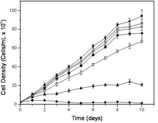
In order to investigate a possible trypanocide or trypanostatic effect of T4C on T. cruzi, 2.56106 epimastigotes per ml were cultured in the presence of different concentrations of T4C, ranging from 0.1 to 1 mM. The growth of epimastigotes on LIT medium with or without 200mM rotenone and 0.5mM antymicin were also followed as controls. The epimastigotes reached the stationary phase on the tenth day of growth, with 946106cells/ml in LIT. T4C showed a dose-dependent inhibition of epimastigote growth at 28uC and pH 7.5, the standard temperature and pH for cultivating these cells. The observed growth differences between the treated cells and the LIT control were significant (p,0.01), and the IC50 was determined to be 0.8960.02 mM (Figure 1), demonstrating an effect of T4C onT. cruziepimastigotes.
It has been suggested that T4C inhibits the growth ofEscherichia coli due to its specific interference with L-proline for protein synthesis by substituting L-proline in the corresponding prolyl-tRNA [27]. It has also been suggested that T4C could inhibit the synthesis of proline, thus enhancing the incorporation of this analogue into proteins [26]. In addition, whenEscherichia coliis grown in a proline-free medium, the cells are able to actively oxidize T4C, and the product of this oxidation can be used as a carbon source for synthesis of the
Figure 1. Growth curve of epimastigotes ofTrypanosoma cruzitreated with T4C at 28uC and 7.5 pH:&0 mM,D0.1 mM,#0.25 mM,
¤0.5 mM,%0.75 mM,m1.0 mM.Inhibition control (N) was performed by incubation the parasites in the presence of 0.5mM antimycin and 200mM rotenone.
ribonucleic acid bases guanine and uracil [27]. In rat liver and kidney cells, T4C is oxidized by mitochondrial proline oxidase to L-thiazoline-4-carboxylic acid, which is then hydrolyzed to N-formyl-cysteine and later hydrolyzed to N-formyl-cysteine and formic acid by a cytosolic enzyme [28]. Likewise, T4C inhibited proline synthesis in the wilted leaves of plants that synthesize and accumulate proline rapidly during water stress conditions [29]. This inhibition had no effect onD1
-pyrrolidine-5-carboxylic acid reductase (the enzyme that convertsD1-pyrrolidine-5-carboxylic acid into proline). On the other hand, T4C had no influence on the incorporation of proline into proteins in plants. In addition, these authors found that concentra-tions between 0.2 and 1 mM T4C inhibited proline oxidation in mitochondria isolated from etiolated barley, and this inhibition was related to the activity of proline dehydrogenase and not the transport of proline into the mitochondrial matrix. All informations suggest that the inhibitory activity of T4C onT. cruzigrowth could be due to any of the mentioned processes in which proline participates.
Interaction with stress conditions
T. cruzi, as well as other parasitic organisms that cycle among different conditions and environments, must deal with radical physical, chemical and physicochemical variations such as temperature, availability of nutrients, oxidative stress and pH, all constituting stress factors. The interaction of T4C with these stress factors could identify a possible role for proline metabolism inT. cruzi biology, so we analyzed this interaction. As proline metabolism is involved in resistance to several stress conditions in a variety of organisms ranging from yeast to plants [30,31,32], we investigated if this amino acid could also be involved in resistance to such unevaluated stress conditions in T. cruzi. We initially evaluated the effects of T4C together with high temperature (33uC and 37uC), acidic pH (5.5 and 6.5), stress from nutrient deficiency (incubation in the presence of PBS supplemented or not with several carbon sources such as amino acids or glucose) and oxidative stress due to different reactive oxygen species (incubation in the presence of H2O2).
1) Temperature stress
As already mentioned,T. cruzigoes through temperature changes throughout its life cycle because the insect vector does not regulate its body temperature and because it alternates between an insect vector and a mammalian host. Therefore, this parasite faces environmental temperature variations and the high temperature of the mammalian body (up to 37uC). Epimastigotes grow optimally at a temperature of 28uC, a condition in which a cell density of 946106cells/ml was reached at the stationary phase. Our data showed, however, that cell growth reached a higher density at 33uC (1206106cells/ml), while cell growth at 37uC was even less than that obtained at 28uC (706106 cells/ml). These results are in opposition to those obtained by [33], in which researchers reported an optimal temperature of epimastigote growth between 20uC and 28uC. The simultaneous effects of T4C and 33uC or 37uC diminished cell density in relation to that observed at 28uC (Figure 1). The IC50 values were calculated, being 0.8360.07 mM and 0.5460.01 mM for 33uC and 37uC, respec-tively (Figures S1, S2, S3, supplementary material). The simultaneous treatment with 0.5 mM of T4C and 37uC evidenced an effect for both variables that was significantly higher than the additive effects of T4C and temperature by themselves (Figure 2a), showing a synergistic interaction between these variables (p,0.05).
2) pH stress
T. cruzialso faces pH variations throughout its life cycle. In the intestinal tract of the insect vector, the pH varies between 5.9 and 8.9, depending on the starvation and blood meal periods [34].
About 2 months after feeding, the pH of triatomine excreta is acidic, and the parasite population in the feces is mostly constituted of metacyclic trypomastigotes. After feeding, the pH of excreta switches to alkaline, and one to five days later, it returns to a slightly acidic pH. During these periods, the population of epimastigotes increases, and the trypomastigotes and spheromas-tigotes forms are less common [35]. In addition, the metacyclic infective forms living in an acidic medium must face a slightly alkaline medium in the blood or inside the cell cytoplasm. Given this situation, it seemed interesting to evaluate how the parasites responded to acidic pH in the presence or absence of T4C. Epimastigotes were grown at pH 5.5, 6.5 and pH 7.5 as a control (i.e., the usual pH of LIT medium). It was found that their growth was strongly inhibited at pH 5.5, giving a maximum cell density of 216106cells/ml in LIT medium, while parasites grown at pH 6.5 achieved similar cell densities (1026106cells/ml at the stationary phase) to those seen at pH 7.5 (946106 cells/ml) (Figure 2b). When the cultures were treated with T4C, the inhibitory effect on parasite growth was enhanced. It was observed that growth inhibition in the presence of T4C at pH 5.5 and 6.5 was stronger than at pH 7.5 (Figure 1). The IC50values were calculated, being 0.5860.03 mM at pH 5.5 and 0.5160.03 mM at pH 6.5 (Figures S4, S5, S6, supplementary material). However, an interaction between the proline analogue and these growth conditions could not be confirmed, since the statistical test used to evaluate synergism was not significant (p.0.05). This result suggests that proline metabolism is not involved in pH stress resistance mechanisms.
3) Nutritional stress
enzymes that are activated under nutritional stress conditions in order to generate free metabolites within the cell [37,15], the viability of epimastigotes after 72 h was very low in PBS conditions.
4) Oxidative stress
In order to study the possible interaction between T4C and oxidative stress, we first evaluated the dose-response of the epimastigotes to different concentrations of H2O2. Cells (5610
6
parasites/mL) were resuspended in PBS and challenged with different hydrogen peroxide concentrations (between 0 and 200mM) at 28uC for 90 min. After removal of the H2O2, cells were re-inoculated in fresh-LIT medium, and their growth was evaluated, as described in Materials and Methods. The concen-tration of H2O2 inhibiting 50% of parasite growth (IC50) was determined to be 160mM under our experimental conditions. To determine the effects of H2O2on T4C treatment, cells were then incubated under oxidative stress conditions (IC50) or in PBS in the presence or absence of 0.5 mM T4C, and parasite growth was followed (Figure 2d). The differences between each treatment (H2O2or T4C) and its respective control were significant (p,0.01).
The number of cells previously challenged with T4C was reduced by 55% with respect to the control, and cells incubated simultaneously in T4C and H2O2 showed a 75% reduction in growth. The protective effect of L-proline against oxidative stress was previously reported by some authors, such as [30] and [38]. Taking into account the effects of T4C on mammalian infection, it is possible that this could cause increased inhibition of the viability of infective forms. It was known that T. cruzi could generate different reactive oxygen species (ROS) by its own aerobic metabolism and the host immune response. These species lead to membrane disruption, inactivation of essential enzymes, mutagenesis and damage to the DNA repair machinery. The importance of these effects is primarily seen when the parasite invades the mammalian cell or when it is exposed to drugs used to treat Chagas’ disease [39]. In the same way, T4C could act as a drug that increases the inhibitory effect of H2O2. In spite of the ROS detoxifying mechanisms inT. cruzi[40,16], the epimastigote survival was low, at 0.5 mM T4C.
From the results presented above, it could be proposed that the enhanced effect of T4C in the presence of H2O2was due to the fact that this reagent affected a proline-dependent oxidative stress Figure 2. Response of epimastigotes at different stress conditions and T4C treatment.Panel a: the parasite density was measured at the 5th. day of growth, submitted or not to thermal stress (33
uC and 37uC) and drug treatment (0.5 mM T4C). Panel b: the parasite density was measured at the 5th. day of growth, submitted or not to pH stress (pH 5.5 or 6.5) and drug treatment (0.5 mM T4C). Panel c) the parasite viability was measured by MTT assay after nutritional stresses performed by 72 h incubation in PBS, PRO and GLC, combined or not with the treatment with drug (0.5 mM T4C). Panel d: the parasites were submitted to oxidative stress by incubation in the presence of 160mM H2O2and the addition or not of drug (0.5 mM T4C). The effect of treatments and controls were measured by growing the parasites in LIT for five day and cell density, quantified.
resistance mechanism. Alternatively, it could be argued that the enhancement of the effects of T4C seen under oxidative stress conditions was due to the fact that T4C made the cells more fragile by partially arresting proline metabolism, which is relevant for energy generation [19] and redox balance, as occurs in other organisms [30,41,38]. To better understand both the involvement of proline metabolism in resistance to oxidative and metabolic stresses and the interaction between these factors and T4C treatment, a three-variable experiment was performed: cell viability was evaluated as a function of oxidative and metabolic stress challenges and T4C treatment. Preliminary results forced us to diminish the metabolic stress by diminishing the starvation time from 72 (the time used above for metabolic stress experiments) to 48 h, since 72 h of starvation combined with oxidative stress and T4C treatment reduced viability to immeasurable levels. The parasites (206106 cells/mL) were incubated for 48 h in PBS, supplemented or not with 3 mM proline. As done previously, cultures maintained in PBS supplemented with 3 mM glucose were also evaluated. All groups were maintained in the presence or absence of 0.5 mM T4C (T4C), and the cultures were then challenged with H2O2, as previously described, or mock challenged with H2O. The cell viability in each case was evaluated by a MTT assay. Before the 3 h challenge (T0), the viability of parasites incubated with carbon sources (proline or glucose) differed significantly (p,0.01) from the viability of those incubated with PBS (Figure 3). As expected, T4C showed a synergistic effect with each nutrient condition: the cell viability was diminished by 75%, 88% and 62% in parasites starved with PBS, PRO and GLC, respectively. After a 3 h mock challenge, cell viability diminished between 33% and 55% with respect to T0, showing significant differences (p,0.01) in all of the treatments. When the T4C-treated parasites were challenged with H2O2, a significant reduction in viability was observed (p,0.01) compared to parasites
that were mock challenged (Figure 3). Interestingly, the difference was not significant between the PRO or GLC groups. This pattern changed, however, when T4C was present, showing a reduction in cell viability after 3 h of incubation with H2O2of 21% for PBS, 57% for PRO and 85% for GLC with respect to parasites not incubated under oxidative stress. Only simultaneous treatment with PRO-T4C and oxidative stress showed a significant difference with respect to PRO and oxidative stress (p,0.05).
The enhanced effect of T4C on the PRO group with respect to the GLC group was expected and could be explained by the fact that T4C interfered with proline rather than glucose metabolism. In order to confirm this, we measured the intracellular free-proline levels in parasites starved for 48 h with PBS or 3 mM proline, in the presence or absence of T4C. Parasites starved with 3 mM glucose were used as controls, as previously described. After 48 h of incubation (T0), the intracellular free proline concentration was twice as high in the PRO group compared to the GLC group (4.52 and 2.26mM/106 cells, respectively) (Figure 4), indicating that when available, this metabolite could be transported and accumu-lated, as previously reported [21]. It is worth stressing that no differences were observed in cell viability between the PRO or GLC groups (p,0.05) (Figure 3). After 48 h of incubation (T0), the concentration of intracellular L-proline in the presence of T4C was maintained (PBS = 0.88mM/106 cells) or reduced (PRO = 2.87mM/106 cells, GLC = 1.29mM/106 cells) with respect to incubation without the analogue (Figure 4). The effect of the oxidative stress on the intracellular proline level was also evaluated by treating the cells with H2O2(or H2O as mock stress) for 3 h. There were significant differences (p,0.01) in the intracellular L-proline concentration between the cells kept under oxidative stress and in the presence of T4C. These two results confirm the synergistic effects of T4C and oxidative stress. Higher sensitivity to H2O2of the cells maintained in PRO/T4C with respect to those Figure 3. The effect of oxidative stress on epimastigotes previously incubated in PBS (PBS), L-proline 3 mM in PBS (PRO), and 3 mM Glucose in PBS (GLC), were treated or not with 0.5 mM T4C (PBS+T4C, PRO+T4C and GLC+T4C).After washing, the parasites were
incubated for 3 h in PBS and stressed with 160mM H2O2. The cells viability was evaluated by MTT assay.
not treated with T4C could be attributed to lower intracellular free proline levels rather than to inhibition of its metabolism.
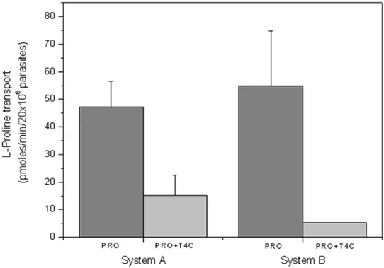
T. cruzitakes up proline through two active transport systems, one of them being a high affinity, low capacity transporter called system A, and the other a low affinity high capacity transporter called system B [21]. To further clarify the fact that L-proline is diminished by the presence of T4C, we performed a proline transport competition assay. Since T4C is a structural analogue of proline, the assay was designed under the hypothesis of a competitive inhibition, thus proline was used at concentrations in the range of the Km for each transport system, and T4C as a competitor was used at near its IC50(0.5 mM) concentration for both assays. As can be observed, both transporters resulted partially inhibited (68% inhibition for system A and 91% inhibition for system B) (Figure 5). In turn, the proline dehydrogenase and proline racemase activities were not inhibited (data not shown).
Effect of T4C on trypomastigotes bursting from infected host cells
In order to evaluate the effectiveness of treating the infected host-cells, the toxicity of T4C on mammalian CHO-K1 cells was determined. Interestingly, the inhibitory T4C concentrations tested on epimastigotes (between 0.1 and 1.0 mM) were not toxic to mammalian cells (IC50= 11.460.36 mM) (Figure 6). It is worth mentioning that epimastigotes ofT. cruziare not infective stages, but they serve as a model to analyze whether similar effects would be seen in the infective trypomastigote stage. Therefore, the effects of the treatment of infected CHO-K1 cells were evaluated using concen-trations up to 10 mM, causing a reduction of 31% and 56% in parasite bursting (Figure 7). The highest T4C concentration used to analyze survival through the intracellular stages of the parasite and the following release of trypomastigotes was 10 mM. The mamma-lian cells were then infected and treated with different concentrations
of T4C (between 1.0 and 10.0 mM). After five days of infection, the number of trypomastigotes that burst into the extracellular control medium was approximately 3.16106cells/ml. The parasite concen-tration in the presence of T4C was reduced to between 1.46106 cells/ml and 2.16106 cells/ml (Figure 7). There were significant differences between drug treatment and the control (p,0.05).
Proline is a relevant metabolite inT. cruzibiology, involved in several processes that are essential for the parasite life cycle [4]. Among them, differentiation in the insect vector stages [20] and inside the mammalian cell [9], as well as energy metabolism [19], seem to be of relevance. Taken together, these data are encouraging for the use of proline analogues able to inhibit the proline transport activity., on one side to identify new lead compounds for Chagas’ disease therapy, and on the other side, to validate the metabolite transporters as drug targets [4,12]. Further experiments are in process to analyze the synergic effects of T4C combined with nifurtimox and benznidazole, the drugs presently in use to treat this infection, with the aim of reducing their doses and perhaps diminishing their undesired side effects.
Materials and Methods
Reagents
L-proline, L-thyazolidine-4-carboxylic acid, rotenone, antimy-cin and MTT (3-(4,5-dimethylthiazol-2-yl)-2,5-diphenyltetrazo-lium bromide) were from Sigma (Saint Louis, MO, USA). L-[2, 3, 4, 5-3H] proline, 100 Ci/mmol was purchased from NEN Life Science Products (Boston, MA USA). RPMI culture medium and Fetal Calf Serum were from Cultilab (Campinas, SP, Brazil). All other reagents were from Amresco.
Cells and parasites
The Chinese Hamster Ovary cell line CHO-K1 was routinely cultivated in RPMI medium supplemented with 10% heat-Figure 4. The intracellular proline concentration was measured as a function of nutrient starvation, oxidative stress and T4C treatment.The epimastigotes were incubated for 48 h in PBS (PBS), 3 mM L-proline (PRO) and 3 mM Glucose (GLC), supplemented or not with 0.5 mM T4C (PBS+T4C, PRO+T4C and GLC+T4C) (T0). After washings, the parasites were incubated along 3 h in PBS in the presence or not of 160mM
inactivated fetal calf serum (FCS), 0.15% (w/v) NaHCO3, 100 units mL21
penicillin and 100mg mL21
streptomycin at 37uC in a humid atmosphere containing 5% CO2. Epimastigotes ofT. cruzi,
CL strain, clone 14 [42], were maintained in the exponential growth phase by subculturing every 48 h in LIT (liver infusion-tryptose) medium supplemented with 10% FCS at 28uC [43]. Figure 5. The effect of 0.5 mM T4C on L-proline uptake in epimastigotes of T. cruzi. Proline transport was measured at proline concentrations corresponding to the Km values for system A (0.31 mM) and system B (1.36 mM).
doi:10.1371/journal.pone.0004534.g005
Figure 6. The effect of T4C on CHO-K1line cells viability.CHO-K1cells were challenged with different concentrations of T4C, and the cell viability was measured by MTT assay.
Trypomastigotes were obtained by infection of CHO-K1 cells with trypomastigotes, as described previously [7,9]. Trypomastigotes were collected in the extracellular medium from five days onward.
Growth inhibition assays
Epimastigotes in the exponential growth phase (approximately 506106 cells/mL) were washed three times by centrifugation and resuspended in phosphate buffered saline (PBS), then cultured in fresh-LIT medium supplemented or not (controls) with different concentrations of T4C ranging from 0.1 to 1.0 mM at 28uC. The assays were carried out in 96 well plates by inoculating 2.56106cells/ mL in 200mL of medium. Cell growth was estimated by absorbance readings at 620 nm every day for ten days. The absorbance was transformed into a cell density value (cells/mL) using a linear calibration equation previously obtained under the same conditions (R2= 0.9981,p,0.05). The concentration of T4C that inhibited 50% of parasite growth (IC50) was determined in the exponential growth phase (sixth day) by adjusting the effect (growth inhibition values) as a function of T4C concentration to a classical sigmoidal equation. As a cell growth inhibition control, growth curves in which 200mM rotenone and 0.5mM antimycin were added to the culture medium were run in parallel for all experiments.
The effect of T4C on growth inhibition under stress conditions
To analyze the combined effect of T4C at different pH values and at different temperatures, the growth curves were generated as described above, with adjustment of the LIT pH to the desired values or by incubating the cultures at 28uC (no stress), 33uC and 37uC. To evaluate the effect of T4C combined with nutritional stress conditions, 206106 parasites/mL were washed and
resus-pended in 0.5 mM T4C in PBS (Phosphate Buffered Saline) or PBS supplemented with 3 mM glucose, 3 mM proline, or nothing (as control) for 72 h in Eppendorf tubes. Subsequently, cell viability was estimated by the MTT assay. To evaluate the combined effect of T4C and oxidative stress, 56106 parasites/mL in the stationary phase were maintained for 1 h and 30 min at 28uC in PBS or with 160mM H2O2in the presence or absence of 0.5 mM T4C. The cells were then collected by centrifugation and resuspended in LIT medium, and after 5 days, the number of cells/mL was determined as described previously [44]. A second experiment was carried out to evaluate the protective effects of L-proline and glucose (used as a control) on parasite viability under oxidative stress in the presence of T4C. In this experiment, 206106epimastigotes/mL were washed twice, resuspended in PBS and pre-incubated for 48 h in PBS in the presence or absence of 3 mM L-proline and 3 mM glucose, with or without 0.5 mM T4C. After pre-incubation, the cells were washed twice, resuspended in PBS with or without 160mM H2O2 and maintained under those conditions at 28uC for 3 h. Cell viability was then estimated using the MTT assay [45]. The L-proline concentration was measured before and after oxidative stress as previously described. To quantify free intracellular L-proline, the parasites were washed in cold PBS, suspended in 1 ml of PBS and lysed by sonication. The lysate was centrifuged (100,000 g, 30 min), and the supernatant was freed of proteins by trichloroacetic acid precipitation followed by centrifugation at 20,000 g for 30 min. Measurements of intracellular L-proline were made using the remaining supernatant solution, as described by Bates [46].
Proline uptake and enzymatic activities
The proline competition assays were performed as previously described [47]. In brief: two-day cultured parasites were washed three times by centrifugation and resuspended in phosphate Figure 7. The effect of T4C on the trypomastigote bursting was evaluated.The trypomastigotes bursted at 5th. day post-infection in cell cultures treated or not with at different concentrations of T4C were counted.
buffered saline (PBS), pH 7.4 at a final density of 2006106cells/ ml and distributed in aliquots of 100ml (containing 206106cells each). Transport assays were initiated by the addition to the assay tubes of 100ml of radioactively traced proline (0.5mCi) to a final concentration of 0.31 mM or 1.36 mM, supplemented or not with 0.5 mM T4C. V0 was measured at 28uC for 30 s, and proline transport was stopped by addition of 800 ml of cold 50 mM proline in PBS, and rapid washing by centrifugation at 10,000 g for 60 s. TheT. cruziproline dehydrogenase activity was assayed as previously described [48]. Proline racemase was assayed inT. cruziextracts by performing a coupled reaction, using D-proline as the enzyme substrate and L-proline dehydrogenase as the revelation system. Both assays were performed in the presence or absence of 0.5 mM T4C.
Effect of T4C on mammalian cell viability
CHO-K1 cells (5.06105cells ml21) were inoculated in 24-well plates in FCS supplemented RPMI medium, as previously described, in the presence of increasing concentrations of T4C or no T4C (controls). The viability of cells was determined by the MTT assay, and the IC50was obtained by fitting the data to a typical dose-response sigmoidal curve.
Effect of T4C on trypomastigote bursting
CHO-K1 cells were grown on cover slips and infected with trypomastigotes in RPMI medium supplemented with 10% FCS. After 3 h at 37uC, free trypomastigotes in the medium were removed by washing with PBS, and the infected cells were maintained at 33uC in RPMI medium supplemented with 2% FCS, with or without different concentrations of T4C. These concentrations of T4C were not toxic to the mammalian cells. The trypomastigotes were collected in the extracellular medium on the fifth day and counted in a hemocytometer [9].
Statistical analysis
A one-way ANOVA followed by Dunnet’s test was used for statistical analysis. To analyze synergism between two independent treatments, a two-way ANOVA was performed as described previously [49]. Apvalue that was less than 0.05 was considered statistically significant.
Supporting Information
Figure S1 Growth curve of epimastigotes of Trypanosoma cruzi treated with T4C at 33uC and 7.5 pH:&0 mM,n0.1 mM,#
0.25 mM,X0.5 mM,%0.75 mM,m1.0 mM. Inhibition control
(’) was performed by incubation the parasites in the presence of 0.5mM antimycin and 200mM rotenone.
Found at: doi:10.1371/journal.pone.0004534.s001 (0.48 MB TIF)
Figure S2 Growth curve of epimastigotes of Trypanosoma cruzi treated with T4C at 37uC and 7.5 pH:&0 mM,n0.1 mM,#
0.25 mM, X 0.5 mM, % 0.75 mM, m 1.0 mM. Inhibition
control (’) was performed by incubation the parasites in the presence of 0.5mM antimycin and 200mM rotenone.
Found at: doi:10.1371/journal.pone.0004534.s002 (0.45 MB TIF)
Figure S3 Sigmoidal dose-response curve of epimastigotes of Trypanosoma cruzi. Percentage of growth inhibition of T. cruzi epimastigotes at different temperatures and T4C. The curves represent the sigmoidal equation at 28uC (—), 33uC (---), and 37uC
(NNNNNNNNNNNNNNNN). It also shows the maximum cell growth obtained in
the control at each condition of temperature (inset).
Found at: doi:10.1371/journal.pone.0004534.s003 (0.53 MB TIF)
Figure S4 Growth curve of epimastigotes of Trypanosoma cruzi treated with T4C at 28uC and 5.5 pH:&0 mM,n0.1 mM,#
0.25 mM,X0.5 mM,%0.75 mM,m1.0 mM. Inhibition control
(N) was performed by incubation the parasites in the presence of 0.5mM antimycin and 200mM rotenone.
Found at: doi:10.1371/journal.pone.0004534.s004 (0.55 MB TIF)
Figure S5 Growth curve of epimastigotes of Trypanosoma cruzi treated with T4C at 28uC and 6.5 pH:&0 mM,n0.1 mM,#
0.25 mM,X0.5 mM,%0.75 mM,m1.0 mM. Inhibition control
(’) was performed by incubation the parasites in the presence of 0.5mM antimycin and 200mM rotenone.
Found at: doi:10.1371/journal.pone.0004534.s005 (0.45 MB TIF)
Figure S6 Sigmoidal dose-response curve of epimastigotes of Trypanosoma cruzi. Percentage of growth inhibition of T. cruzi epimastigotes at different temperatures and T4C. The curves represent the sigmoidal equation at pH 7.5 (’), 6.5 (---), and 5.5 (NNNN). It also shows the maximum cell growth obtained in the control at each condition of pH (inset).
Found at: doi:10.1371/journal.pone.0004534.s006 (0.53 MB TIF)
Acknowledgments
We are indebted to Prof. Silvia R. B. Uliana for critically reading this manuscript and Pablo S. Bogdanic for stimulating fruitful discussions. AM and IYA are postdoctoral fellows, and LSP is a post-graduate fellow from the FAPESP. AMS is fellow from the CNPq.
Author Contributions
Conceived and designed the experiments: AM LSP AMS. Performed the experiments: AM IYA LSP AMS. Analyzed the data: AM IYA AMS. Wrote the paper: AM AMS.
References
1. WHO (2002) Control of Chagas diseases. World Hearth Organ Tech Rep Ser 905: 64.
2. Tempone AG, Sartorelli P, Mady C, Fernandes F (2007) Natural products to anti-trypanosomal drugs: an overview of new drug prototypes for American Trypanosomiasis. Cardiovasc Hematol Agents Med Chem 5: 222–235. 3. Urbina JA (2002) Chemotherapy of Chagas disease. Curr Pharm Des 8:
287–295.
4. Silber AM, Colli W, Ulrich H, Alves MJ, Pereira CA (2005) Amino acid metabolic routes inTrypanosoma cruzi:possible therapeutic targets against Chagas’ disease. Curr Drug Targets Infect Disord 5: 53–64.
5. Brener Z (1973) Biology ofTrypanosoma cruzi. Annu Rev Microbiol 27: 347– 382.
6. De Souza W (1984) Cell biology ofTrypanosoma cruzi. Int Rev of Cytology 86: 197.
7. Almeida-de-Faria M, Freymuller E, Colli W, Alves MJ (1999)Trypanosoma cruzi: characterization of an intracellular epimastigote-like form. Exp Parasitol 92: 263–274.
8. Tyler KM, Engman DM (2001) The life cycle ofTrypanosoma cruzi revisited. Int J Parasitol 31: 472–481.
9. Tonelli RR, Silber AM, Almeida-de-Faria M, Hirata IY, Colli W, et al. (2004) L-proline is essential for the intracellular differentiation ofTrypanosoma cruzi. Cell Microbiol 6: 733–741.
10. Alves MJ, Colli W (2007)Trypanosoma cruzi: adhesion to the host cell and intracellular survival. IUBMB Life 59: 274–279.
11. Zilberstein D (1993) Transport of nutrients and ions across membranes of trypanosomatid parasites. Adv Parasitol 32: 261–291.
12. Landfear SM (2008) Drugs and transporters in kinetoplastid protozoa. Adv Exp Med Biol 625: 22–32.
13. Pereira CA, Alonso GD, Paveto MC, Flawia MM, Torres HN (1999) L-arginine uptake and L-phosphoarginine synthesis in Trypanosoma cruzi. J Eukaryot Microbiol 46: 566–570.
15. Pereira CA, Alonso GD, Ivaldi S, Silber AM, Alves MJ, et al. (2003) Arginine kinase overexpression improvesTrypanosoma cruzisurvival capability. FEBS Lett 554: 201–205.
16. Miranda MR, Canepa GE, Bouvier LA, Pereira CA (2006)Trypanosoma cruzi: Oxidative stress induces arginine kinase expression. Exp Parasitol 114: 341–344. 17. Rohloff P, Rodrigues CO, Docampo R (2003) Regulatory volume decrease in Trypanosoma cruziinvolves amino acid efflux and changes in intracellular calcium. Mol Biochem Parasitol 126: 219–230.
18. Rohloff P, Montalvetti A, Docampo R (2004) Acidocalcisomes and the contractile vacuole complex are involved in osmoregulation in Trypanosoma cruzi. J Biol Chem 279: 52270–52281.
19. Sylvester D, Krassner SM (1976) Proline metabolism in Trypanosoma cruzi epimastigotes. Comp Biochem Physiol B 55: 443–447.
20. Contreras VT, Salles JM, Thomas N, Morel CM, Goldenberg S (1985) In vitro differentiation ofTrypanosoma cruziunder chemically defined conditions. Mol Biochem Parasitol 16: 315–327.
21. Silber A, Tonelli R, Martinelli M, Colli W, Alves M (2002) Active transport of L-proline inTrypanosoma cruzi. J Eukaryot Microbiol 49: 441–446.
22. Nomura M, Nakamori S, Takagi H (2003) Characterization of novel acetyltransferases found in budding and fission yeasts that detoxify a proline analogue, azetidine-2-carboxylic acid. J Biochem 133: 67–74.
23. Deutch CE (2004) Oxidation of 3,4-dehydro-D-proline and other D-amino acid analogues by D-alanine dehydrogenase from Escherichia coli. FEMS Microbiol Lett 238: 383–389.
24. Andreasson C, Neve EP, Ljungdahl PO (2004) Four permeases import proline and the toxic proline analogue azetidine-2-carboxylate into yeast. Yeast 21: 193–199.
25. Ratner S, Clarke HT (1937) The action of formaldehyde upon cysteine. . J Am Chem Soc 59: 200.
26. Unger L, DeMoss RD (1966) Metabolism of a proline analogue, l-thiazolidine-4-carboxylic acid, byEscherichia coli. J Bacteriol 91: 1564–1569.
27. Unger L, DeMoss RD (1966) Action of a proline analogue, l-thiazolidine-4-carboxylic acid, inEscherichia coli. J Bacteriol 91: 1556–1563.
28. Bohler S, Wagner K, Bassler KH (1989) Metabolism of L-thiazolidine-4-carboxylic acid. Infusionstherapie 16: 82–86.
29. Elthon TE, Stewart CR (1984) Effects of the proline analog L-thiazolidine-4-carboxylic acid on proline metabolism. Plant Physiol 74: 213–218.
30. Chen C, Wanduragala S, Becker DF, Dickman MB (2006) Tomato QM-like protein protectsSaccharomyces cerevisiaecells against oxidative stress by regulating intracellular proline levels. Appl Environ Microbiol 72: 4001–4006. 31. Cuin TA, Shabala S (2007) Compatible solutes reduce ROS-induced potassium
efflux in Arabidopsis roots. Plant Cell Environ 30: 875–885.
32. Tuteja N (2007) Mechanisms of high salinity tolerance in plants. Methods Enzymol 428: 419–438.
33. Asin S, Catala S (1995) Development ofTrypanosoma cruziinTriatoma infestans: influence of temperature and blood consumption. J Parasitol 81: 1–7. 34. Kollien AH, Grospietsch T, Kleffmann T, Zerbst-Boroffka I, Schaub GA (2001)
Ionic composition of the rectal contents and excreta of the reduviid bugTriatoma infestans. J Insect Physiol 47: 739–747.
35. Kollien AH, Schaub GA (1998)Trypanosoma cruziin the rectum of the bug Triatoma infestans:effects of blood ingestion by the starved vector. Am J Trop Med Hyg 59: 166–170.
36. Cazzulo JJ (1994) Intermediate metabolism inTrypanosoma cruzi. J Bioenerg Biomembr 26: 157–165.
37. Cazzulo JJ, Stoka V, Turk V (1997) Cruzipain, the major cysteine proteinase from the protozoan parasiteTrypanosoma cruzi. Biol Chem 378: 1–10. 38. Krishnan N, Dickman MB, Becker DF (2008) Proline modulates the intracellular
redox environment and protects mammalian cells against oxidative stress. Free Radic Biol Med 44: 671–681.
39. Turrens JF (2004) Oxidative stress and antioxidant defenses: a target for the treatment of diseases caused by parasitic protozoa. Mol Aspects Med 25: 211–220.
40. Fairlamb AH, Blackburn P, Ulrich P, Chait BT, Cerami A (1985) Trypanothione: a novel bis(glutathionyl)spermidine cofactor for glutathione reductase in trypanosomatids. Science 227: 1485–1487.
41. Monaghan PJ, Leys D, Scrutton NS (2007) Mechanistic aspects and redox properties of hyperthermophilic L-proline dehydrogenase fromPyrococcus furiosus related to dimethylglycine dehydrogenase/oxidase. FEBS J 274: 2070–2087. 42. Brener Z, Chiari E (1963) Morphological variations observed in different strains
ofTrypanosoma cruzi. Rev Inst Med Trop Sao Paulo 19: 220–224.
43. Camargo EP (1964) Growth and differentiation inTrypanosoma cruzi. I. Origin of metacyclic trypanosomes in liquid media. Rev Inst Med Trop Sao Paulo 12: 93–100.
44. Finzi JK, Chiavegatto CW, Corat KF, Lopez JA, Cabrera OG, et al. (2004) Trypanosoma cruzi response to the oxidative stress generated by hydrogen peroxide. Mol Biochem Parasitol 133: 37–43.
45. Molinari BL, Tasat DR, Palmieri MA, O’Connor SE, Cabrini RL (2003) Cell-based quantitative evaluation of the MTT assay. Anal Quant Cytol Histol 25: 254–262.
46. Bates LS (1973) Rapid determination of free proline for water-stress studies. . Plant Soil 39: 205–207.
47. Silber AM, Rojas RL, Urias U, Colli W, Alves MJ (2006) Biochemical characterization of the glutamate transport inTrypanosoma cruzi. Int J Parasitol 36: 157–163.
48. Lamour N, Riviere L, Coustou V, Coombs GH, Barrett MP, et al. (2005) Proline metabolism in procyclicTrypanosoma bruceiis down-regulated in the presence of glucose. J Biol Chem 280: 11902–11910.