TFIIS-Dependent Non-coding Transcription
Regulates Developmental Genome
Rearrangements
Kamila Maliszewska-Olejniczak1, Julita Gruchota1, Robert Gromadka1, Cyril Denby Wilkes2, Olivier Arnaiz2, Nathalie Mathy2, Sandra Duharcourt3, Mireille Bétermier2, Jacek
K. Nowak1*
1Institute of Biochemistry and Biophysics, PAS, Warsaw, Poland,2Institute for Integrative Biology of the Cell (I2BC), CNRS, CEA, Université Paris Sud, Gif-sur-Yvette, France,3Institut Jacques Monod, CNRS, UMR 7592, Université Paris Diderot, Sorbonne Paris Cité, Paris, France
*jknowak@ibb.waw.pl
Abstract
Because of their nuclear dimorphism, ciliates provide a unique opportunity to study the role of non-coding RNAs (ncRNAs) in the communication between germline and somatic line-ages. In these unicellular eukaryotes, a new somatic nucleus develops at each sexual cycle from a copy of the zygotic (germline) nucleus, while the old somatic nucleus degenerates. In the ciliateParamecium tetraurelia, the genome is massively rearranged during this pro-cess through the reproducible elimination of repeated sequences and the precise excision of over 45,000 short, single-copy Internal Eliminated Sequences (IESs). Different types of ncRNAs resulting from genome-wide transcription were shown to be involved in the epige-netic regulation of genome rearrangements. To understand how ncRNAs are produced from the entire genome, we have focused on a homolog of the TFIIS elongation factor, which regulates RNA polymerase II transcriptional pausing. Six TFIIS-paralogs, represent-ing four distinct families, can be found inP.tetraureliagenome. Using RNA interference, we showed thatTFIIS4, which encodes a development-specific TFIIS protein, is essential for the formation of a functional somatic genome. Molecular analyses and high-throughput DNA sequencing uponTFIIS4RNAi demonstrated that TFIIS4 is involved in all kinds of genome rearrangements, including excision of ~48% of IESs. Localization of a GFP-TFIIS4 fusion revealed that TFIIS4 appears specifically in the new somatic nucleus at an early developmental stage, before IES excision. RT-PCR experiments showed that TFIIS4 is necessary for the synthesis of IES-containing non-coding transcripts. We propose that these IES+ transcripts originate from the developing somatic nucleus and serve as pairing substrates for germline-specific short RNAs that target elimination of their homologous sequences. Our study, therefore, connects the onset of zygotic non coding transcription to the control of genome plasticity inParamecium, and establishes for the first time a specific role of TFIIS in non-coding transcription in eukaryotes.
OPEN ACCESS
Citation:Maliszewska-Olejniczak K, Gruchota J, Gromadka R, Denby Wilkes C, Arnaiz O, Mathy N, et al. (2015) TFIIS-Dependent Non-coding Transcription Regulates Developmental Genome Rearrangements. PLoS Genet 11(7): e1005383. doi:10.1371/journal. pgen.1005383
Editor:Douglas Chalker, Washington University in St. Louis, UNITED STATES
Received:December 16, 2014
Accepted:June 22, 2015
Published:July 15, 2015
Copyright:© 2015 Maliszewska-Olejniczak et al. This is an open access article distributed under the terms of theCreative Commons Attribution License, which permits unrestricted use, distribution, and reproduction in any medium, provided the original author and source are credited.
Data Availability Statement:The TFIIS4-RNAi DNA-seq and RNA-seq datasets have been deposited in the GenBank Sequence Read Archive (accession numbers SRP047508 and SRX1022957). IES retention scores may be obtained via ParameciumDB (http://paramecium.cgm.cnrs-gif.fr/ download/). The data concerning microarray transcriptome analysis upon TFIIS4 silencing have been deposited at the Gene Expression Omnibus (GEO) under the accession number GSE64682.
Author Summary
Paramecium tetraureliaprovides an excellent model for studying the mechanisms involved in the production of non-coding transcripts and their mode of action. Different types of non-coding RNAs (ncRNAs) were shown to be implicated in the programmed DNA elimination process that occurs in this organism. At each sexual cycle, during devel-opment of the somatic nucleus from the germline nucleus, the genome is massively rear-ranged through the reproducible elimination of germline-specific sequences including thousands of short, single copy, non-coding Internal Eliminated Sequences (IES). Here, we demonstrate, using RNA interference, that theTFIIS4gene encoding a development-specific homolog of RNA polymerase II elongation factor TFIIS, is indispensable for ncRNA synthesis in the new somatic nucleus. TFIIS4 depletion impairs the assembly of a functional somatic genome and affects excision of a large fraction of IESs, which leads to strong lethality in the sexual progeny. We propose that TFIIS4-dependent ncRNAs pro-vide an important component of the molecular machinery that is responsible for develop-mental genome remodeling inParamecium.
Introduction
Recent progress in high-throughput transcriptome analysis has led to a constantly growing cat-alog of non-coding transcripts (ncRNAs, for review see [1,2]). The prevalence of a variety of short (~20–35 nt; sRNAs) and long ncRNAs (>~200 nt; lncRNAs) has been reported in
numerous organisms. However, ncRNAs differ not only in their size but also in their genomic context, cellular function and, finally, biosynthesis pathway. Large intervening/intergenic ncRNAs (lincRNAs) can be transcribed as distinct transcription units. Genomic regulatory ele-ments also give rise to ncRNAs: transcription start site-associated short RNAs (TSSa-RNAs) or promoter upstream transcripts (PROMPTs) are produced from promoter regions, and eRNAs from enhancer regions. Regulatory short micro-RNAs (miRNAs) or longer ncRNAs can be transcribed from gene introns. Endogenous ncRNA molecules, at least partially complemen-tary to known protein-coding transcripts, were also discovered and named natural antisense transcripts (NATs). Long ncRNAs may as well be produced from transcriptionally active pseu-dogenes, and can in turn yield endogenous short interfering RNAs (siRNAs) or miRNAs. Moreover, piwi-interacting RNAs (piRNAs), inDrosophilaas in mammals, are derived from heterochromatin domains containing mostly transposable elements and degenerate transpo-sons, and are involved in transposon silencing. To sum up, virtually all kinds of genomic regions are reported to have some transcriptional activity. For instance, around 80% of the human genome was shown to display transcriptional activity, while only a few percent consist of annotated coding regions [3]. The function of numerous ncRNAs, including those attributed to promiscuous transcription of non-coding genomic regions, still needs to be determined.
Most ncRNA production has been attributed to RNA polymerase II, although miRNAs are also transcribed by RNA polymerase III [4]. Moreover, the fidelity of transcriptional initiation by RNA polymerase II is postulated to be quite lowin vivoand up to 90% of polymerase II initi-ation events may correspond to“transcriptional noise”[5], which makes it difficult to distin-guish between background and functional RNAs. Some long ncRNAs were reported to be processed post-transcriptionally as mRNA transcripts–they are spliced (NeST, ANRIL) or polyadenylated (HOTTIP) [6]. Among the known sRNAs, endogenous siRNAs and miRNAs originate from double-stranded RNA precursors and are processed by enzymes displaying RNase III activity. One important question that needs to be answered is whether the synthesis
to JKN,http://www.ncn.gov.pl/), the Polish Ministry of Science and Higher Education (#IP2010 028470 to JKN,http://www.nauka.gov.pl/), the French National Research Agency (collaborative projects ANR-10-BLAN-1603“GENOMAC”and ANR-12-BSV6-0017 “INFERNO”,http://www.agence-nationale-recherche. fr/), the ARC Foundation for Cancer Research (#SFI20121205487 to MB,http://www.fondation-arc. org/) and the Centre National de la Recherche Scientifique (CNRS; an ATIP Plus grant to SD,http:// www.cnrs.fr/). KMO, CDW and MB received exchange grants from the CNRS and the Polish Academy of Sciences (http://www.pan.pl/) in the framework of the CNRS GDRE“Paramecium Genome Dynamics and Evolution”. KMO was supported by PhD fellowship from the School of Molecular Biology—Institute of Biochemistry and Biophysics, PAS (http://www.ibb.waw.pl/) and exchange visit grant from the European Science Foundation (http://www.esf.org/,“Frontiers of Functional Genomics”program). The project was carried out in the framework of the CNRS-supported PICS project“Epigenetic targeting of programmed DNA elimination in the ciliate Paramecium tetraurelia” and the European Science Foundation COST action BM1102“Ciliates as model systems to study genome evolution, mechanisms of non-Mendelian inheritance, and their roles in environmental adaptation”(http:// www.cost.eu). The funders had no role in study design, data collection and analysis, decision to publish, or preparation of the manuscript.
of the precursor transcripts that give rise to different kinds of ncRNAs in eukaryotes requires a particular composition of the transcriptional machinery as reported in plants, where the spe-cialized RNA polymerase IV synthesizes siRNAs and RNA polymerase V produces nascent RNAs that act as a scaffold to allow siRNAs to interact with chromatin [7].
Paramecium tetraureliaand other ciliates provide excellent models for studies of non-cod-ing RNA synthesis pathways, since genome-wide transcription leadnon-cod-ing to different classes of ncRNA molecules has been reported in these unicellular organisms. Small ncRNAs and longer non-coding transcripts have been implicated in the epigenetic programming of developmental genome rearrangements that take place during assembly of the somatic genome from the germ-line genome [8].P.tetraureliahouses its somatic genome in its macronucleus (MAC), which is responsible for gene expression. Two diploid micronuclei (MICs), transcriptionally inactive during vegetative divisions, harbor the germline genome and are used for the sexual exchange of DNA. At each sexual cycle, the maternal MAC is destroyed and a new MAC differentiates from a copy of the germline nucleus. During this process, the genome is massively endo-repli-cated (from 2n to 800n) and rearranged. Genome rearrangements include the imprecise elimi-nation of repeated DNA elements (transposons, minisatellites) and the precise excision of over 45,000 short, single-copy Internal Eliminated Sequences (IESs) distributed both in gene-con-taining and non-coding regions [9]. IESs are removed by means of an extremely precise mecha-nism leading to the reconstitution of functional genes, which is crucial for the development of the functional new MAC and the progeny survival after sexual events (reviewed in [10,11]). It was shown that PiggyMac (Pgm), a potentially catalytically active domesticatedpiggyBac trans-posase, is indispensable for DNA rearrangements and involved in DNA cleavage at IES ends [12]. The extremities ofParameciumIESs carry very loosely conserved inverted repeats, each containing one invariant TA dinucleotide, and these signals are not sufficient to define sequence-specific excision sites across the genome [9].
answered, and make the catalog of components involved in this pathway–including proteins and RNA molecules—far from complete.
In this study, we addressed the question of which factors are involved in the production of ncRNAs inParamecium. Since IESs are distributed throughout the genome, the“scanning”
model requires ncRNA synthesis from the entire germline and somatic genomes, both from coding and non-coding regions. As a starting point, we postulated that non-coding transcrip-tion would involve specialized components of the RNA synthesis machinery, which would ini-tiate transcription genome-wide, for instance by changing the promoter specificity of the RNA polymerase complex or its sensitivity to termination signals. A good candidate for such a tran-scriptional modulator is the TFIIS general transcription factor, which was shown in yeast to interact closely with Rpb1, the large subunit of RNA polymerase II (Pol II) [21]. Indeed, TFIIS plays a key role in unblocking the arrested polymerase that has backtracked along the DNA template, which leads to the displacement of the 3’end of the nascent RNA from the Pol II active site. TFIIS stimulates cleavage of the nascent transcripts extruded from the active center by enhancing the intrinsic endonucleolytic activity of the polymerase, consequently allowing RNA synthesis to resume [22]. TFIIS, therefore, stimulates transcription elongation by short-ening the duration of Pol II transcriptional pausing and facilitating transcription through the nucleosomal barrier [23]. In addition, TFIIS was also shown to play a role in the formation or/ and stabilization of Pol II preinitiation complex [24,25]. TFIIS was also demonstrated to be a polymerase III general transcription factor inS.cerevisiae[26] and mammals [27], as it was dis-covered that TFIIS is associated with class III genes and with SINE elements. Here, we report the identification of sixP.tetraureliagenes encoding TFIIS homologs, which could be grouped in four distinct families. A whole-genome survey of the transcriptome during autogamy, a self-fertilization process [28], led us to focus on theTFIIS4gene, which is not transcribed during vegetative growth and is specifically induced during autogamy. We provide evidence for a novel function of a TFIIS homolog in the regulation of developmentally programmed genome rearrangements. Using a combination of molecular analyses and high-throughput DNA sequencing, we show that TFIIS4 is indispensable for the synthesis of IES-containing non-cod-ing transcripts in the new developnon-cod-ing MAC. We propose that TFIIS4-dependent zygotic ncRNAs complete the model for RNA-mediated regulation of programmed genome remodel-ing inParamecium.
Results
A multigenic
TFIIS
family in
Paramecium tetraurelia
In order to find putativeParameciumTFIIS homologs, BLASTp and tBLASTn searches were performed against a library of predicted proteins and theP.tetraureliasomatic genome, respectively (ParameciumDB, [29]), using the sequence of Dst1p, the sole TFIIS present inS.
cerevisiae[30]. We identified six genes encoding TFIIS-like proteins, representing four evolu-tionarily distant TFIIS families that share only 23 to 40% overall amino acid identity. Families 1 and 2 each harbor two close paralogs that were issued from ancestral whole genome duplica-tions (WGD) of theParameciumgenome [31], and therefore constitute pairs of ohnologs. According to their evolutionary history, and following the Paramecium Gene Nomenclature Guidelines (see ParameciumDB),Paramecium TFIISgenes were namedTFIIS1aand 1c,
TFIIS2aand2b,TFIIS3andTFIIS4(Fig 1A).
characteristic for domain I [32] with an unstructured region between domains I and II and a 3-helix bundle followed by other helixes in domain II. Domain II is tethered to domain III through a short linker region of approximately 30 amino acids predicted to form a helix. Domain III contains a predicted zinc ribbon motif which could be stabilized by a tetrad of zinc-chelating cysteine residues [21,33]. A highly conserved DE dipeptide indispensable for TFIIS protein function [34,35] is placed between two predicted beta-sheets. Based on their con-served domain organization, therefore, allP.tetraureliaTFIIS proteins seem to be active TFIIS factors.
The presence of four distant TFIIS families inP.tetraureliais unusual compared with other species. The emergence of these four families seems to be at least as old as the speciation of the
Parameciumgenus. Indeed, allParameciumspecies that have been sequenced so far also exhibit these four TFIIS families (S2 Fig). Four genes, one of each encoding TFIIS1, TFIIS2, TFIIS3 and TFIIS4 proteins, are present inP.caudatum[36] andP.multimicronucleatum. In species from theP.aurelia complex–P.primaurelia,P.biaurelia,P.sexaurelia[37],P.octaurelia
andP.tredecaurelia–the exact number of WGD paralogs found in each family varies from one species to the other.TFIIS1is present in two or three copies;TFIIS2andTFIIS3are present in one or two copies, while in all speciesTFIIS4is encoded by a single gene (Michael Lynch’s lab data from ParameciumDB). The fact that all four TFIIS families have been conserved during
Parameciumevolution may indicate that each family has a specific function that cannot be replaced by another family.
Expression patterns of
TFIIS
genes reveal a strong induction of
TFIIS4
during autogamy
We investigated the expression of all sixTFIISgenes during the sexual cycle ofP.tetraurelia. Expression profiles were extracted from published microarray data [28] and showed that
TFIIS1a,1cand3are expressed at significant levels during vegetative growth and also during autogamy (Fig 1B). In contrast, little or no expression is detected in vegetative cells forTFIIS2a,
2band4, while these genes are specifically induced during autogamy. All expression patterns were confirmed using northern blots for an independent autogamy time-course experiment–
seeS3 Fig. The different expression patterns ofTFIISgenes makeP.tetraureliaa promising model for the study of potentially divergent roles of TFIIS proteins. Interestingly, among autogamy up-regulated genes,TFIIS4is the most highly induced. Its transcription increases sharply early during autogamy, at the time when only vegetative and meiotic cells can be detected in the samples and remains high throughout autogamy. In all subsequent functional analyses, therefore, we paid closer attention to this gene.
Fig 1. The multigenic TFIIS family inParamecium tetraureliaand its expression profiles.(A) Domain organization and neighbor-joining tree ofP.tetraureliaTFIIS proteins. The evolutionary history was inferred in MEGA4 [76] based on the alignment of entire protein sequences (342 positions in the final dataset) and the following parameters: deletion of gaps in pairwise sequence comparisons, uniform rates among sites, bootstrap 1000 (bootstrap values displayed next to the branches), Poisson correction. The scale is in the units of the number of amino acid substitutions per site. Accession numbers in ParameciumDB: TFIIS1a— GSPATP00003556001, TFIIS1c—GSPATP00008714001, TFIIS2a—PTETP1100023001, TFIIS2b— GSPATP00003298001, TFIIS3—GSPATP00019582001, TFIIS4—GSPATP00025792001. The Dst1 protein fromS.cerevisiaewas used as an outgroup. (B) Mean expression signals obtained in microarray experiment “Paramecium tetraureliaautogamy series 1”from [28]. Values obtained forTFIIS2awere recalculated using the signals obtained only for microarray probes covering the corrected gene annotation. V: vegetative; -3.5: meiosis; 0: 50% of cells with fragmented MAC; 5 to 20: 5 to 20 hours after“0”time point. Y-axis shows mean signals.
Transient specific localization of TFIIS4 in the developing MAC during
autogamy
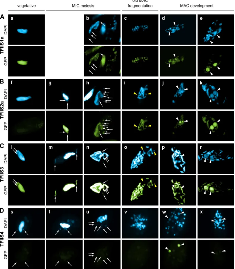
During sexual processes, coding and non-coding transcription take place in the three different types of nuclei that coexist in the cytoplasm ofP.tetraurelia. Gene transcription progressively switches from the old MAC fragments to the developing new MACs [38], which undergo genome rearrangements. The MIC is transcribed specifically during meiosis to give rise to scnRNAs [15]. Constitutive generalized non-coding transcription takes place in the maternal MAC to produce the protective transcripts that antagonize scnRNAs [18]. During MAC devel-opment, short iesRNAs are produced from putative precursor transcripts synthesized in the new developing MAC [17]. Finally, nascent non-coding transcripts produced from the new developing MAC were proposed to serve as substrates for the pairing of MIC-restricted scnRNAs to guide DNA elimination in the developing new MACs [13,18]. In order to gain insight into the role of each TFIIS family in these different nuclear compartments, GFP fusions were constructed for each of the six proteins and expressed under the control of their respective endogenous regulatory regions. The cellular localization of GFP-TFIIS proteins was monitored by injecting each transgene into the macronucleus of vegetative cells and following the GFP fluorescence during vegetative growth and throughout autogamy (Fig 2). All proteins were shown to be nuclear, but they localized to different nuclear compartments.
TFIIS1a is present in the vegetative MAC (Fig 2A). During autogamy the GFP fluorescence progressively shifts from the fragmented old MAC to the developing new MAC (Fig 2B–2E). Localization of its close paralog TFIIS1c is similar, although it seems to disappear more abruptly from the fragmented old MAC (S4C Fig), before it eventually accumulates in the new MACs (S4D and S4E Fig). In conclusion, proteins from the TFIIS1 family shift from the old to the new MAC during autogamy. This localization pattern may reflect a possible role of TFIIS1a and 1c in mRNA synthesis and gene expression in the somatic nucleus.
Protein fusions encoded by the autogamy up-regulated genesTFIIS2aand2baccumulate during meiosis, both in the old MAC and the meiotic MICs (Fig 2G). GFP fluorescence was also detected in all meiotic products (Figs2H,S4G and S4H, respectively). Both TFIIS2 fusions eventually concentrate in the developing new MACs (Figs2J, 2KandS4J, S4K). TFIIS3 follows the same localization pattern with clear presence in the vegetative MAC. The TFIIS2 and TFIIS3 families are present wherever transcription takes place—in the old and new MACs and in meiotic MICs—and may be associated with coding or non-coding transcription. In particu-lar, the presence of TFIIS2 and TFIIS3 in the MICs during meiosis suggests their possible involvement in the non-coding transcription that gives rise to scnRNAs.
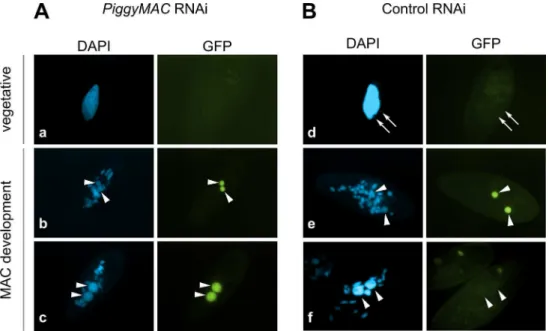
Consistent with the expression pattern ofTFIIS4, no fluorescence was observed in vegetative cells injected with aGFP-TFIIS4fusion transgene (Fig 2Ds). During autogamy, trace amounts of the protein were detected in the old MAC during MIC meiosis (Fig 2T), then GFP fluores-cence accumulated in the new MACs at early stages of MAC development (panel w), and diminished at later stages (panel x). No staining of the MICs was detected at the crescent stage and after meiotic divisions (panel t and u). This very peculiar localization may reflect a specific transcription-related function connected with the DNA elimination process that starts by the time TFIIS4 appears in the new MAC. In this respect, we noted that the presence of
cells, in which DNA elimination is inhibited [12]. Under these conditions, GFP-TFIIS4 per-sisted in the new MACs until the latest stages of autogamy (Fig 3). Taken together, these obser-vations suggest that GFP-TFIIS4 accumulates in the new MACs before IES excision, and disappears once IESs have been removed.
Expression of
TFIIS4
is essential for the successful completion of
autogamy
ParameciumTFIIS factors exhibit different expression and localization patterns during vegeta-tive growth and sexual processes. To check if any of them have an essential function during autogamy, we silenced eachTFIISgene, by feedingParameciumcultures on dsRNA-overpro-ducing bacteria to trigger RNA interference [39]. ForTFIIS2aand2b, we also performed dou-ble silencing experiments by mixing induced bacteria designed to silence individual genes. The effect of each RNAi was first examined for ~8 vegetative divisions by monitoring cell division rate and general morphology, as described in [40]. None of the silencing experiments gave an obvious phenotype during this period of vegetative growth. In a second step, autogamy was induced by starvation, and the survival of sexual progeny was checked following transfer of individual autogamous cells to standard medium (Table 1). Inactivation of individual genes from families 1, 2 or 3 did not produce any visible phenotype. For autogamy up-regulated
TFIIS2aand2bgenes, we did not observe any phenotype when both genes were silenced together. It may indicate that TFIIS2 proteins are not essential or display functional redun-dancy with other TFIISs, for example TFIIS3, which shows a similar localization pattern. It is of course possible that someTFIISgenes were not completely silenced by the RNAi method used in our study and, therefore, that no phenotype was revealed in our screen. We should also note that, in other model systems, a mutation ofTFIISvery often does not give strong pheno-types: in the yeastsS.cerevisiaeandS.pombe, null mutants in the single copyTFIISgene are viable under standard laboratory conditions, but sensitive to the nucleoside analog 6-azauracil [41,30]. In contrast, single RNAi againstParamecium TFIIS4led to strong lethality in post-autogamous progeny, with only 15% normally growing survivors. Most of the remaining sur-viving progeny was sick, grew slowly, failed to divide normally, and finally died after a few divi-sions. We conclude thatTFIIS4shows a clear-cut RNAi phenotype during autogamy and a specific localization of its encoded protein in the developing new MAC. Cytological observa-tion of DAPI-stained cells confirmed that TFIIS4 depleobserva-tion does not impair the differentiaobserva-tion of new MACs, which are formed and amplify their DNA normally (S5 Fig).
TFIIS4 is required for excision of a subset of IESs
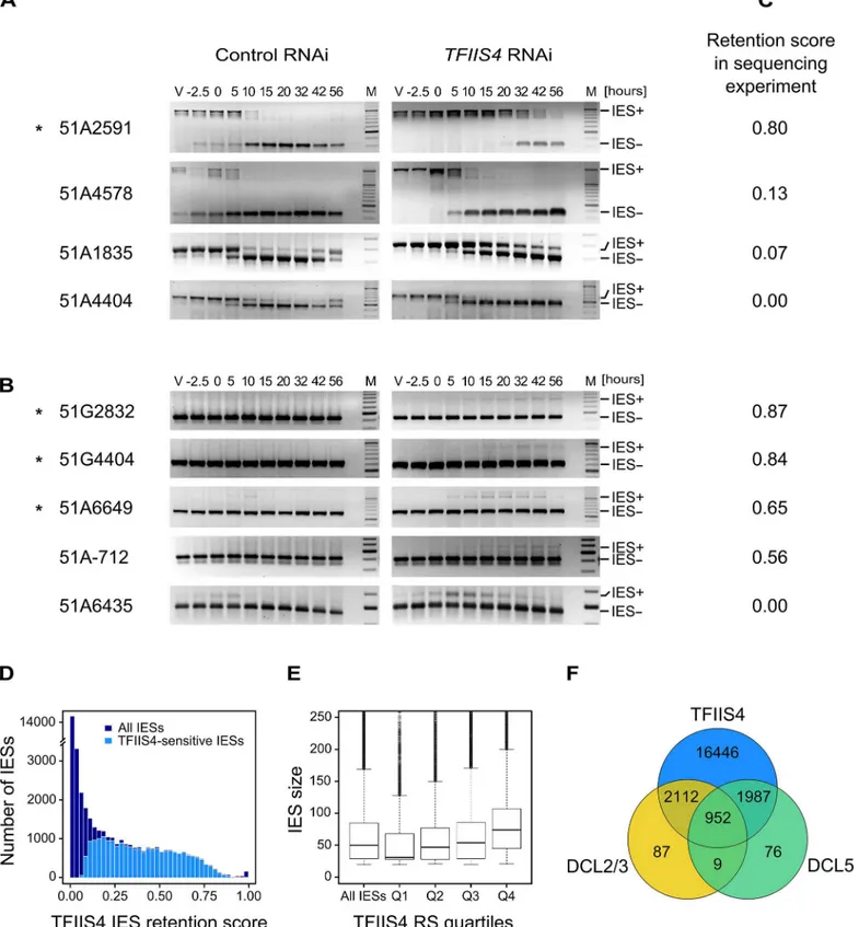
Because half of the genes inP.tetraureliaare interrupted by at least one IES [9], the develop-ment of a functional new MAC depends upon the completion of IES excision. We therefore tested the excision of several known IESs by PCR, using primers located in the flanking MAC sequences upstream and downstream of each particular IES. In this experiment, we used a
vegetative cell is also present in the middle of panel b and on the left of panel h). All other panels show successive stages of autogamy: panels g, m and t– meiotic crescent stage; panels b and n–first meiotic division; panels h and u–cells with 8 haploid nuclei resulting from meiosis II; panels c, i, o and v– fragmentation of old MAC; panels d, j, p, and w–early MAC development; panels e, k, r and x–late MAC development. Note that panel b contains not only one meiotic cell (on the left) but also one vegetative cell (in the middle) and two cells with their fragmented old MAC (at the top and on the right). In all panels, white arrows point at MICs (some were omitted when MICs were not clearly distinguishable by DAPI staining), white arrowheads indicate new MACs. Yellow arrowheads in panels i and o point to division products of the zygotic nucleus. (A) A GFP-TFIIS1a fusion localizes to old, then new MACs. (B) A GFP-TFIIS2a fusion localizes to old MAC during meiosis, then to new MACs and is present in meiotic MICs. (C) As in B for a GFP-TFIIS3 fusion. GFP-TFIIS3 cannot be seen in division products of the zygotic nucleus. (D) A GFP-TFIIS4 fusion is essentially restricted to the new MACs specifically during early MAC development. Very weak GFP signal is visible in the old MAC during meiosis.
strain carrying a somatic deletion of part of surface antigen geneA, in which a region contain-ing three IESs is absent from the maternal MAC [42]. We extracted genomic DNA and total RNA samples during an autogamy time-course of this strain silenced either for a non-essential control gene or forTFIIS4(S6A Fig). The efficiency ofTFIIS4silencing was confirmed by northern blot hybridization of total RNA (S6B Fig), and genomic DNA was used to monitor genome rearrangements at the molecular level. In the control experiment, the use of aΔAstrain allowed us to detectde novoIES excision junctions for this locus (Fig 4A). In theTFIIS4RNAi, we observed a strong delay in excision of IES 51A2591 and very low amounts of excision prod-ucts (IES-) relative to the control RNAi. Excision was also delayed to some extent for IESs 51A4578 and 51A1835, whereas we observed a normal elimination profile for 51A4404. Other
Fig 3. Localization of the GFP-TFIIS4 fusion protein uponPiggyMacRNAi.New developing macronuclei (new MAC) are indicated by white arrowheads, while white arrows point at MICs that are clearly visible only in panel d. Panel a and d show vegetative cells, b and e: early MAC development, c and f: late MAC
development. (A) Cells silenced forPiggyMac. The efficiency ofPiggyMacsilencing was confirmed by the observation of 100% lethality in the sexual progeny. (B) Control experiment, in which the nonessentialICL7 gene was silenced. The silencing ofICL7gene does not interfere with autogamy (seeTable 1) and does not influence the localization of TFIIS4 relative to cells grown in standardK.pneumoniaemedium.
doi:10.1371/journal.pgen.1005383.g003
Table 1. RNAi-screening for essentialTFIISgenes.
Targeted gene TFIIS1a TFIIS1c TFIIS2a TFIIS2b TFIIS2aandTFIIS2b TFIIS3 TFIIS4 ND7 ICL7 none
Progeny % wild type 94 91 94 86 89 90 15 97 97 87
% sick 1 0 1 2 3 2 10 0 0 2
% death 5 9 5 12 8 8 75 3 3 11
Total cells 192 192 192 192 192 192 493 282 210 222
Number of experiments 3 3 3 3 3 3 13 6 7 4
Survival test of post-autogamous cells submitted to RNAi against allTFIISgenes and control non-essential genes—ICL7andND7. The last column“none” corresponds to the control grown in standard non-feedingK.pneumoniaemedium. For each condition, the number of replicate experiments is indicated in the last line. In one replicate experiment, wild-type survivors were systematically tested for MAC regeneration (as previously described [52]) and all turned out to be true postautogamous cells.
IESs located outside the region of theΔAmacronuclear deletion were also tested (Fig 4B). For these IESs, due to the presence of rearranged DNA in the old MAC, we could only monitor IES retention during autogamy. Based on the persistence of the IES+ form at late time-points, exci-sion of IESs 51G2832, 51G4404, 51A6649 and 51A-712 was found to be inhibited inTFIIS4 -silenced cells, while another IES (51A6435) seemed to be eliminated normally. Interestingly, all the known maternally controlled IESs that we tested [43] are affected by silencing of theTFIIS4
gene (indicated by aninFig 4A and 4B). In particular, we confirmed the retention of IES
51G4404 by Southern blot hybridization (Fig 5A). In conclusion, our molecular results indicate that excision of some IESs is inhibited byTFIIS4-silencing to various extents, while other IESs are eliminated normally.
To get a quantitative and genome-wide insight into the dependence of IES excision upon TFIIS4, we performed high-throughput sequencing of DNA extracted from a nuclear prepara-tion enriched for new MACs ofTFIIS4-silenced autogamous cells (obtained from an indepen-dent RNAi experiment), as described in [9]. As a control we used sequencing data for the DNA sample obtained from autogamous cells of the same strain, but with no silencing ([19], see
Materials & Methodsfor details). In both datasets, IES retention scores were determined for each IES by calculating the ratio of IES-containing reads (IES+) over the sum of: (i) the number of reads that map to the IES excision junction (IES- reads), and (ii) the number of IES-contain-ing reads (IES+). Hence a retention score of 0 means complete excision whereas a score of 1 means complete retention of the IES as described in [19]. The results are in good agreement with the above molecular data: IESs shown to be retained by PCR or Southern blot hybridiza-tion exhibit higher retenhybridiza-tion scores in the sequencing experiment (Fig 4C). To ensure that the IES retention observed in the TFIIS4 RNAi sample is indeed due to the silencing of this gene, we performed statistical comparison of IES retention scores between control condition and TFIIS4-RNAi (for details seeMaterials and methods). This statistical analysis revealed that ~21,500 IESs (48%) are sensitive toTFIIS4silencing, with a very wide distribution of IES reten-tion scores (Fig 4D). Among the set of TFIIS4-dependent IESs, we found all five maternally controlled IESs that were identified in previous experiments [43] and all IESs that were shown to be dependent upon the presence of the WG/GW-repeat protein Nowa1/2 [17,44]. Moreover, almost all TFIIS4-sensitive IESs (96%) can be found in a larger set of IESs dependent on the putative histone methyltransferase Ezl1 [19] (Table 2). TFIIS4-dependent IESs are character-ized by higher average retention scores inEZL1-silenced cells relative to IESs that do not depend upon TFIIS4. However, we did not observe a strong correlation of IES retention scores betweenTFIIS4andEZL1silencing experiments (S7A Fig). addition, we noticed a correlation between IES size and IES sensitivity toTFIIS4silencing: IESs with higher retention scores (i.e. strongly dependent upon TFIIS4) tend to be longer (Fig 4E). However, because of a wide distri-bution of the TFIIS4 retention scores for all IES size groups (S7B Fig), the parallel increase of the retention score with increasing IES size is not as obvious as reported forEZL1RNAi [19]. The apparent overlap between the requirements for TFIIS4, Nowa1/2 and Ezl1 for IES excision
corresponding to each IES- form are amplified mostly from the fragments of the old MAC. Oligonucleotide sequences are listed inS2 Table. (C) IES retention scores calculated from the genome-wide sequencing of DNA extracted from purified nuclei of cells silenced forTFIIS4during an independent RNAi
experiment (87% lethality in post-autogamous progeny). (D) Superimposed histogram of TFIIS4 retention scores for all IESs (dark blue) and for IESs that are significantly retained in TFIIS4-depleted cells (light blue). Around 25,000 IESs are not significantly affected by the inactivation ofTFIIS4and a large fraction of IESs exhibits a retention score equal to 0. For TFIIS4-dependent IESs, retention scores are almost uniformly distributed between 0.1 and 0.7. (E) The graph shows a positive correlation between IES size and retention score inTFIIS4RNAi. The box plot displays the IES size distribution for all IESs and for each of TFIIS4retention score (RS) quartiles. The median retention score (horizontal line inside the box) and the first (top of box) and third (bottom of box) quartiles are shown. Range of RS for particular quartiles are as follows: Q1: [0–0.01[; Q2: [0.01–0.12[; Q3: [0.12–0.39[; Q4: [0.39–1.00]. The medians are significantly different between all the groups (p<2e-40). (F) Venn diagram of significantly retained IESs afterTFIIS4,DCL5orDCL2/3silencing. Almost all IESs that are dependent upon Dcl2/3 or Dcl5 for their excision are also dependent upon TFIIS4.
suggests that TFIIS4 may be implicated in the control of IES excisionviathe same RNA-related pathway as the one in which nucleosomes are marked by methylation.
Involvement of TFIIS4 in imprecise DNA elimination
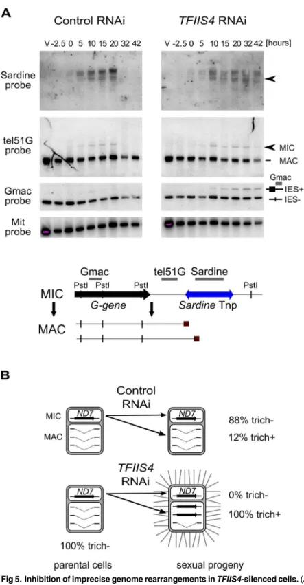
In addition to the precise excision of single-copy IESs, genome rearrangements include the elimination of repeated DNA sequences such as transposable elements. Two families ofTc1/
mariner-related transposons were identified in the part of MIC-specific sequences that are removed imprecisely during MAC development:SardineandThon[9]. We first used Southern blot hybridization with a specific probe to monitor the transient amplification and the elimina-tion ofSardinetransposons from the developing MAC during autogamy (Fig 5A). InTFIIS4 -silenced cells, we observed an accumulation of the signal corresponding toSardine transpo-sons, indicating at least a partial block of transposon elimination. One copy of theSardineis located downstream of a telomere addition site and its elimination is associated with chromo-some fragmentation. Using a macronuclear subtelomeric probe (tel51G, see [12]), we con-firmed that retention of this copy of theSardineuponTFIIS4silencing correlates with the persistence of non-fragmented forms of the chromosome (Fig 5A). We also used our genome-wide sequencing data to estimate the fraction of repeated sequences that require TFIIS4 for elimination. Since the germline reference genome is not available forP.tetraurelia, we used an unrearranged version of the genome, previously assembled from the sequencing experiment following PGM depletion, as our reference [9]. We mapped the sequencing reads from the con-trol sequencing (wild type genome) as well as PGM- and TFIIS4-knockdowns on this unrear-ranged reference genome and measured the complexity of the regions present in the PGM and TFIIS4 samples but not in the control sample (see legend ofS3 Tablefor the entire procedure). We found that 64% of the MIC-restricted sequences need TFIIS4 for their elimination (S3 Table). We conclude, therefore, that TFIIS4 is necessary for removal of some, but not all repeti-tive sequences. A true micronuclear assembly along with its annotation would be required for further analysis of the role of TFIIS4 in imprecise DNA elimination during MAC development.
MAC version obtained by telomere addition downstream of theGgene (lower band); Gmac hybridizes with fragments containing (upper band) or not (lower band) IES 51G4404. The same blot was hybridized with a mitochondrial DNA probe (Mit probe) as a loading control. (B) Phenotypic test of the ability to discharge trichocysts in the sexual progeny of cells carrying a macronuclear deletion of theND7gene, followingICL7or TFIIS4gene silencing.TFIIS4silencing restores a wild-type trich+ phenotype, most probably due to lack of inheritance of the macronuclear deletion.
doi:10.1371/journal.pgen.1005383.g005
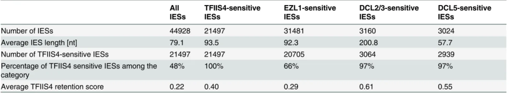
Table 2. Global analysis of genome rearrangements inTFIIS4silencing—comparison withEZL1,DCL2/3andDCL5.
All IESs
TFIIS4-sensitive IESs
EZL1-sensitive IESs
DCL2/3-sensitive IESs
DCL5-sensitive IESs
Number of IESs 44928 21497 31481 3160 3024
Average IES length [nt] 79.1 93.5 92.3 200.8 57.7
Number of TFIIS4-sensitive IESs 21497 21497 20705 3064 2939
Percentage of TFIIS4 sensitive IESs among the category
48% 100% 66% 97% 97%
Average TFIIS4 retention score 0.22 0.40 0.29 0.61 0.55
IESs retained in different samples (in column) are described according to their size and their presence in the TFIIS4-dependent IES set. Almost all IESs retained in other RNAi experiments are also retained inTFIIS4silencing, with high retention score. Apart from Dcl5-dependent IESs, IESs retained followingTFIIS4RNAi are larger (on average) than the overall set.
Imprecise DNA elimination is also involved in the maternal inheritance of somatic dele-tions, as was demonstrated for the macronuclear deletion of theND7gene [45]. TheND7gene encodes a trichocyst discharge protein that is non-essential during autogamy. Its micronuclear version harbors one TFIIS4-independent IES. We tested the inheritance of a macronuclear
ND7deletion during autogamy ofTFIIS4-silenced cells. In a control RNAi we observed that 88% of sexual progeny (35 cells out of 40) retained the mutant phenotype, which is detectable only when all copies of theND7gene are deleted from the new MAC (Fig 5B). Following
TFIIS4silencing, all post-autogamous progeny (30 cells out of 30) switched back to a wild-type phenotype, indicating that inheritance of theΔND7macronuclear deletion is strongly inhib-ited. Although it does not allow precise quantification, this experiment indicates that TFIIS4 is involved in the maternal inheritance of imprecise somatic deletions.
TFIIS4 is involved in IES transcription in the new MAC
According to the localization of a GFP fusion, TFIIS4 appears in the new MAC at an early developmental stage, which possibly coincides with the activation of global transcription in the new MAC and may precede the start of IES excision [38,46]. Given these observations, we con-sidered two possibilities: TFIIS4 is required for the synthesis of non-coding IES transcripts prior to IES excision, or for the start of coding transcription in the new MAC.
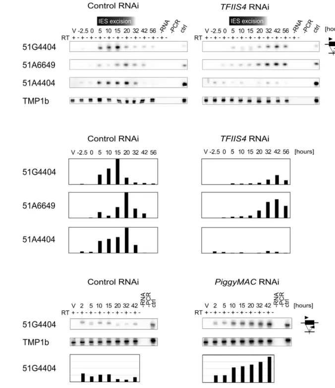
We first examined whether TFIIS4 plays any role in the production of IES transcripts in the developing new MAC. Indeed, at early stages of MAC development, IES sequences are still present in the yet non-rearranged genomic DNA and a large fraction of genes, therefore, can-not produce functional mRNAs. IES transcripts were detected as soon as the new MACs were observed for one particular IES, 51G4404 [18], suggesting that they likely originate from the developing new MAC before IES excision. However, because cells at different autogamy stages coexist at each time-point, the exact origin of IES transcripts–and their putative cellular func-tion—has remained unclear. We hypothesized that, during this period of time, non-coding IES-containing (IES+) transcripts may be produced in a TFIIS4-dependent manner. To test this hypothesis, we performed RT-PCR experiments for three IESs: the maternally controlled IESs 51G4404 and 51A6649, which belong to the set of TFIIS4-dependent IESs, and the non-maternally controlled IES 51A4404, which does not depend upon TFIIS4 for excision. We used total RNA samples isolated during the autogamy time-course experiments described above (Figs4,5andS6), in whichTFIIS4or a control gene were silenced.
In the control RNAi, IES+ transcripts were detected for all three IESs starting from T5 until T20-T32 (Fig 6A and 6B), which coincides with the early stages of MAC development, when IES excision takes place (seeFig 4A). UponTFIIS4silencing, practically no transcripts were detected before T20 during autogamy and only very delayed transcription was observed for IESs 51G4404 and 51A6649 starting from T32 until T56 (Fig 6A and 6B). For IES 51A4404, transcripts were hardly detectable at any autogamy time-point. We conclude, therefore, that the synthesis of IES+ transcripts during IES excision is strongly repressed in TFIIS4-depleted cells for all tested IESs. Interestingly, excision of IESs 51G4404 and 51A6649 is strongly inhib-ited byTFIIS4-silencing (seeFig 4B) and, as a consequence, these IESs are amplified together with MAC-destined DNA during autogamy. The detection of higher amounts of their corre-sponding transcripts at late time-points may result from the retention of these IESs in the genome of the new MAC, when somatic mRNA transcription eventually starts in this nucleus. Alternatively, it may also be explained by the fact that RNAi-mediated silencing ofTFIIS4
Fig 6. Detection of IES-containing (IES+) transcripts.(A) RT-PCR and Southern blot detection of IES-containing transcripts (IES+) in a control culture (cells silenced forICL7gene expression) and inTFIIS4-silenced cells. Autogamy stages are marked as inS6 Fig: V–vegetative cells, -2.5–cells during meiosis, 0 to 56–autogamy stages in hours. Time-window when IES excision take place based on PCR shown inFig 4is indicated. PCR primers were located within each tested IESs: 51G4404, 51A6649 and 51A4404. The TMP1b panel shows the RT-PCR signal obtained for the constitutively expressed gene encoding trichocyst matrix protein TMP1b. (B) Histograms showing the normalization of IES+ signals shown in (A) with TMP1b mRNA. (C) Detection of IES-containing transcripts (IES+) with PCR primers located within IES 51G4404 in a control experiment, in which theND7gene was silenced, and in PiggyMac-silenced cells. SeeS8 Fig, panel B for details about autogamy stages.
the possibility that probably all IESs—maternally or non-maternally controlled, located in dif-ferent regions of the genome—are transcribed during genome rearrangements in a TFIIS4-de-pendent manner. However, all IESs do not require the presence of TFIIS4 to be excised. Based on the study of a GFP-TFIIS4 fusion, TFIIS4 shows a specific, but transient, localization in the new MAC. IES+ transcripts are detected as a peak during macronuclear development. This similar timing and the specific localization of TFIIS4 strongly suggest that the IES+ transcripts that are detected in our RT-PCR experiments mostly originate from the new MAC. To verify this hypothesis, we monitored IES transcription in cells silenced forPiggyMacexpression, in which all IESs are retained in the developing MAC. Consistent with the persistence of a GFP-TFIIS4 fusion in the new MACs ofPGM-silenced cells until late stages of autogamy (Fig 3), we observed an accumulation of IES+ transcripts relative to a control silencing (Fig 6C). Taken together these data indicate that TFIIS4-dependent IES transcripts are produced in the new developing MAC before IES excision.
No significant role of TFIIS4 in the synthesis of other developmental
transcripts
Two other types of regulatory ncRNAs were previously reported to participate in the control of genome rearrangements: protective maternal MAC transcripts, which are an RNA copy of the rearranged somatic genome, and the deletion-inducing scnRNAs, which are produced from the non-rearranged germline genome during MIC meiosis [15,18]. We were able to exclude any role of TFIIS4 in the biosynthesis of either type of ncRNA. Indeed, similar levels of consti-tutive maternal MAC transcripts were detected by RT-PCR in a control RNAi and in aTFIIS4
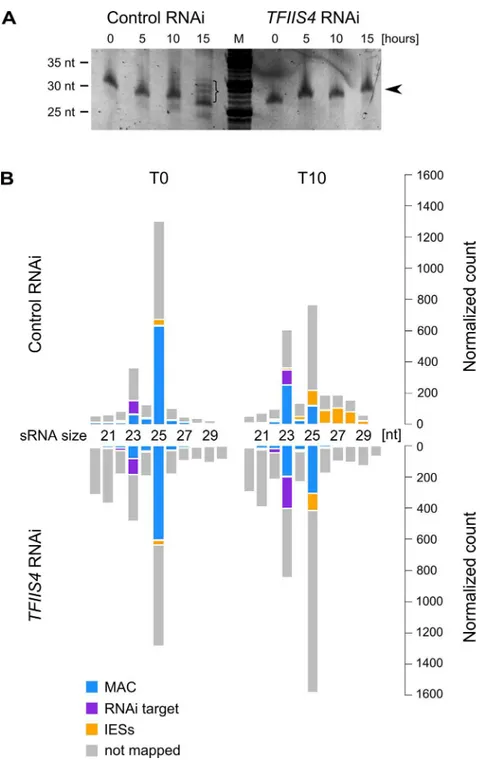
RNAi (S8A Fig). We did not detect any difference either in the global amounts of 25-nt scnRNAs between the two conditions, as revealed by SYBR Gold-staining of polyacrylamide gels (Fig 7A). These data are fully consistent with the absence of TFIIS4 from the MICs, and with a role of TFIIS4 downstream the synthesis of maternal MAC transcripts and scnRNAs. As
Fig 7. Analysis of sRNA populations inTFIIS4-silenced cells.(A) Total RNA samples corresponding to the T0, T5, T10 and T15 time-points from the above experiment were run on a denaturing 15%
polyacrylamide-urea gel. After electrophoresis the gel was stained with SYBR Gold (Invitrogen). M: DNA Low Molecular Weight Marker (USB). Arrowhead points to the ~25 nt signal that was shown to correspond to the fraction of scnRNAs [15]. In the control, at the T15 time-point, additional bands corresponding to 26–30 nt iesRNAs are present (indicated by a bracket). InTFIIS4-silenced samples iesRNAs can clearly not be seen. (B) Small RNA libraries corresponding to the T0 and T10 time-points from the above experiment were sequenced and mapped to the reference genomes (P.tetraureliaMAC reference genome and MAC+IES reference genome). The top panel corresponds to a control culture (cells silenced forICL7gene expression), while results forTFIIS4-silencing are shown below. Histograms show normalized number of sRNA reads that match to: the target silencing regions (ICL7orTFIIS4gene, respectively)–in purple; the rest of MAC genome–in blue; all annotated IESs–in yellow; all other not mapped sRNA–in gray.
S8C) One explanation may be that TFIIS4-dependent IES transcription in the new MAC vides precursors for Dcl5-dependent iesRNA synthesis. Alternatively, iesRNAs may be pro-duced from excised IESs and their production would be inhibited due to a block in excision of TFIIS4-dependent IESs. It is important to note that the disappearance of iesRNAs cannot be the sole reason for defective IES excision uponTFIIS4-silencing, since we observe much stron-ger phenotype inTFIIS4-RNAi than inDCL5-RNAi–both in the IES retention and the cell lethality.
Finally, we investigated the possible role of TFIIS4 in the transcription of protein-coding genes known to be involved in IES excision and focused onNOWA1/2andPiggyMac. Previous studies using GFP fusion transgenes introduced in the old MAC indicated that expression of
NOWA1andPGMoriginates, at least in part, from the old MAC [12,44]. Therefore, we did not expect that depletion of TFIIS4, which localizes preferentially in the new developing MAC dur-ing autogamy, could lead to a strong effect onNOWA1/2andPGMexpression. Indeed, we observed by northern blot hybridization that aTFIIS4RNAi does not cause any dramatic change in the level ofNOWA1/2mRNA relative to a control RNAi (S9A Fig). We obtained the same result forPiggyMacmRNA (S9A Fig). At the protein level, we confirmed that a Pgm-GFP fusion is detected similarly to the control in TFIIS4-depleted cells (S10 Fig). Taken together, there is no reason to believe that the defect in IES excision observed inTFIIS4RNAi is due to depletion in Nowa1/2 or PiggyMac. The role of TFIIS4 in coding transcription was studied at the genome-wide level by performing a single microarray hybridization experiment using RNA samples extracted during vegetative growth and at five points during the autogamy time-course shown inS6 Fig. We focused on the ~5000 genes showing the most significant changes in their expression during autogamy under standard conditions [28]. We did not notice impor-tant global changes in the variations of mean transcript levels between the control andTFIIS4
RNAi experiments (S9B Fig), especially for the early activated (maximum induction at the T-2.5 and T0 time-points), the late autogamy genes (induced at T10 and T20) and for those genes from the intermediate induction cluster that show gradual induction. A group of genes from the intermediate induction cluster exhibited a maximal induction peak at T5 in the control, but seemed to have a delayed pattern of induction in theTFIIS4RNAi, reaching a maximal mRNA level only at T20. At this stage, however, closer examination of microarray expression patterns for individual genes is not possible since variations of the signals calculated from a single hybridization experiment are not statistically significant. Additional replicate experiments will be required to strengthen the statistical significance of our microarray data and to identify a potential set of genes with altered expression inTFIIS4-RNAi.
Discussion
TFIIS4 couples transcription and DNA elimination in
P
.
tetraurelia
The functional analysis of TFIIS4 inP.tetraureliaestablished the role of a TFIIS homolog in the control of developmentally programmed DNA elimination. Our data indicate that TFIIS4 influences all kinds of genome rearrangements: it stimulates the precise excision of a large group of IESs, favors the elimination of multicopy transposons and the inheritance of macro-nuclear deletions. We show here that all three tested IESs are transcribed in a TFIIS4-depen-dent manner by the time DNA elimination takes place in the new MAC, and that IES
the dependence upon TFIIS4 seems to exhibit an IES size bias: excision of less than 30% of the shortest IESs (26–32 bp) requires the presence of TFIIS4 whereas up to ~60% of the IESs larger than 100 bp depend upon TFIIS4 for their excision. Our study, therefore, provides the first example of the participation of a TFIIS homolog in both the control of non-coding transcrip-tion and the regulatranscrip-tion of programmed genome rearrangements.
Using genome-wide microarrays, we obtained no convincing evidence that TFIIS4 is involved in the induction of mRNA synthesis during sexual processes, even though we cannot completely exclude this possibility. Northern blot hybridization performed for two essential IES excision genes—PGMandNOWA1/2–confirmed that normal expression patterns are observed inTFIIS4-silenced cells. In particular,PGMmRNA displayed a wild-type“ intermedi-ate induction profile”, with a maximum induction peak around the time when IES excision starts (T5, seeFig 4A and 4B) and a decrease at later time-points (S9A Fig). This observation stands in contrast to previous work, which suggested that inhibition of genome rearrangements may cause dramatic mRNA accumulation for genes from the“intermediate induction cluster”
[19,47]. In particular,PGMtranscripts were found to accumulate in cells depleted for the essential Pgm partner Ku80c, suggesting the existence of a transcriptional feedback loop depending upon the completion of genome rearrangements. In contrast, no accumulation of
PGMtranscripts was observed at late autogamy time-points in theTFIIS4RNAi. This differ-ence may indicate that none of the TFIIS4-dependent IESs is involved in the control of this putative transcriptional feedback loop.
TFIIS4-dependent zygotic transcription and the model for
RNA-mediated regulation of programmed genome rearrangements
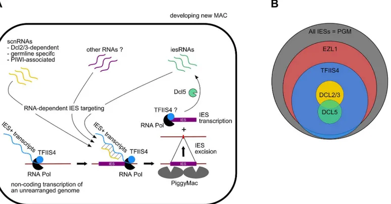
The present study confirms the existence of IES-containing transcripts inP.tetraureliaand provides the first evidence that IES transcripts originate from the developing new MAC. Our work shows thatTFIIS4mRNA starts to accumulate at early time-points of autogamy–as soon as meiotic cells can be detected. Yet, we note that the GFP fluorescence is still very weak at the stage and increases only in the new MACs. Two alternative explanations can be proposed for this delay: either TFIIS4 protein production is delayed relative to mRNA synthesis or protein is expressed but is diluted in the entire cell and cannot be detected. We cannot therefore definitely exclude the possibility that TFIIS4 plays a role in the processes that precede new MAC develop-ment, especially because some amounts of the GFP-TFIIS4 localize to the old MAC. For the moment, however, we found no evidence for its involvement in scnRNA synthesis or selection. We showed nevertheless that TFIIS4 is involved in synthesis of zygotic IES+ transcripts. We propose therefore that TFIIS4-dependent nascent zygotic transcripts are pairing substrates for IES-specific scnRNAs in the new MAC [13]. In the current version of the genome scanning model (Fig 8A), the 25-nt scnRNAs produced in meiotic MICs by the Dicer-like proteins Dcl2/3 [15,17] are transferred from the old MAC, in which they have become enriched for germline-specific sequences, to the developing new MAC, in which they are thought to pair to homologous nascent transcripts. According to the model, the pairing of scnRNAs to zygotic nascent transcripts leads to loading of chromatin modifications and, eventually, allows the tar-geting of DNA elimination. However, recent discoveries [17,19] indicate that the genome scan-ning model with a central role of scnRNAs does not explain the entire complexity of genome rearrangements inParamecium. High-throughput analysis of IES retention after RNAi knock-down of particular genes allows us, nevertheless, to draw some conclusions regarding a possible interplay of TFIIS4 with other factors.
nearly all IESs requiring scnRNAs (~3,200 Dcl2/3-dependent IESs) or iesRNAs (~3,000 Dcl5-dependent IESs) also depend upon TFIIS4 for their excision (Figs4Fand8B), and the observation that these two partially overlapping IES subsets are among the most strongly dependent upon TFIIS4 (Table 2andS11A and S11B Fig), is consistent with a model in which both types of sRNAs interact with TFIIS4-dependent IES+ nascent transcripts. However, the current genome scanning model, including scnRNAs and iesRNAs, explains the control of excision for only around 12% of all IESs, which is a few times less than those anticipated to be maternally controlled [43]. Thus, we cannot exclude that a novel class of sRNAs of yet
unknown origin, which would be independent on Dicer-like proteins Dcl2/3 and Dcl5 (as pro-posed in [19]), interacts with TFIIS4-dependent zygotic transcripts to promote IES excision. Different types of sRNAs synthesized through Dicer-independent pathways were reported in other organisms, including the germline-specific piRNAs in metazoans (reviewed in [49]) or the disiRNAs of the filamentous fungusNeurospora crassa, all of which are associated with DNA methylation [50].
Practically all TFIIS4-dependent IESs are also dependent on Ezl1, a histone-methyl transfer-ase required for excision of two-thirds of IES sequences [19]. Both proteins have the strongest impact on the longest IESs, which have a higher probability of being covered by a nucleosome. It may be a sign of some functional link between TFIIS4-dependent transcription and the mechanisms necessary for recognition of most (but not all) regions that need to be marked by H3K9/K27 trimethylation for their elimination. An alternative hypothesis would be that TFIIS4-dependent IES transcription plays another role, which would be unrelated to the pair-ing of sRNAs, for instance by openpair-ing up chromatin and allowpair-ing access of the Pgm complex to its target sites. This may be achieved by a similar mechanism to that reported for class
Fig 8. Proposed role of TFIIS4 in RNA-dependent DNA elimination.(A) Possible role of TFIIS4 in the new developing MAC. Description in the text. (B) Summary of the impact ofTFIIS4,EZL1,DCL2/3orDCL5silencing of on IES excision. The area of each circle is proportional to the fraction of IESs that significantly depend on each factor.
switch recombination (CSR), through which the constant regions of antibodies are exchanged (reviewed in [51]): in this system, a nascent RNA synthesized at the recombination locus forms a hybrid RNA-DNA R-loop structure that displaces the complementary DNA strand, provid-ing a recombination substrate. Non-codprovid-ing transcription was also shown to have an impact on V(D)J recombination, which also plays a role in generating the diversity of the immune response. In this process, transcription is believed to affect recombination by altering chroma-tin structure (for review see [52]).
Finally, a large fraction of IESs (52%) do not require TFIIS4-dependent transcription to be excised properly. Most of these IESs are among the shortest ones, which appeared in the genome a relatively long time ago (see [9]). We suggest that these IESs have evolved to become independent of their own transcription for efficient excision–they give rise to scnRNA [17] and probably also to IES+ transcripts from the new MAC, but the presence of these RNAs is not necessary for their elimination. We may therefore consider a general model, in which foreign DNA sequences inserted recently in the germline genome, like transposable elements, are rec-ognized and eliminated from the somatic genome using a specialized RNAi mechanism requir-ing sRNAs and TFIIS4-dependent zygotic nascent transcripts (as proposed in [13]). The physical elimination of foreign DNA depends on the PiggyMac domesticated transposase [12] and on components of the non-homologous end-joining (NHEJ) repair pathway [47,53] (dis-cussed in [10,11]). Over time, these sequences would have evolved to yield IESs, by shortening in size and eventually becoming independent from the sRNA machinery. How current IESs are recognized and targeted for excision still remains an open issue, but deciphering the underlying molecular mechanisms will certainly provide a better understanding of other developmentally programmed chromatin diminution systems that were reported in numerous eukaryotes [54].
A novel function in non-coding transcription for a specialized TFIIS factor
TFIIS is conserved in most eukaryotes and functional homologs are also found inArchaea
(GreA and GreB proteins) and in some viral genomes [55]. In yeast, plants and worm, TFIIS factors are encoded by a single gene, while two genes can be found in the genomes of Trypano-somaandDrosophila. Three genes are present in vertebrate genomes [56], which may be explained by whole-genome duplications that played an important role in vertebrate evolution [57]: retention of these three copies was proposed to be correlated with high organism com-plexity. During evolution, four TFIIS families have emerged inParamecium, independently from the multigeneTFIISfamilies found in other eukaryotic species [58], including the ciliate
Oxytricha trifallax(S2 Fig). AllP.tetraureliaproteins contain the three canonical TFIIS domains and represent the most divergent group of TFIIS factors encoded by a single genome. This study shows, for the first time in a unicellular organism, that TFIIS factors may be special-ized with regard to their expression patterns and localization, even though future in-depth studies will be required to unravel their exact respective functions. TFIIS1 and TFIIS3 seem to be linked to expression of the somatic genome, while TFIIS2 and TFIIS3 might be required for general transcription of the germline genome during meiosis. Finally, TFIIS4 is specifically expressed during sexual processes and is responsible for zygotic non-coding transcription, therefore playing an essential role during MAC development and assembly of the new somatic genome.
[61] and of the function of TCEA3, which is highly enriched in mouse embryonic stem cells and regulates their pluripotent differentiation [62]. Thus,Parameciumprovides a promising system for the functional analysis of TFIIS function during development. Execution of develop-mental programs in eukaryotes involves several ncRNAs and involves epigenetic programming of the genome (reviewed in [63]). Our work on TFIIS4 inParameciumdemonstrates, for the first time, a role of a TFIIS homolog as an essential factor for the production of regulatory non-coding transcripts, and establishes a novel connection between non-non-coding transcription and the control of genome plasticity.
Materials and Methods
Paramecium
strains, cultivation and autogamy
All experiments were carried out withParamecium tetraureliastrain 51new [64]. In large-scale silencing experiments, a 51ΔAΔND7strain carrying an injection-induced macronuclear dele-tion of the surface antigenAgene [42] and a silencing-induced macronuclear deletion of the
ND7gene [45] was used. In microinjection experiments, strain 51nd7-1was used as described previously [47].
Parameciumcell cultivation and autogamy were carried out as described previously at 27°C [65]. For standard cultivation, cells were grown in a Wheat Grass Powder medium (WGP, Pines International, Lawrence, KS, USA) inoculated the day before withKlebsiella pneumoniae, and supplemented just before use with 0.8μg/mlβ-sitosterol (Merck) [66].
DNA and RNA extraction
Genomic DNA and total RNA were extracted from ~400,000Parameciumcells during vegeta-tive growth and at different time points of the autogamy time-course, as described in [12].
Northern and Southern blot hybridization
For northern blots, 20μg of denatured total RNA were loaded on a 1% agarose gel.
Electropho-resis, blotting and hybridization were performed as described previously [40], or using the NorthernMax-Gly Kit (Ambion) as recommended by the supplier. Southern blot hybridization was performed as in [12]. Electrophoresis ofPstI-digested genomic DNA (2μg per lane) or
RT-PCR products were carried out in 0.8%–2% agarose gels (Resolva GQT–for smaller prod-ucts, Basica LE GQT for larger fragments (Prona)) in 0.5x TBE buffer, and transferred to Hybond N+ or Hybond XL membranes (GE Healthcare) in 0.4 N NaOH. Double-stranded probes were labeled by random priming with [α-32P] dATP (3000 Ci/mmol, Hartmann Ana-lytic). Oligonucleotide probes were labeled with [γ-32P] ATP (3000 Ci/mmol, Hartmann Ana-lytic) using T4 polynucleotide kinase. Southern blots were hybridized at 60°C and washed in 0.2x SSC and 0.1% SDS at 60°C prior to image plate exposure. Northern blots were hybridized at 42°C in Ultrahyb buffer (Ambion) and washed as recommended by the supplier. All radioac-tive signals were quantified using ImageJ. Hybridization probes are described inS1andS2
Tables.
Construction of GFP fusions
usage [44]. Then, the putative promoter ofTFIIS4(bp 151832..151714) was inserted between theSalI andXbaI sites of the plasmid. The other N-terminal GFP fusions (pGFP-TFIIS1a, pGFP-TFIIS1c, pGFP-TFIIS2a, pGFP-TFIIS2b, pGFP-TFIIS3) were obtained by an overlap-ping PCR method [67]. In general, each construct contained the putative promoter, coding sequence and putative terminator region of the appropriateTFIISgene (exact coordinates of cloned genomic fragments are given inS1 Table). For each construct, DNA fragments repre-senting the endogenous promoter, the EGFP coding sequence and theTFIIScoding sequence with its putative terminator region were amplified separately. Each PCR product was designed to contain a 50-bp overlap with its adjacent fragment(s), so that all fragments could hybridize in the proper order to assemble the desired sequence. Annealed fragments were amplified with external primers containing overhangs with restriction sites, and then cloned between theXhoI andPstI sites of the pCRscript vector (Invitrogen). Platinum Taq polymerase (Invitrogen) was used in all PCR reactions and at each step PCR products were purified using the Invisorb Frag-ment CleanUp kit (Stratec). All constructs were checked by Sanger sequencing (IBB, PAS).
Injection of GFP fusion transgenes
Before microinjection, all plasmids were purified using a QIAfilter Plasmid Maxi Kit (Qiagen) and linearized within the vector sequence. They were filtered through a 0.22μm Ultrafree-MC
filter (Millipore), precipitated with ethanol and dissolved in filtered water to a final concentra-tion of 5μg/μL. Linearized plasmids carrying GFP fusion transgenes were microinjected into
the MAC of vegetative 51nd7-1cells, as described previously [44]. Briefly,Parameciumcells were microinjected in Dryl solution containing 0.2% bovine serum albumin, under a paraffin oil film, while they were visualized with a phase-contrast inverted microscope. All observations were performed using a Nikon Eclipse E800 or a Zeiss Axioplan 2 epifluorescence microscope.
Gene inactivation by RNAi
All RNAi plasmids are derivatives of vector L4440 [68] and carry a fragment of the target gene inserted between two convergent T7 promoters (inserts used in this study are listed inS1 Table). Additionally, thePiggyMacRNAi plasmid PGM-1 [12] was used. Control RNAi plas-mids were: p0ND7c [45] and pICL7a [69], which target the non-essentialND7andICL7a
genes, respectively. In all feeding experiments, the efficiency ofND7silencing was confirmed by the lack of trichocyst discharge in the presence of picric acid.ICL7silencing was checked by transferring cells in AED 0.5% buffer containing Ca2+, and observation of a failure in cell short-ening and backward swimming behavior [70]. Silencing media were prepared basically as described in [71] and [40], by inoculating precultures of the appropriate bacterial strains into WGP medium containing 0.1 mg/mL ampicillin. Following 6–8 hrs of shaking at 37°C, bacte-rial cultures were diluted six-fold into the same medium containing 0.4 mM IPTG to induce dsRNA synthesis. After overnight induction at 37°C, all silencing media were supplemented with 0.8μg/mLβ-sitosterol (Merck) before use.
Microarray analysis of gene expression
Cell lysis and purification of new developing macronuclei
As described in [9], a fraction enriched in late new developing macronuclei was obtained through different centrifugation steps from 3.8 L of autogamous cells (at a concentration of ~2000 cells/ml) submitted toTFIIS4RNAi in the independent experiment from the one used for PCR assays. After dialysis, 4.4μg of DNA was obtained. Southern-blot detection of the
retention of IES 51G4404 was performed by32P-labelling of the Gmac probe [12], which corre-sponds to MAC sequences just downstream of IES 51G4404 within the surface antigenG51
gene. The contamination with bacterial DNA was estimated by hybridization of the same blot with a32P-labelled 23S rDNA probe fromK.pneumoniae[9].
Genome-wide analysis of IES retention
The DNA obtained from the nuclear fraction enriched for late developing MACs was submit-ted to paired-end sequencing using an Illumina HiScan SQ next-generation sequencer. The average shotgun library fragment length was 250 bp and the read length equaled 101 nt (Gen-Bank Sequence Read Archive SRP047508). After quality filtering and removal of adapters, Illu-mina reads were processed as described elsewhere [48], and aligned to the reference genomes (P.tetraureliaMAC reference genome and MAC+IES reference genome) using BWA [72] with default parameters. Alignments were indexed with Samtools [73].
For each sample, IES retention scores (RS) were determined as described in [19]. For each IES that was previously identified in [9], the number of reads that contain the IES sequence (symbolized IES+) and the number of reads that contain only the macronuclear IES excision junction consisting of a TA dinucleotide (IES-) were determined. Only reads with unambigu-ous alignments were counted. Each read was counted only once to avoid over-counting owing to paralogous matches. Reads were only counted at IES ends, to avoid length biases resulting from IES length variation. The fraction of IES+ reads/(IES+ and IES-) reads gives the RS.
Then, we compared the RS of a given IES to the control RS observed for the control DNA sequencing (as described in [19]) to make sure that the observed retention can be attributed to
TFIIS4silencing. First we calculated the confidence interval (alpha = 0.95) of the control RS value, using the Pearson-Klopper exact method as implemented by the R binom package ver-sion 1.0–5 [74]. Then we tested for higher retention in the experiment, thanks to a frequency comparison test (based on a binomial law of probability) between the experimental RS and the upper bound of the confidence interval in the control. Resulting p-values were adjusted for multiple testing using the Benjamini &Hochberg method [75]. IESs for which the frequency comparison test gives an adjusted p-value lower than 0.05 are considered significantly retained in the sample.
sRNA sequencing
Reference genomes
The following reference genomes [9] were used in the IES analyses and for read mapping: MAC reference (strain 51):
http://paramecium.cgm.cnrs-gif.fr/download/fasta/assemblies/ptetraurelia_mac_51.fa
MAC+IES reference (strain 51):
http://paramecium.cgm.cnrs-gif.fr/download/fasta/assemblies/ptetraurelia_mac_51_with_ies.fa
PGM contigs:
http://paramecium.cgm.cnrs-gif.fr/download/fasta/assemblies/ptetraurelia_PGM_k51_ctg.fa
Macronuclear DNA reads for PiggyMac-depleted cells [9], Ezl1-depleted cells and control DNA-seq [19], Dcl2/3-depleted cells and Dcl5-depleted cells [17] were obtained from the European Nucleotide Archive (Accession number ERA137420, ERA309409) and the GenBank Sequence Read Archive (Accession number SRX387766, SRX387766), respectively.
RT–PCR detection of non-coding RNAs
Total RNA samples were treated with RNase-free DNaseI (Ambion) for 30 min at 37°C, then extracted with acid phenol pH 4.3 (Sigma) and precipitated with ethanol. Fiveμg of RNA was
reversed-transcribed using RevertAid H Minus Reverse Transcriptase (Thermo Scientific) according to the supplier’s instructions, using random hexameric primers (Thermo Scientific). IES-specific PCR primers were designed to amplify fragments of the maternally controlled IESs 51G4404 and 51A6649, as well as the non-maternally controlled IES 51A4404. Conditions of PCR amplification using DreamTaq DNA Polymerase (Thermo Scientific) were adjusted in order not to saturate the amplification reactions, which were subsequently blotted and visual-ized by Southern blot hybridization using specific IES probes. Normalization was performed relative to the cDNA of the constitutively expressedT1bgene, which encodes a trichocyst matrix protein (TMP1b). Oligonucleotide sequences are listed inS2 Table.
Supporting Information
S1 Fig. Alignment and conservation of predicted structural domains forP.tetraurelia
TFIIS proteins.Full protein sequences were aligned using T-Coffee [77] with default parame-ters and corrected manually. The alignment was colored using Boxshade athttp://www.ch. embnet.org/software/BOX_form.html(grey: similar residues; black: identical residues; fraction of aligned residues that must agree for shading: 0.4). Only regions encompassing the conserved TFIIS domains are shown, since the region between domains I and II did not give significant alignment. The structural annotation below the alignment (structural features of the protein represented by filled rectangles) is based on the structure ofS.cerevisiaeDst1p [21,32]. The Zn finger-forming conserved cystein residues and the DE dipeptide are highlighted in green and pink, respectively. The secondary structure prediction forP.tetraureliaTFIIS proteins was run using PSIPRED from the PRALINE package [78]: red and yellow open rectangles indicate the prediction of alpha helices in domains I and II or in the linker region, respectively. Blue open rectangles designate predicted beta-strands in domain III. Similar prediction results were obtained using NPS@ [79]. Abbreviations and accession numbers are as follows: Pt: Parame-cium tetraurelia—accession numbers as in the legend ofFig 1A; Tt:Tetrahymena thermophila
TFIIS—XP_001032085.3; Ot:Oxytricha trifallax22233_0_g55—Contig22233_0_g55(protein), 1015_0_g5—Contig1015_0_g5(protein), 14486_0_g34—Contig14486_0_g34(protein) (OxyDB); Mc:Moneuplotes crassusconN1—AAG00939; Im:Ichthyophthirius multifiliis—
IMG5_116810 (IchDB); Lm:Leishmania majorLm-TFIIS1-1—CAJ04034, Lm-TFIIS2-1—