Rev Odontol UNESP. 2014 Sep.-Oct.; 43(5): 314-318 © 2014 - ISSN 1807-2577 Doi: http://dx.doi.org/10.1590/rou.2014.050
Relationship among local and functional factors
in the development of denture stomatitis in
denture wearers in northern Brazil
Relação entre fatores locais e funcionais no desenvolvimento de estomatite protética
em usuários de dentadura no norte do Brasil
Lurdete Maria Rocha GAUCH
a*, Fabíola SILVEIRA-GOMES
a, Simone Soares PEDROSA
a,
Renata Antunes ESTEVES
a, Silvia Helena MARQUES-DA-SILVA
aaUFPA – Universidade Federal do Pará, Belém, PA, Brasil
Resumo
Objetivo: O objetivo deste estudo foi avaliar a relação entre fatores funcionais e qualitativos no desenvolvimento da estomatite protética (EP) (de acordo com a classificação de Newton) em usuários de dentadura acrílica
residentes no norte do Brasil. Material e método: Um total de 99 pacientes, que usavam dentadura superior de
resina acrílica parcial ou total, foi incluído neste estudo. Os participantes preencheram um formulário de dados epidemiológicos, que incluiu gênero, idade, fatores locais (hábitos de higiene, retirar a dentadura ao dormir, uso de colutório, condição atual da dentadura, idade da dentadura) e fatores funcionais (dimensão vertical de repouso, dimensão vertical de oclusão, oclusão, retenção, estabilidade estática e dinâmica). Para detectar leveduras, amostras foram coletadas da superfície interna da dentadura e da mucosa palatal em contato com esta. Posteriormente, as amostras foram cultivadas em ágar Sabouraud dextrose, observando-se características macro e microscópicas.
Resultado: No presente estudo, não foi encontrada relação significante entre gênero e início da doença. Baseada na classificação de Newton, 36,3% dos pacientes apresentaram EP e 89,0% foram colonizados por leveduras; destes indivíduos, 50% tiveram lesões tipo I, 33,3% tiveram lesões tipo II e 16,6% tiveram lesões tipo III. Todos os fatores locais e qualitativos, exceto o uso de colutório, foram clinicamente relevantes para o desenvolvimento da doença. Conclusão: Estomatite protética em usuários de dentadura do norte do Brasil foi multifatorial, associando fatores locais, funcionais e microbiológicos.
Descritores: Dentaduras; estomatite; higiene bucal; Candida.
Abstract
Objective: The aim of this study was to evaluate the relationship among functional and qualitative factors in the development of denture stomatitis (DS) (according to Newton’s classification) in acrylic-based denture wearers
residents from northern Brazil. Material and method: A total of 99 patients who wore partial or total acrylic
resin-based upper dentures were included in this study. The subjects completed an epidemiological data form that includes the patient’s gender, age, local factors (hygiene habits, remove denture to sleep, use of mouthwash, present condition of the denture, age of the denture) and functional factors (vertical dimension at rest, vertical dimension of occlusion, occlusion, retention, and static and dynamic stability). To detect yeasts, samples were collected from the inner surface of the dentures and from the palatal mucosa in contact with it. Subsequently, the samples were
cultured on Sabouraud dextrose agar, observing macro and microscopic characteristics. Result: In the present
study, we did not find any significant relationship between the gender and disease onset. Based on the Newton classification, 36.3% of the patients presented with DS and 89.0% were colonized by yeasts; of these subjects, 50% had type I lesions, 33.3% had type II lesions, and 16.6% had type III lesions. All of the qualitative and local factors, except the use of mouthwash, were clinically relevant to the development of disease. Conclusion: Denture stomatitis in denture users in northern Brazil was multifactorial, involving local, functional and microbiological factors.
INTRODUCTION
The rehabilitation of totally or partially edentulous patients requires them to carefully adhere to prescribed clinical and laboratory regimens so that the dentures can integrate more harmoniously, thereby restoring the stomatognathic system function and aesthetics and preserving the oral mucosa and underlying bone structures1. Iatrogenic factors, such as trauma
caused by ill-fitting dentures, poor hygiene, and inadequate occlusal dimensions, facilitate the onset of pathological processes in the oral cavity, the most common of which is denture stomatitis (DS)2,3. Other factors also contribute to the
onset of disease, such as a change in the resin polymerization (although the criteria for the liquid-to-powder proportions recommended by the manufacturer and polymerization cycles are followed); these areas are sites of disease onset because of the pores that remain within the resin due to compression and roughness of the surface, which favors the adherence and colonization of microorganisms4,5. Dağistan et al.6 described
DS as an inflammatory process that primarily involves the palatal mucosa (PM) when it is fully or partially covered by dentures, affecting 60-100% of acrylic denture users. Barbeau et al.7 noted that the etiology of DS is multifactorial
and includes advanced age, decline in the defense mechanisms of the immune system, systemic diseases, smoking, the use of dentures while sleeping, poor oral hygiene resulting in the accumulation of plaque on the denture, poorly fitting dentures, and functional factors related to the occlusion. Pattanaik et al.1
reported that DS may be triggered by an allergy to residual resin monomers and is always associated with yeast from the genus Candida, particularly Candida albicans, which is a dimorphic fungus that has two major forms: a yeast form (commensal) and a hyphal form (pathogenic). C. albicans is frequently found in patients who wear full or partial dentures, immunocompromised patients, patients who have undergone antibiotic therapy, and patients who take medications that induce xerostomia8.
Pattanaik et al.1 reported that because the etiology of DS is
multifactorial, the treatment is complex and must include the use of ef ective antifungals, denture removal while sleeping, and ei cient control of bioi lm. Patients with DS typically present
the clinical signs described by Newton9, which may include
unusual symptoms such as pain, halitosis, or an itching and burning sensation. h ese symptoms are ot en associated with
C. albicans spp. that express high levels of exoenzymes, which predominantly include proteinases that facilitate adhesion modulated by host factors such as saliva, pH, and other microorganisms in the oral environment10. In the present study
we evaluated functional factors (such as vertical dimension at rest (VDR), vertical dimension of occlusion (VDO), occlusion, retention, and static and dynamic stability) and microbiological in the thermopolymerized acrylic resin-based denture wearers for possible correlations between these variables and the onset of DS.
MATERIAL AND METHOD
Patients
h e present study included 99 patients who wore partial (class I of Kennedy11 up to four tooth) or total upper acrylic resin-based
dentures and were examined at the School of Dentistry, Federal University of Para in 2012. h is investigation was approved by the Research Ethics Committee at the Evandro Chagas Institute (CEP/IEC 032/10). All of the study participants signed an informed consent form. h e subjects also i lled out a form that included epidemiological data such as gender, age, local factors (i.e., hygienic habits, use at night, use of mouthwash, present denture condition, denture age), and functional factors (i.e., VDR, VDO, occlusion, retention, and static and dynamic stability). h e hygiene evaluation was based on the presence of bioi lm, for which the patient’s hygiene was considered unsatisfactory when there was bioi lm on the denture surface. h e present condition of the dentures was considered unsatisfactory when there were fractures, loss of structures and/or teeth, or the presence of stains or wear. h e VDR evaluation was performed using a Willis gauge (Jon Ltd., São Paulo, Brazil) (with the patient at rest), of which the horizontal shat s were used to measure the distance from the base of the nose to the lower base of the chin, with its vertical shat leaning against the chin of the patient. With the gauge still in position, the patients were asked to occlude, and the measurement thus obtained corresponded to the VDO. For occlusion and the open- and closed-mandibular movements, the laterality and protrusion were evaluated and was deemed satisfactory in patients who had denture stability during these movements (i.e., bilateral balanced occlusion). h e retention and dynamic stability were considered satisfactory when there were no complaints of denture displacement during the normal function of the stomatognathic system (e.g., speech, swallowing, articulatory phonetics, or facial expressions). For retention and static stability, gentle vertical and horizontal tension was applied to the incisors in the premolar region, and slight pressure was applied using i ngers placed against the sot tissue at the Denture Base (DB). h e absence of movement and/or dislocation of the dentures were considered satisfactory for this examination.
To diagnose DS, the following criteria proposed by Newton9
were considered: type I, slight color change of the Palatal Mucosa (PM) to a punctate hyperemia; type II, dif use hyperemia; and type III, granular hyperemia.
h e exclusion criteria eliminated people with diabetes or autoimmune diseases and those who used corticosteroids.
Mycological Examination
To determine whether yeasts were present, sterile swabs (Jiangsu Suyun Medical Materials Co, Ltd, China) were used to collect samples from the PM and the inner surface of the DB.
h e samples were cultured on Sabouraud dextrose agar (SDA)
performed on a smear of the colony to ensure that there was no bacterial contamination and to conirm the yeast isolation. Identiication at genus level was performed according to Sidrim, Rocha12.
Statistical Analysis
he Bioestat version 5.3 sotware (Maumirauá Institute, Belém, Brazil) was used. Descriptive statistical inference of the results presented in this work were performed using nonparametric and chi-square tests, with a signiicance level of p ≤ 0.05.
RESULT
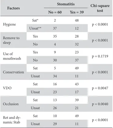
Of the 99 patients enrolled (all between 29 and 83 years of age), 64 (64.6%) were female and 35 (35.4%) were male. Eighty-seven (87.8%) participants wore total dentures, and 12 (12.1%) wore partial free-end dentures. he average age of dentures was 5.3 years. We found no evidence that gender was associated with disease (p = 0.4613). Yeasts were isolate both from DB (58/99; 58.6%) as PM (39/99; 39.4%). hirty-six (36.3%) patients showed signs of DS, 32 (32/36; 89.0%) of whom were colonized by yeasts, and for four of them (11.0%), yeasts were not isolated. Of the patients with DS, 18 (18/36; 50%) had type I lesions, 12 (12/36; 33.3%) had type II lesions, and six (6/36; 16.6%) had type III lesions. Upon analysis of the DS patients who were colonized, we observed that both the DB and the PM of 24 (66.6%) of these subjects were colonized. he factors related to the occlusion and denture stability were signiicantly related to the development of DS (Table 1). he results revealed that all the local factors analyzed in this study inluenced the development of DS, except for the use of mouthwash (p = 0.1719). Ater analyzing the qualitative and functional factors of the dentures, we observed that it was not possible to correlate the disease with a single factor, as presented in Table 1. he statistical tests indicated that the age of the prosthesis was directly proportional to the onset of DS (p=0.0055) (Table 2).
DISCUSSION
In our study, there was no association between gender and DS, which is inconsistent with the indings of da Silva et al.13 and
Arnaud et al.14, who reported a signiicant association in females.
We observed that the prevalence of DS was 36/99 (36.4%), which is similar to the results reported by Arnaud et al.14, who examined
174 patients wearing acrylic-based dentures, 35% of whom had DS. However, our results are inconsistent with the indings reported by Dağistan et al.6, who examined 70 patients and found
that 70% had DS.
he factors that contribute to DS are variable and have been associated with both local and systemic components15. Because
of their chemical and physical properties, the use of poly(methyl methacrylate) dentures facilitates the colonization of various microorganisms including Candida spp., which are normal fungal commensals, and members of the normal oral cavity microbiota, which can become pathogenic when local and systemic changes
promote their proliferation16. Immunocompromised patients
undergo changes in their oral environment that afect their immune response, resulting in the inability of these oral tissues to support the use of dentures3. Jeganathan, Lin17 and
Budtz-Jørgensen18 reported that the interaction between Candida and
oral bacteria promote the onset of the disease when combined with local factors such as temperature, pH, adhesive capacity of these microorganisms, and systemic components.
he present study evaluated several local, qualitative factors such as oral and denture hygiene, the use of mouthwash, nighttime denture use, age of the dentures and functional factors such as VDO, VDR, occlusion, retention, and static and dynamic stability. We noted that these factors alone could not lead to disease onset, but when combined with yeast presence, they promoted disease onset, which is consistent with the indings reported by Naik, Pai16.
Cruz et al.19 conducted a clinical trial comparing the eicacy
of chemical and chemomechanical methods to clean dentures
Table 1. Qualitative local and functional values associated with denture stomatitis
Factors
Stomatitis Chi-square test No = 60 Yes = 39
Hygiene
Sat* 2 48
p < 0.0001
Unsat** 37 12
Remove to sleep
Yes 35 28
p < 0.0001
No 4 32
Use of mouthwash
Yes 9 23
p = 0.1719
No 30 37
Conservation
Sat 5 49
p < 0.0001
Unsat 34 11
VDO
Sat 16 43
p = 0.0047
Unsat 23 17
Occlusion
Sat 13 39
p = 0.0040
Unsat 26 21
Ret and dy-namic Stab
Sat 10 49
p < 0.0001
Unsat 29 11
* Sat = satisfactory; ** Unsat = unsatisfactory.
Table 2. Relationship between denture age and denture stomatitis
Age Stomatitis Chi-square test Yes No Total
0 - 48 months 17 42 59
p = 0.0055
49- 96 months 7 11 18
More than 96 months 15 7 22
in denture wearers. he researchers reported that chemical methods alone do not reduce the amount of bacterial bioilm on dentures and that the chemomechanical method is more efective at removing bioilm. heir data reinforce our indings that mouthwash use did not prevent colonization by yeasts, although in the present study was not conducted in vitro analysis of the action of mouthwash on Candida colonization of dentures. Identiication of strains at genus level was performed according to Sidrim, Rocha12. However, our data disagree with the indings
reported by Orsi et al.20 and Işeri et al.21, who evaluated the action
of antiseptics on denture surfaces and concluded that these agents are efective in eliminating Candida from the resin surface. Salerno et al.15 stated that poor hygiene is one factor that promotes
disease onset; they concluded that good hygiene alone alleviates the symptoms of DS and that the control of denture hygiene is essential for preventing relapse ater antifungal treatments. Hygiene is therefore an important prophylactic measure against oral candidiasis manifestations, such as DS.
Ferreira et al.22 reported a highly signiicant association
between the failure of a patient to remove his/her dentures before sleep and the development of disease. hese data are consistent with our indings that wearing dentures while sleeping promotes disease onset (Table 1). Contrary to our results, da Silva et al.13
evaluated local factors in 102 patients and found that denture removal before sleep was not relevant to the onset of DS. Hadžić et al.23 analyzed the denture age and colonization and
noted that age was a facilitating factor for colonization because of wear, roughness, and plaque accumulation, which is consistent with our indings (Table 2). We found that the denture condition is as important as its age, which was also noted by Naik, Pai16.
A study conducted by Garcia, Souza24 evaluated the necessity of
replacing or relining dentures 4 years ater installation. hese authors noted that this procedure was not necessary in most cases ater 1 year but was necessary ater 3 years of use. Based on these indings, we grouped the age of the dentures into 4-year spans and found that the age of the dentures was directly proportional to the onset of DS.
A study conducted by Bomim et al.25 demonstrated that
problems related to denture occlusion were signiicantly associated with the development of DS because of the increased likelihood of trauma that would cause tissue damage. hese data are consistent with our indings that all of the functional factors promoted the onset of DS (Table 1). Overall, instructions regarding oral hygiene, denture maintenance, and regular visits to the dental surgeon are essential in maintaining the overall health of the oral cavity. Because dental surgeons can diagnose DS in their clinical practice and then either provide therapy or refer the individuals for treatment to resolve the causative factors, these patients are able to achieve increased comfort and a better quality of life.
CONCLUSION
Denture stomatitis in denture users in northern Brazil was multifactorial, involving local, functional and microbiological factors.
ACKNOWLEDGEMENTS
Post-graduation Program in Biology of Infectious and Parasitic Agents - Federal University of Pará.
REFERENCES
1. PattanaikS, VikasBVJ, PattanaikB, SahuS, LodamS. Denture stomatitis: a literature review.J Indian Acad Oral Med Radiology.2010July-September; 22: 136-40. http://dx.doi.org/10.5005/jp-journals-10011-1032.
2. CastroAL, FuruseTA, Gaetti-Jardim JúniorE, CastroEVFL, JardimPTC, ParoMLC. Denture stomatitis induced by the improper use of complete dentures: a case report. Rev Odontol Araçatuba. 2006Jul-Dez; 7: 87-90.
3. Dar-OdehNS, ShehabiAA. Oral candidosis in patients with removable dentures.Mycoses. 2003June; 46(5-6): 187-91. http://dx.doi.org/10.1046/j.1439-0507.2003.00871.x. PMid:12801360
4. FalcãoAFP, SantosLB, SampaioNM. Candidiasis associated with dental prosthesis.Sitientibus.2004Jan-Jun; 30: 135-46.
5. Serrano-GrangerC, Cerero-LapiedraR, Campo-TraperoJ, Del Río-HighsmithJ. In vitro study of the adherence of Candida albicans to acrylic resins: relationship to surface energy.Int J Prosthodont. 2005September-October; 18(5): 392-8. PMid:16220804.
6. DağistanS, AktasAE, CaglayanF, AyyildizA, BilgeM. Differential diagnosis of denture-induced stomatitis, Candida, and their variations in patients using complete denture: a clinical and mycological study.Mycoses. 2009May; 52(3): 266-71. http://dx.doi.org/10.1111/j.1439-0507.2008.01592.x. PMid:18643887
7. BarbeauJ, SéguinJ, GouletJP, de KoninckL, AvonSL, LalondeB, et al. Reassessing the presence of Candida albicans in denture-related stomatitis.Oral Surg Oral Med Oral Pathol Oral Radiol Endod. 2003January; 95(1): 51-9. http://dx.doi.org/10.1067/moe.2003.44. PMid:12539027
8. ScalercioM, ValenteT, IsraelM, RamosME. Denture stomatitis associated with candidiasis: diagnosis and treatment. Rev Gaúcha Odontol. 2007Out-Dez; 55(4): 395-8.
9. NewtonAV. Denture sore mouth: a possible etiology.Br Dent J.1962; 112: 357-60.
10. ElguezabalN, MazaJL, DorronsoroS, PontónJ. Whole saliva has a dual role on the adherence of Candida albicans to polymethylmetacrylate.Open Dent J. 2008; 2(1): 1-4. http://dx.doi.org/10.2174/1874210600802010001. PMid:19088875
11. KennedyE.Partial denture construction.Dent Items Interest.1925; 47(1): 23-5.
13. da SilvaHF, Martins-FilhoPR, PivaMR. Denture-related oral mucosal lesions among farmers in a semi-arid Northeastern Region of Brazil.Med Oral Patol Oral Cir Bucal. 2011September; 16(6): e740-4. http://dx.doi.org/10.4317/medoral.17081. PMid:21196849
14. ArnaudRR, SoaresMSM, SantosMGC, SantosRC. Denture stomatitis: prevalence and correlation with age and gender.Rev Bras Ciênc Saúde.2012Mar; 16(1): 59-62.
15. SalernoC, PascaleM, ContaldoM, EspositoV, BusciolanoM, MililloL, et al. Candida-associated denture stomatitis.Med Oral Patol Oral Cir Bucal. 2011 March; 16(2): e139-43. http://dx.doi.org/10.4317/medoral.16.e139. PMid:20711156
16. NaikAV, PaiRC. A study of factors contributing to denture stomatitis in a north Indian community.Int J Dent. 2011; 2011: 589064. http://dx.doi. org/10.1155/2011/589064. PMid:22194746
17. JeganathanS, LinCC. Denture stomatitis—a review of the aetiology, diagnosis and management.Aust Dent J. 1992April; 37(2): 107-14. http://dx.doi. org/10.1111/j.1834-7819.1992.tb03046.x. PMid:1294074
18. Budtz-Jørgensen E. Ecology of Candida-associated denture stomatitis. Microb Ecol Health Dis. 2000; 12(3): 170-85. http://dx.doi. org/10.1080/089106000750051846.
19. CruzPC, AndradeIM, PeraciniA, Souza-GugelminMC, Silva-LovatoCH, de SouzaRF, et al. The effectiveness of chemical denture cleansers and ultrasonic device in biofilm removal from complete dentures.J Appl Oral Sci. 2011November-December; 19(6): 668-73. http://dx.doi.org/10.1590/S1678-77572011000600021. PMid:22231005
20. OrsiIA, JuniorAG, VillabonaCA, FernandesFH, ItoIY. Evaluation of the efficacy of chemical disinfectants for disinfection of heat-polymerised acrylic resin.Gerodontology. 2011December; 28(4): 253-7. http://dx.doi.org/10.1111/j.1741-2358.2010.00400.x. PMid:20609007
21. IşeriU, UludamarA, OzkanYK. Effectiveness of different cleaning agents on the adherence of Candida albicans to acrylic denture base resin.Gerodontology. 2011December; 28(4): 271-6. http://dx.doi.org/10.1111/j.1741-2358.2010.00379.x. PMid:21554382
22. FerreiraRC, MagalhãesCS, MoreiraAN. Oral mucosal alterations among the institutionalized elderly in Brazil.Braz Oral Res. 2010July-September; 24(3): 296-302. PMid:20877966.
23. HadžićS, DedićA, Gojkov-VukelićM, PašićE, OžegovićL, BešlagićE.Influence of Candida infection on denture stomatitis.Acta Med Acad. 2009;
38(1): 6-10.
24. GarciaAR, SouzaV. Necessity of relining free-end extension partial dentures.Rev Odontol UNESP.1992; 21: 333-8.
25. BomfimIPR, SoaresDG, TavaresGR, SantosRC, Araújo TP, PadilhaWWN. Prevalence of oral mucosa lesions in denture wearers.Pesqui Bras Odontopediatria Clín Integr.2008Jan-Abr; 8(1): 117-21.
CONFLICTS OF INTERESTS
he authors declare no conlicts of interest.
*CORRESPONDING AUTHOR
Lurdete Maria Rocha Gauch. Programa de Pós-graduação em Biologia de Agentes Infecciosos e Parasitários, Instituto de Ciências Biológicas, UFPA – Universidade Federal do Pará, Av. Augusto Corrêa, 1, 66000-000, Belém, PA, Brazil. e-mail: [email protected]