7
p-ISSN:2231-6140, e-ISSN:2395-7859 Original ArticleClinical Study of Epidemiology and Histopathological Correlation of Lid
Tumours in Indian Population.
Shivani Acharya1*, Ruchika Pattanaik2, Deepak.C.Mehta3, Ruchi Kabra4
1
Senior Resident Doctor, Department of Ophthalmology, GMERS Medical College, Sola Civil Hospital, Ahmedabad
2
MS Ophthalmology
3
Dean, GMERS Medical College, Himmatnagar
4
Assistant Professor, Oculoplasty Unit, M & J Western Regional Institute Of Ophthalmology, Civil Hospital, Ahmedabad
ABSTRACT
Objective: To study the presentation, pattern of distribution and correlate histopathological examination of lid tumor. Materials and Methods: A prospective study of 50 patients presenting to tertiary referral care centre with benign or malignant tumors between July 2010 and June 2012 was carried out. All the cases were biopsied for histopathological examination (HPE). Patients were followed up at 1 month and 6 monthly intervals to observe regrowth or recurrence. Results: Benign tumours presented at a younger age (mean age 37.24 years) Malignant tumours presented at the mean age of 51.60 years and were more common among females (male: female ratio 1:1.67). Amongst benign tumours, nevus and pyogenic granuloma were most common (24.60%). Amongst malignant tumors, Meibomian gland carcinoma was commonest -12 cases (50%). 2 cases benign on clinical examination were malignant on HPE, 3 cases malignant clinically were benign on HPE. Conclusion: Many benign lesions have a tendency to masquerade as malignant lesions. Thus all lid lesions should be subjected to histopathological examination to discern not only the diagnosis but also the management.
Keywords: Biopsy, Histopathology, Lid tumors.
Introduction:
The eyelids are a highly specialised region of the ocular adnexa consisting of multiple tissue types, all having the potential to give rise to a spectrum of benign and malignant tumours. Eye lid tumours are by far the most common neoplasm encountered in clinical ophthalmic practice. They are estimated to represent more than 90% of all ophthalmic tumours. Approximately 5% to 10% of all skin cancers occur in the eyelid1. Vast majority of these tumors are inflammatory or non malignant neoplasm. It becomes imperative to bear in mind that malignancies can
mimic a host of benign tumours. Hence many a times the conclusion of diagnosis becomes based on the expertise of a histopathological *
Corresponding Author:
Dr Shivani Acharya,
8
p-ISSN:2231-6140, e-ISSN:2395-7859 Original Article examination of the excised specimen of the tumour mass or the whole tumour itself.Tumours of the eyelids can be classified based on tissue of origin such as tumours of the epidermis/dermis, tumours of melanocytic origin, those of glandular, neural, vascular, metastatic, xanthomatous, histiocytic, and inflammatory origin1. A high index of suspicion is required when there is a slowly enlarging lump, loss of eye lashes, prominent blood vessels, pigmentation or recurrent blepharitis1.
World-wide the incidence of lid malignancies is increasing and a varied distribution has been observed, much of which is under characterised. Worldwide studies performed so far viz Iowa study spanning a 38 year period, a study in southern Taiwan over a period of 5 years2 and many more show that amongst benign tumours, seborrheic keratosis, epithelial cysts are commoner entities and amongst the malignant tumours, basal cell carcinoma is the most prevalent followed by sebaceous gland and thenceforth, squamous cell carcinoma. These studies also describe the sex distribution of the individual tumours and the predilection of eyelids and site for the tumours described.
Studies conducted in India, including study by Abdi UN; Tyagi V; Maheshwari V; Gogi R; Tyagi SP3 of 207 cases over a period of 34 years and another by Dr Mukesh Sharma covering 135 cases of eyelid tumours4 treated at department of ophthalmology S.M.S hospital, Jaipur between 1999 and 2006 revealed that amongst the malignant tumours, Indian population shows a predilection for sebaceous gland tumours. Each of the nationwide or international studies duly emphasise the role of concurrent histopathological confirmation as most of the malignant tumours tend to masquerade the benign lesions.
Aims and objectives:
1. To study histopathopathological examination of lid tumor biopsy. 2. To study the incidence of lid tumours in western region of India. 3. To study the presentations of benign and malignant lid tumours. 4. To characterise a pattern of distribution of lid tumours.
Materials and Methods:
This study was conducted in tertiary referral care centre of Western India between the period of June 2010 to June 2012. 50 cases were included in the study. A detailed study of the clinical history and examination was performed. All benign tumours cystic, vasculogenic, non cystic like epidermal inclusion cyst, pyogenic granuloma5, epidermal nevus, keratocanthoma , neurofibroma and all malignant tumours like squamous cell carcinoma, basal cell carcinoma, meibomian gland carcinoma , sebaceous gland carcinoma and lymphoma were included in the study.
9
p-ISSN:2231-6140, e-ISSN:2395-7859 Original Article incisional biopsy was done.An initial clinical impression was drawn following which other investigational modalities in form of ultrasonographic scanning, ultrasonic bio microscopy, CT SCAN and MRI were done in relevant cases especially in the cases where an intraocular/ intra orbital extension was suspected.
In most cases frozen section biopsy6 was used to confirm the tumour free margins. These cases underwent a primary lid reconstruction in the same sitting. In other cases histopathology report was awaited and secondary lid reconstructions were performed. Methods thus employed in cases of large defects also included Tenzel advancement flaps, Cutler Beard flap reconstruction7 with autogenous cartilage, Composite Grafting viz pedicle flap from lower to upper lid. In relevant cases, radiotherapy and chemotherapy was also employed. Cases were followed up on day one, one week, fifteen days, one month and there after six monthly. Observation for any regrowth or recurrence of symptoms or signs was noticed on each follow up.
Results:
Image 3: Basal Cell Carcinoma with Image 4: Histopathological Picture of Lid Reconstruction Basal Cell Carcinoma
10
p-ISSN:2231-6140, e-ISSN:2395-7859 Original Article Out of 50 cases 24 cases were malignant (48%) and 26 benign (52%). Benign tumours presented at a younger age (mean age 37.24 years) and were more common among males (male: female ratio 1.36:1). Malignant tumours presented at the mean age of 51.60 years and were more common among females (male: female ratio 1:1.67).An overall predilection for right eye and upper lid in both eyes was found.
Amongst benign tumours, cystic lesions like nevus and pyogenic granuloma were most common (24.60%).
Amongst malignant tumors, Meibomain gland carcinoma (MGC) was commonest (50%). Signs of distant metastasis were seen in 2 cases, one MGC and one Basal cell carcinoma (BCC). Lymph node positivity was seen in 14 cases, maximally with Squamous cell carcinoma (SCC).
In this study, 2 cases benign on clinical examination were malignant on HPE.3 cases, malignant on clinical examination were benign on HPE.
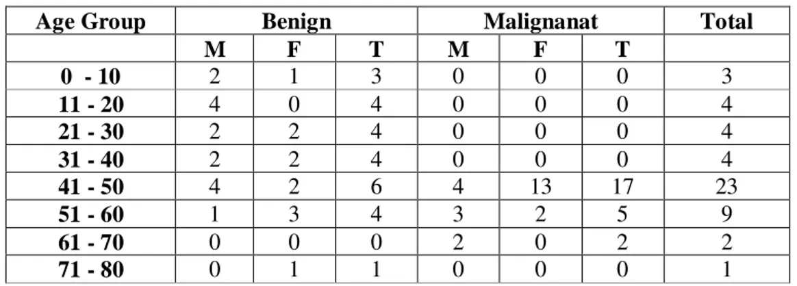
Table 1: Age wise tumor distribution
Age Group Benign Malignanat Total
M F T M F T
0 - 10 2 1 3 0 0 0 3
11 - 20 4 0 4 0 0 0 4
21 - 30 2 2 4 0 0 0 4
31 - 40 2 2 4 0 0 0 4
41 - 50 4 2 6 4 13 17 23
51 - 60 1 3 4 3 2 5 9
61 - 70 0 0 0 2 0 2 2
71 - 80 0 1 1 0 0 0 1
For benign tumours, most common age group for males was in 30-40 whereas in females, age group 40-50 was common. For malignant cases, most common age group for females and males was 50-60.
Image 5 Graph showing age wise tumor distribution
0 2 4 6 8 10 12 14 16
(71-80)
(61-70)
(51-60)
(41-50)
(31-40)
(21-30)
(11-20)
11
p-ISSN:2231-6140, e-ISSN:2395-7859 Original Article Table 1 Eye and Eyelid distributionAmongst the cases of meibomian gland carcinoma right eye was more common in females and left eye in males. A higher incidence of upper lid involvement was seen. Basal cell carcinoma had lower eye lid predilection.
Image 6 Graph showing eye and eyelid distribution:
Discussion:
Our study of 50 patients included 24 males and 26 females. There were 24(48%) malignant cases and 26(52%) benign cases. A male preponderance with a male: female (15vs11) 1.36:1 was found for benign tumours whereas for the malignant cases females outnumbered males (15vs9) 1.67:1. Mean age at presentation for benign tumours was 37.24years and for malignant cases was 51.60years. For benign tumours, most common age group for males was in 30-40 whereas in females age group 40-50 was commoner. For malignant cases, most common age group for females and males was 50-60. A predilection for right eye (RE: LE-1.47:1) as an overall incidence was observed amongst the tumours.
In a similar study a retrospective analysis of 135 eyelid tumors treated at department of ophthalmology S.M.S hospital, Jaipur4 from 1999-2006 , malignant eyelid tumors were 54 (40%)in number. Female: male ratio was 1.3:1 in malignant cases. In this study benign tumours were 60% with a female: male ratio of 1.2:1. In another study by Abdi UN; Tyagi V; Maheshwari V; Gogi R; Tyagi S, a retrospective analysis of 207 cases, a slight preponderance of males with male/female ratio of 1.3:1. Malignancy was noticed in 85 cases (41.1%) 3.
Worldwide studies have reported that amongst western population, in study of eyelid tumors in southern Taiwan2: a 5-year survey, conducted between January 1994 and
6
2
3
5 9
3
7
5
1
3
1
3
0 1 2 3 4 5 6 7 8 9 10
RIGHT EYE LEFT EYE UPPER LID LOWER LID
SCC
MGC
BCC Right eye Left eye Upper lid Lower lid Total
SCC 6 2 3 5 8
MGC 9 3 7 5 12
12
p-ISSN:2231-6140, e-ISSN:2395-7859 Original Article December 1998 including 144 cases, about half of the tumors were located in the upper eyelids and the other half in the lower eyelids. There were 18 cases (12.5%) of malignant tumors, In Iowa, in a study spanning a 38-year period between 1932 and 1969, 892 lid lesions were processed through the pathology laboratory. Of these lesions, 76 per cent were benign3.In our study amongst the malignant tumours meibomian gland carcinoma was found to be maximum in no 12 cases (50%), followed by squamous cell carcinoma 8 cases (33%) and then basal cell carcinoma (17%)
In India in a study in central India by Sameer S Jahagirdar, Tushar P Thakre, Satish M Kale, Hemant Kulkarni, Manju Mamtani where a series of 27 cases of eyelid malignancies were analysed sebaceous cell carcinoma (~37%) was almost as prevalent as basal cell carcinoma (~44%)8. In another study by Dr Mukesh Sharma et al, Sebaceous gland carcinoma (SGC) constituted 44.4% (24) of all malignancies similar to our findings4. Worldwide incidence of basal cell carcinoma has been found to be the highest. In a study of Eyelid tumors in southern Taiwan a 5-year survey conducted between January 1994 and December 1998 revealed that among the 144 cases, 18 cases (12.5%) of malignant tumors, including 14 basal cell carcinomas, three sebaceous carcinomas, and one squamous cell carcinoma2. Nearly three-quarters of malignant eyelid tumors in this series were basal cell carcinomas, which are similar to the proportion of skin tumors and eyelid tumors in other series .The second most common malignant tumour in this series was sebaceous carcinoma, followed by squamous cell carcinoma. In the study in Iowa among the malignant tumors, the vast majority (80.4 per cent) were basal cell carcinoma3.
In the malignant tumours, in meibomian gland carcinoma male: female ratio was equal; there was 1:3 in basal cell carcinoma and squamous cell carcinoma. Mean age of meibomian gland tumours in males was 52.5 and females was 49.6. Amongst the cases of meibomian gland carcinoma right eye was more common in females and left eye in males. A higher incidence of upper lid involvement was seen. Basal cell carcinoma has lower eye lid predilection.
Kass and Hornblass performed a meta-analysis of the 13 case series of sebaceous carcinoma; according to their data, the incidence in Western populations is from 0.2% to 0.7% among all eyelid tumors and 1-5.5% among eyelid malignancies9. In a more recent analysis of eyelid malignancies other than basal cell or squamous cell carcinoma in Florida, the rate of sebaceous carcinoma in Caucasians was 0.5 per (million)10. However, reports of the incidence of sebaceous carcinoma is much higher in China; Ni reports an incidence of 33% among all eyelid (malignancies) 9. Sebaceous carcinoma occurs more commonly in women than men (reported between 57% and 77% of all patients) and average age of detected disease is between 60 and 69 years of age. Shields et al recently reported their experience in 60 cases; the upper eyelid was involved in 75% of cases, lower eyelid (22%), caruncle (2%), and bulbar conjunctiva (2% = 1 case) 9. Of all cases 28 (51.9%) were seen on upper eyelid and 24 (44.4%) occurred on the lower eyelid, both lids were involved in 3 (5.5%) cases. Sebaceous gland carcinoma occurred predominantly on upper eyelid and rest of the malignant lesions had predilection for lower lid similar to our study.
13
p-ISSN:2231-6140, e-ISSN:2395-7859 Original Article benign tumour was nevus and pyogenic granuloma (24.60%) and then sebaceous cyst. One unusual case of pseudocarcinomatous hyperplasia was also reported. Right eye was again common among benign tumours and upper lid predilection as also observed.In Iowa, in a study spanning a 38-year period between 1932 and 1969, 892 lid lesions were processed through the pathology (laboratory) 3. Of these lesions, 76 per cent were benign; the most common tumours were seborrheic keratosis (23.8 per cent), benign epithelial cyst (21.9 per cent), chalazion (16 per cent), inflammatory dermatosis and nevus (each about 12 per cent), and xanthelasma (4.4 per cent). In a study in Taiwan benign tumors included 38 nevi, 15 squamous papillomas, 13 cysts, 11 verrucae, 10 seborrheic keratoses, four hemangiomas, and others. Dr Mukesh et al found that benign lesions were more common till third decade out of the 81 benign tumors the commonest was papilloma (20 cases;24.6%), followed by dermoid cyst (19 cases; 23.4%), granuloma(14 cases; 17.2%), hemangioma (11 cases;13.6%), naevus (7 cases; 8.6%),neural tumors(5 cases; 6.1%), keratoacanthoma (2 cases; 2.4%), lymphangioma (2 cases; 2.4%) and one case was of implantation cyst4. Abdi U; Tyagi N et al found that benign lesions common ones were vascular tumours (21.3%), neural tumours (18.0%), dermoid cysts (16.4%), squamous cell papilloma (13.1%) and naevi (12.3%), unlike ours where nevus was found to be the commonest benign lesion2.
Conclusion:
Lid tumours have myriad presentations. Many benign lesions have a tendency to masquerade as malignant lesions. These tumours thus are a clinicopathologic challenge for the ophthalmologist. The tumour distribution stays largely undercharacterised and under described especially in western part of the country.
Ours being a tertiary care institute malignant and late presenting tumours are observed more often than not. All lid lesions should be subjected to histopathological examination to discern not only the diagnosis but also the management.
References:
1. Malignant and Benign Eyelid lesions in San Francisco: American journal of clinical medicine winter 2011, vol-8 (1): 40 - 46.
2. Chang, Cheng-Hsien, et al. "Eyelid tumors in southern Taiwan: a 5-year survey from a medical university." The Kaohsiung journal of medical sciences 19.11 (2003): 549-553.
3. Abdi U, Tyagi N, Maheshwari V, Tumours of the eyelid: a clinicopathologic study. J Indian Med Assoc 1996; 94:405-9.
4. Dr. Mukesh Sharma et al: Eye Lid Tumors: A Retrospective Analysis of 135 Cases at A Referral Centre in Western India 2006; JEMDS Dec 2014.
5. Malik SRK, Sood GC, Aurora AL: Granuloma pyogenicum. Br J Ophthalmology 48:502, 1964.
14
p-ISSN:2231-6140, e-ISSN:2395-7859 Original Article 7. Fischer, Thomas, et al. "Experience in upper eyelid reconstruction with theCutler-Beard technique." Annals of plastic Surgery 47.3 (2001): 338-342.
8. Jahagirdar SS, Thakre TP, Kale SM, Kulkarni H, Mamtani M. A clinicopathological study of eyelid malignancies from central India. Indian J Ophthalmol 2007;55:109-12 9. Hornblass A., Lauer S.A.: Sebaceous carcinoma of the eyelids ophthalmology 2005
;111:1641
10.Shields JA, Demirci H, Mar BP, et al: Sebaceous carcinoma of the eyelids: personal experience with 60 cases. Ophthalmology 2004; 111:2151-2157.
11.Straatsma, Bradley R. "Meibomian gland tumors." AMA Archives of Ophthalmology 56.1 (1956): 71-93.