RBCCV 44205-1648
DOI 10.5935/1678-9741.20140082
Mitral annulus morphologic and functional
analysis using real time tridimensional
echocardiography in patients submitted to
unsupported mitral valve repair
Análise morfológica e funcional do anel mitral com o uso da ecocardiograia tridimensional em tempo
real em indivíduos submetidos à plástica mitral sem o uso de anéis protéticos
Marco Antônio Vieira Guedes
1, MD, PhD; Pablo Maria Alberto Pomerantzeff
1, MD, PhD; Carlos
Manuel de Almeida Brandão
1, MD, PhD; Marcelo Luiz Campos Vieira
1, MD, PhD; Flávio
Tarasoutchi
1, MD, PhD; Pablo da Cunha Spinola
1, MD, PhD; Fábio Biscegli Jatene
1, MD, PhD
1Instituto do Coração do Hospital das Clínicas da Faculdade de Medicina da USP (InCor HC-FMUSP), São Paulo, SP, Brazil.
This study was carried out at the Instituto do Coração do Hospital das Clíni-cas da Faculdade de Medicina da USP (InCor HC-FMUSP), São Paulo, SP, Brazil.
Financial support Fapesp - Fundação de Amparo à Pesquisa do Estado de São Paulo (Project 06/50454-4).
Correspondence address: Marco Antônio Vieira Guedes Instituto do Coração
Av. Dr. Enéas de Carvalho Aguiar, 44 Bloco II 2º andar - Sala 13 – Pinheiros, São Paulo, SP, Brazil - Zip code: 05403-900
E-mail: [email protected]
Article received on March 17th, 2014 Article accepted on June 22nd, 2014 Abstract
Introduction: Mitral valve repair is the treatment of choice
to correct mitral insuficiency, although the literature related to
mitral valve annulus behavior after mitral repair without use of
prosthetic rings is scarce.
Objective: To analyze mitral annulus morphology and func
-tion using real time tridimensional echocardiography in individu
-als submitted to mitral valve repair with Double Telon technique.
Methods: Fourteen patients with mitral valve insuficiency
secondary to mixomatous degeneration that were submitted to mitral valve repair with the Double Telon technique were in
-cluded. Thirteen patients were in FC III/IV. Patients were eval
-uated in preoperative period, immediate postoperative period, 6 months and 1 year after mitral repair. Statistical analysis was made by repeated measures ANOVA test and was considered statistically signiicant P<0.05.
Results: There were no deaths, reoperation due to valve dys
-function, thromboembolism or endocarditis during the study.
Posterior mitral annulus demonstrated a signiicant reduction in immediate postoperative period (P<0.001), remaining stable
during the study, and presents a mean of reduction of 25.8% comparing with preoperative period. There was a signiicant reduction in anteroposterior and mediolateral diameters in the immediate postoperative period (P<0.001), although there was
a signiicant increase in mediolateral diameter between imme
-diate postoperative period and 1 year. There was no difference in mitral internal area variation over the cardiac cycle during the study.
Conclusion: Segmentar annuloplasty reduced the posteri
-or component of mitral annulus, which remained stable in a 1-year-period. The variation in mitral annulus area during car
-diac cycle remained stable during the study.
Descriptors: Mitral Valve. Mitral Valve Annuloplasty. Mi
INTRODUCTION
Epidemiological data have demonstrated that mitral insuf-iciency secondary to prolapse of the valve, from moderate to severe extent, is the main valve disease in the United States[1] and gives rise to the second most common form of surgically treated heart valve disease in Europe[2]. In Brazil, prolapse of the mitral valve was found to be the etiology of 25.9% of the patients undergoing mitral valvuloplasty, over the course of 12 years of experience at the Heart Institute of Hospital das Clíni-cas, University of São Paulo Medical School[3].
Previous studies have demonstrated that conserving the mitral valve is better than replacing it[4]. In the technique of quadrangular resection of the posterior cuspid, with plication of the corresponding ring and edge-to-edge suturing of the cuspids, the use of prosthetic rings is still a matter for discus-sion[5]. In Brazil, Pomerantzeff et al.[6] developed a technical modiication in which threads with “pledgets” on a Telon lap were used for segmental plication of the posterior ring corresponding to the segment removed from the cuspid, without using a prosthetic ring. This technique, named the “double Telon technique”, has presented excellent long-term results, with low morbidity and mortality rates[7,8].
Three-dimensional is the diagnostic technique that has contributed most to knowledge of the anatomy and function-ing of the mitral valve[9]. New discoveries regarding the sad-dle shape of the mitral valve ring[10] and its dynamics during
Abbreviations, acronyms & symbols
AP Anteroposterior AP Anteroposterior diameter CI Circularity index
FC Functional class
ML Mediolateral
the cardiac cycle[11] have provided great advances in the tech-niques for mitral valve conservation. The aim of the present study was to analyze the morphology and functioning of the mitral valve ring in individuals who underwent mitral val-vuloplasty by means of the double Telon technique, using real-time three-dimensional echocardiography.
METHODS
This study was conducted at the Heart Institute, Hospital das Clinicas, Faculty of Medicine, University of São Paulo for Heart Valve Surgery Unit and the Echocardiography Unit, with support from CEPEC (Echocardiography Research Center). After the study was approved by the Ethics Commit-tee of Hospital das Clinicas, Faculty of Medicine, University of São Paulo and obtaining a written post-informed consent, between May/2006 and August/2008 we included 14 consec-utive patients with mitral insuficiency secondary to mitral valve prolapse of degenerative etiology, due to elongation or tendon chordal rupture related to the mitral valve posterior cusp, who underwent mitral valve repair with the Double Tef-lon technique (Figure 1). Patients with associated valvular heart disease or submitted to previous heart surgery were ex-cluded from the study. In this population, the age ranged be-tween 39 and 75 years, with average of 61±11 years. Among all individuals, 10 were male and 4 were female. The average weight and height of patients was 75.6±10.9 kg and 1.69±0.1 Resumo
Introdução: A plastia valvar mitral é o tratamento de esco
-lha para a insuiciência mitral, porém, a literatura é escassa em relação ao comportamento do anel mitral após a plástica mitral sem utilização de anéis protéticos.
Objetivo: Realizar a análise morfofuncional do anel mitral
de indivíduos submetidos à plastia valvar mitral pela Técnica de Duplo Telon, sem utilização de anel protético, por meio da ecocardiograia tridimensional em tempo real.
Métodos: Foram incluídos 14 pacientes com insuiciência
mitral mixomatosa submetidos à plástica mitral pela técnica de Duplo Telon. Treze pacientes encontravam-se em classe III/IV. Os pacientes foram avaliados nos períodos pré-operatório,
pós--operatório imediato, 6 meses e 1 ano. Foi utilizado teste de aná
-lise de variância de medidas repetidas para o estudo estatístico, sendo considerado estatisticamente signiicante P<0,05.
Resultados: Não houve óbito, reoperação por disfunção
valvar, tromboembolismo ou endocardite durante o estudo. A planimetria posterior do anel mitral demostrou uma redução signiicativa (P<0,001) no pós-operatório imediato, que se man
-teve estável durante o estudo, apresentando redução média de 25,8% com 1 ano em relação ao pré-operatório. Houve uma re
-dução signiicativa dos diâmetros ântero-posterior e médio-la
-teral no pós-operatório imediato (P<0,001), porém, houve um
aumento signiicativo no diâmetro médio-lateral entre pós-ope
-ratório imediato e 1 ano. Não houve diferença na variação da área interna mitral ao longo do estudo.
Conclusão: A anuloplastia segmentar reduziu signiicativa
-mente o componente posterior do anel mitral, permanecendo estável no período de um ano. A variação da área valvar durante o ciclo cardíaco permaneceu estável durante o estudo.
Descritores: Valva Mitral. Anuloplastia da Valva Mitral.
m, respectively. Body surface area ranged between 1.64 and 2.10 m2, with a mean of 1.85±0.17 m2. In the investigation of personal history, 11 patients had hypertension; two patients had diabetes mellitus, two from chronic renal failure requir-ing dialysis, three had dyslipidemia, and two had coronary ar-tery disease. The additive EuroSCORE ranged between 0 and 6, being that 11 cases presented additive EuroSCORE from 0 to 3 and the other 3 cases showed additive EuroSCORE between 3 and 6. Regarding the functional class (FC) in the preoperative period, one patient was in FC II, 12 in FC III and one in FC IV.
Among the 14 patients, eight had atrial ibrillation in the preoperative period. All patients were operated by the same surgical team. In one patient, plication of the free edge of the anterior mitral valve cusp was performed, as a technique asso-ciated with the repair. Regarding the location of mitral valve disease, 12 patients had involvement of the P2 segment, one patient had involvement of the P1 segment, and one patient had associated involvement of A2 and P2 segment.
Cord rupture were found in 10 patients, it was found string stretching in one patient; stretching and cord snapping found in two patients; calciication of the ring and cord rupture found in one patient. Two patients had coronary heart disease as an associated diagnosis. Of these patients, one patient had distal coronary lesion, not treatable surgically. One patient underwent revascularization of the left marginal branch.
Patients were evaluated in the preoperative period (up to 30 days before surgery), in the postoperative period (be-tween 5 and 30 days after surgery), 6 months (be(be-tween 6 and 7 months after surgery) and 1 year (between the 12th and 15th month after surgery). In order to perform the test, the IE-33 (Philips Medical Systems, Andover, MA, USA) was used. Echocardiographic images were obtained using the matrix transducer positioned in the acoustic parasternal and apical windows.
The three-dimensional echocardiographic data were ana-lyzed in a workstation, using speciic software QLAB 5.0 and QLAB 6.0 (Philips Medical Systems, Andover, MA, USA). With the acquisition of three-dimensional data, the image was cut and reconstructed to visualize the cardiac structures within the pyramid or the whole heart block.
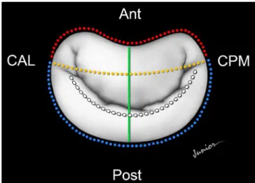
Mitral annulus morphology was evaluated through an-teroposterior diameter (AP), mediolateral (ML), annulus circunference, anterior and posterior mitral planimetry mea-surements (Figure 2).
Circularity index was obtained through the relationship of AP and ML diameter. Mitral annulus function was estimat-ed between the difference of mitral valve area during maxi-mum sistole and maximaxi-mum diastole, in relationship to mitral valve area obtained at maximum sistole, described as valve area reduction during cardiac cycle.
In order to analyze the behavior of the group considering the conditions studied, we used the technique of Analysis of
Variance for Repeated Measures. For the study of reproduc-ibility of echocardiographic measurements we used the in-traclass coeficient correlation. It was considered statistically signiicant P<0,05. The software SPSS version 15.0. (Inc, Chicago) was used for this analysis.
Fig. 1 - Intraoperative aspect of the mitral valve after completion of the mitral valve repair technique “Double Telon”.
We can observe the pledgets anchored in Telon strips and the suture edge to edge of the cusps.
Fig. 2 - Illustration of the echocardiographic variables. Anterior perimeter=red line; posterior perimeter=blue line; anteroposterior diameter=green line; mediolateral diameter=yellow line.
RESULTS
During the study period, no death, endocarditis, reop-eration for valve dysfunction or thromboembolism were observed. In terms of physical activity in the postoperative period, 12 patients were in functional class I and two in func-tional class II, one year after surgery. In the immediate post-operative period, 14 patients had mild mitral insuficiency. There was no signiicant change in the degree of mitral regur-gitation after valvuloplasty during the study.
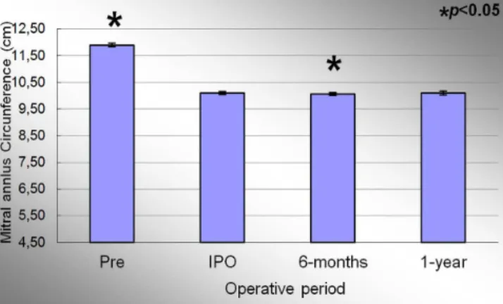
The mean circumference of the mitral annulus in the preoperative period, immediate post-operative period, 6 months and 1 year were 11.90±0.16cm; 10.10±0.13cm; 10.06±0.13cm and 10.10±0,13cm, respectively. Signiicant effect of condition evaluation was observed during the study period (P<0.001). At the end of the study, there was a de-crease of 15.1% of the mitral annulus circumference when comparing the averages of the preoperative and 1 year pe-riod. Figure 3 represents the evolution of the mitral annu-lus circumference during the study. There was a decrease of 15.1% in the measurements when comparing the preopera-tive and IPO period (P<0.001). There was a 0.4% decrease in the circumference of the mitral annulus between the periods IPO and 6 months period (P=0<001). There was no differ-ence between the IPO and 1 year period (P=1.0).
The anterior mitral annulus perimeter in the preoperative, immediate postoperative, 6 months and 1 year period were 4.93±0.06cm; 4.92±0.06cm; 4.91±0.06cm and 4.93±0.07cm, respectively. Signiicant effect of condition evaluation was ob-served during the study period (P<0.001). There was no differ-ence in the average of the anterior perimeter in the preopera-tive and 1 year period. Figure 4 represents the evolution of the anterior perimeter of the mitral annulus during the study. When comparing the preoperative and immediate post-operative pe-riod there was no signiicant difference in anterior mitral
ann-lus perimeter (P=0.17). There was a decrease in 0.2% between IPO and 6 months period (P<0.004). There was no statistical difference when comparing the IPO and 1 year period.
The posterior perimeter of the mitral annulus in the pre-operative, immediate post-pre-operative, 6 months and 1 year period were 6.97±0.13cm; 5.17±0.10cm, 5.15±0.11 and 5.17±0.11cm, respectively. Signiicant effect of condition evaluation was observed during the study period (P<0.001). At the end of the study, there was a decrease of 25.8% on the posterior perimeter of the mitral annulus betwwen preoper-ative and 1 year period. Figure 5 represents the evolution of the posterior mitral annulus perimeter throughout the study. When comparing the preoperative and IPO period, posterior mitral annulus decrease 25.8% (P<0.001), the same decrease value found when comparing the preoperative and 1 year pe-riod. There was a decrease in 0.2% between the IPO and 6 months period (P<0.003). There was no statistical difference when comparing the IPO and 1 year period (P=1.0).
Fig. 3 - Evolution of the circumference of the mitral valve annulus during the study.
Values =mean±standard deviation.
*P<0.05 compared to the immediate postoperative period.
Fig. 4 - Evolution of the anterior perimeter of the mitral valve annulus during the study.
Values =mean±standard deviation.
*P<0.05 compared to the immediate postoperative period.
Fig. 5 - Evolution of the posterior perimeter of the mitral valve annulus during the study.
Values =mean±standard deviation.
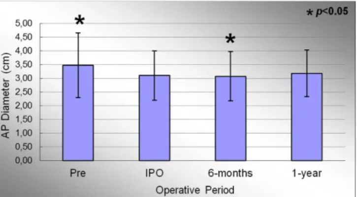
The anteroposterior (AP) diameter of the mitral an-nulus in the preoperative, immediate post-operative, 6 months and 1 year period were 3.47±1.18cm; 3.10±0.90cm; 3.07±0.90cm and 3.18±0.85cm, respectively. Signiicant ef-fect of condition evaluation was observed during the study period (P<0.001). At the end of the study, there was a de-crease in AP diameter of 8.3% compared to the preoperative period. Figure 6 represents the evolution of AP diameter throughout the study. When comparing the preoperative and IPO period, there was evidenced a signiicant reduction in mean of this variable (P<0.001). When comparing the IPO and 6 months period, it was observed a slight further reduc-tion of 1.0% in the mitral annulus AP diameter, statistical-ly signiicant (P=0.012). The comparison between the IPO and 1 year period showed an increase in measures of this variable throughout the study, but without statistical signif-icance (P=0.051).
Mediolateral (ML) diameter of the mitral annulus in the preoperative, immediate post-operative, 6 months and 1 year period were 3.26±1.22cm, 2.87±1.19cm; 2.84±1.19cm and 2.98±0.17cm, respectively. Signiicant effect of condition evaluation was observed during the study period (P<0.001). At the end of the study, there was a decrease of 8.6% in the ML diameter of the mitral annulus when comparing the aver-ages of the preoperative period. Figure 7 shows the evolution of the ML diameter the mitral valve annulus during the study. When comparing the preoperative and IPO period, There was evidenced a signiicant reduction in the average of this variable (P<0.001). When comparing the IPO and 6 months period, a slight further reduction of 1.0% was observed in the ML diameter, statistically signiicant (P=0.003). The
com-parison between the means of IPO and 1 year period showed an increase in measures of this variable by 3.8%, statistically signiicant (P=0.004).
Mitral annulus circularity index in preoperative, im-mediate post-operative, 6 months and 1 year period were 0.93±0.07, 0.91±0.13, 0.90±0.14 and 0.91±0.13, respective-ly. Signiicant effect of condition evaluation was not observed during the study period (P=0.59). The comparison between the means of POI and 1 year periods showed no difference between the means in these periods (P=0.29).
The reduction fraction of mitral internal area during cardiac cycle (variation of the internal area) in the preop-erative period, immediate post-oppreop-erative, 6 months and 1 year were 35.75±9.14%, 33.80±8.59%, and 33.90±8.91% 30.92±8.31%, respectively. Signiicant effect of condi-tion evaluacondi-tion was not observed during the study period (P=0.296). The comparison between the means of IPO and 1 year period showed a decrease of this variable by 8.5%, but without statistical signiicance (P=0.060).
The values of the intraclass correlation coeficient of the values obtained in the analysis of the posterior mitral annu-lus perimeter approached unity (1.0) in all analyzes (0.998 P<0.001).
Table 1 describes a subgroup analysis of the mitral valve annulus morphology variables taking into account the pres-ence of atrial ibrillation during the study. There was no statistically signiicant difference in the behavior of the sub-groups studied throughout the study, and the comparison of variable means in different times evaluated: circumference of the mitral annulus, anterior and posterior perimeter, AP and ML diameter, and circularity index.
Fig. 6 - Evolution of antero-posterior diameter of the mitral valve annulus during the study. Values =mean±standard deviation.
*P<0.05 compared to the immediate postoperative period.
Fig. 7 - Evolution of the medial-lateral diameter of the mitral valve annulus during the study.
Values =mean±standard deviation.
DISCUSSION
The mitral valve system is a complex structure. Clinical use of three-dimensional echocardiography has contributed signiicantly towards understanding its functioning and anat-omy, especially with regard to the mitral valve ring[11].
Fundaró et al.[5] published a review of the most import-ant studies that had analyzed the clinical results from annu-loplasty techniques without a prosthetic ring. They classiied the techniques as either mural or commissural annuloplasty. Mural annuloplasty techniques were subdivided into semicir-cular plication, when shortening of the entire posterior seg-ment of the mitral ring is performed; and segseg-mental plication, when plication is performed on the mitral ring correspond-ing to the segment of the posterior cuspid that is removed through quadrangular resection.
They found that the best immediate and late results oc-curred among patients with degenerative etiologies who had undergone segmental plication or semicircular reduction. In the immediate evaluation, the patients who had undergone
these techniques presented low rates of residual mitral insuf-iciency, between 1 and 2%. In most of the studies reviewed, no early structural failure of the valve repair was observed. Medium-term evaluations showed good results. The actuarial ive-year survival rate was approximately 90%. Moreover, the best survival results free from reoperation were found among patients who had undergone segmental plication or semicircular reduction, especially among those with degen-erative mitral insuficiency. The actuarial survival curves free from thromboembolism and endocarditis presented ex-cellent results. The authors concluded that the techniques of segmental and semicircular plication may be valid and safe options, especially for patients with prolapse of the posterior cuspid in association with slight dilatation of the mitral ring, thus reviving the doubts in relation to the need to use pros-thetic rings[5].
Brandão et al.[8] obtained excellent clinical results from mitral valvuloplasty by means of the double Telon tech-nique, over a 10-year follow-up period. The actuarial sur-vival rate was 94.1±3.6%, the sursur-vival rate free from throm-Table 1. Subgroup analysis regarding the presence of atrial ibrillation.
Variable CAM Ant Annulus Post Annulus AP Annulus ML Annulus CI Period Preoperative IPO 6-months 1-year Preoperative IPO 6-months 1-year Preoperative IPO 6-months 1-year Preoperative IPO 6-months 1-year Preoperative IPO 6-months 1-year Preoperative IPO 6-months 1-year AF (n=8) 11.90±0.18 10.03±0.08 10.00±0.07 10.03±0.08 4.91±0.07 4.90±0.06 4.89±0.06 4.91±0.06 6.99±0.12 5.13±0.06 5.11±0.05 5.13±0.05 3.76±1.25 3.31±1.01 3.27±1.01 3.33±1.01 3.53±1.30 3.22±1.26 3.17±1.26 3.29±1.28 0.93±0.07 0.95±0.10 0.95±0.11 0.97±0.11 No AF (n=6) 11.90±0.16 10.19±0.13 10.15±0.15 10.19±0.15 4.96±0.05 4.95±0.06 4.93 ±0.06 4.95± 0.06 6.94±0.13 5.23±0.13 5.21±0.14 5.23±0.13 3.07±1.05 2.82±0.71 2.79±0.71 2.98±0.62 2.90±1.10 2.41±1.01 2.39±1.02 2.56±0.93 0.94±0.07 0.84±0.16 0.84±0.16 0.84 ± 0.13
PA 0.471 0.845 0.553 0.303 0.166 0.143 PB 0.051 0.172 0.086 0.347 0.264 0.155 P
boembolism was 97.3±1.5% and the survival rate free from reoperation was 99.2±0.8%.
A study on cadavers in which 712 valves resected from patients with mitral prolapse were examined showed that the mean ring circumference was 12.3 cm, whereas it was 9.8 cm in patients without annular dilatation[12]. In a study using three-dimensional echocardiography, Sonne et al.[13] found great variation in the measurements of the mitral ring cir-cumference among 123 normal individuals, with a mean of 10.5±1.4 cm and a range from 7.0 to 14.0 cm. In the present study, the mean circumference of the mitral ring before the operation was 11.9±0.16 cm, thus showing that the popu-lation studied had slight annular dilatation. This inding is probably related to the fact that most of the patients present-ed rupture of the tendinous cords as the genesis of their mi-tral insuficiency. After the surgical intervention, there was a signiicant reduction of 15.1% in the mitral circumference, attaining a mean value of 10.10±0.13, which then remained stable over the course of the follow-up.
The mitral ring was divided into two portions, taking the axis of the mediolateral (ML) diameter into consideration. An-atomically, the anterior portion of the mitral ring is composed of a ibrous portion that is located between the right and left trigones of the mitral ring, and two bilateral muscle portions that line between the ML axis and the corresponding trigone. In a study on cadavers, Hueb et al.[14] found that the anterior intertrigonal distance of the mitral ring was greater in patients with dilated myocardiopathy. Suri et al.[15] compared the ante-rior intertrigonal distance between patients with degenerative mitral insuficiency and normal individuals, using transesoph-ageal three-dimensional echocardiography, and showed that there were no signiicant alterations of the anterior intertrigo-nal distance in these cases. These indings corroborate the idea that the mitral ring has different behavior according to the eti-ology of the mitral insuficiency. In the present study, planim-etry on the anterior portion of the mitral ring showed that there was no signiicant variation over the study period.
The posterior segment of the mitral valve is formed by the muscle portion of the ring. In situations of degenerative etiol-ogy, there is annular dilatation corresponding to the posterior ring. Mihalatos et al.[16] demonstrated that the degree of an-nular dilatation is directly related to the intensity of the mi-tral regurgitation, especially in patients with mimi-tral prolapse and mitral functional insuficiency. Suri et al.[15] found a mean posterior ring size that was greater than what we found in our study, and this is compatible with the predominance of ruptur-ing of the tendinous cords in the present study. The segmental annuloplasty technique used in the present study signiicantly reduced the size of the posterior mitral ring, by 25.8%. This reduction remained stable over the course of the study period, and no annular redilatation was observed over this period. The double Telon technique consists of plication of the anterior portion of the mitral ring alone, without interfering with its
anterior portion. Therefore, the maintenance of the measure-ments of the anterior mitral ring and the reduction in the pos-terior ring that was found are compatible with the segmental annuloplasty technique that was applied in these cases.
Kwan et al.[17,18] demonstrated that during the cardi-ac cycle, the variation in valve area is directly related to the increase in anteroposterior (AP) diameter of the mitral valve. This was not observed in relation to the ML diame-ter. The mitral valve becomes latter at maximum systole, with increases in AP diameter and in the nonplanar angle, thus acquiring its greatest valve area. We analyzed the di-ameters at maximum systole and found values similar to those found by Kwan et al.[18] in normal individuals. These indings conirm that the population studied here presented slight annular dilatation.
In pathological states, the annular dilatation seems not to be due to distension of the mitral ring ibers but, rather, due to an increase in the nonplanar angle, thereby modifying the shape of the mitral ring. This increase in the nonplanar angle gives the valve a more lattened appearance and seems to in-terfere more with the AP diameter than with the ML diameter. In our study, we found that there were signiicant reduc-tions in the measurements of the AP and ML diameters in the immediate postoperative period, compatible with the segmental annuloplasty. In comparing this time with one year after the operation, we found that these measurements had increased slightly: in absolute amounts, a mean of 0.8 mm for the AP diameter and a mean of 1.1 mm for the ML diameter. This change was probably related to the patients’ hemodynamic status. Despite these slight increases from im-mediately after the operation to one year after the operation, we found that overall, there was a reduction of approximately 8% in the AP and ML diameters, from the preoperative diam-eters to the diamdiam-eters at the end of the study period.
The measurements of the internal area of the circumfer-ence obtained through planimetry can be made both at the end of the systole phase and at the end of the diastole phase, thus enabling comparison of the variation of the internal area during the cardiac cycle. This methodology for measur-ing the valve area presents the limitation of bemeasur-ing a two-di-mensional measurement of a three-ditwo-di-mensional structure. It consists of a projection of the three-dimensional mitral ring structure into a transverse plane. Therefore, simple changes to the nonplanar angles would have an impact on the accura-cy of the method.
In relation to the mitral ring area and the magnitude of the variation of this area during the cardiac cycle, the results described in the literature have been diverse, particularly in clinical studies. We believe that these indings relate main-ly to lack of standardization of the times at which measure-ments were made in different studies, thus causing dificulty in making comparisons between them. Moreover, anatomi-cal evaluations of the mitral ring through echocardiography make use of the insertion site of the mitral cuspid, which does not necessarily relect the exact intramuscular location of the mitral ring. Nonetheless, the dynamic nature of the mitral ring during the cardiac cycle has been well established, both in experimental studies and in clinical studies. In these stud-ies, the maximum reduction of the mitral ring size in normal individuals during the cardiac cycle has been shown to be between 22-35%[17,18].
Analyses on mitral ring dynamics in relation to valvu-loplasty using a prosthetic ring has given rise to divergences in the literature. Okada et al.[20] observed a variation in valve area during the cardiac cycle of 26±4%, among patients who underwent implantation of a lexible Duran ring (Duran; Medronic Heart Valve Division, Minneapolis, MN, USA). However, there was no variation in valve area when a rigid Carpentier ring was implanted (Carpentier-Edwards [C-E] Physio; Edwards Lifesciences Corp, Irvine, CA, USA), thus demonstrating behavior that was more physiological than that of the lexible ring. Gillinov et al.[21] evaluated the Cos-grove partial lexible ring (CosCos-grove-Edwards annuloplasty band; Edwards Lifesciences Corp, Irvine, CA, USA) and showed that this prosthesis maintained the saddle shape of the mitral ring, and also presented variation in the valve area of 28±11%, ive years after implantation.
Implantation of prosthetic rings interferes with the saddle shape of the ring. Mahmood et al.[19] showed that implantation of a complete ring interfered with the nonplanar angle, both in patients with ischemic and in patients with degenerative mitral insuficiency. Furthermore, different behavior was observed according to the type of partial ring used in the study, thus sug-gesting that morphological analysis on the mitral valve after implantation might inluence the choice of device.
Komoda et al.[22] showed that there was a reduction in the contraction of the base of the left ventricle after ixation of the
mitral ring by means of prosthetic rings. Moreover, in a study using magnetic resonance, it was demonstrated that mitral plastic techniques that did not involve using prosthetic rings did not interfere with the contraction of either the mitral valve or the base of the left ventricle, as observed six months after the surgical intervention. In the present study, we observed that after the surgical intervention, there was no signiicant reduc-tion in mitral ring performance, thus showing that the segmen-tal annuloplasty did not have any signiicant impact on mitral ring function and that the ring continued to function in a stable manner over the course of the study, such that the mitral ring maintained dynamics that were more physiological.
Barlow disease is generally found in young patients and is characterized by myxomatous degeneration affecting the entire valve, thereby resulting in excess of tissue in the cus-pids and leading to redundant tissue, with prolapse in dif-ferent segments of the valve. Surgical intervention is gener-ally required in the ifth or sixth decade of life. Because of the long course of this disease, it is usually associated with signiicant dilatation of the mitral ring. On the other hand, ibroelastic deiciency is found in patients over the age of 60 years who present a rapid course of mitral valve disease. Fibroelastic deiciency is a disease that essentially affects the tendinous cords and not the cuspids of the mitral valve, and it predisposes towards rupture of the tendinous cords, generally in a single segment of the valve. This condition can be diag-nosed by means of preoperative echocardiography indings that suggest that the valve is of normal size, with thin cuspids and little excess tissue, seen in association with rupturing of the tendinous cords, generally in the P2 segment of the mitral valve. Although Barlow disease and ibroelastic deiciency present different characteristics, these conditions cannot be distinguished in approximately 20% of the patients[23]. In our series, we found echocardiographic characteristics that were compatible with ibroelastic deiciency in the majority of our patients. Our patients presented prolapse of a single segment, affecting the P2 segment in 80% of the cases, in association with slight dilatation of the mitral valve.
We believe that the decision to use prosthetic rings should be correlated with ventricular function and the size of the mitral ring. It is possible that patients with normal ventricular function and slight annular dilatation (the characteristics ob-served in cases of ibroelastic deiciency) would beneit from correction of their mitral insuficiency through techniques that do not involve using prosthetic rings. On the other hand, patients with ventricular dysfunction and signiicant dilata-tion of the mitral ring might beneit from techniques involv-ing use of prosthetic rinvolv-ings for remodelinvolv-ing and stabilizinvolv-ing the mitral ring, because their disease affects not only the mitral valve but also the left ventricle.
Authors’ roles & responsibilities
MAVG Manuscript writing or critical review of its content
PMAP Analysis and/or interpretation of data; inal approval of the
manuscript; study design; implementation of projects and/ or experiments; manuscript writing or critical review of its content
CMAB Analysis and/or interpretation of data; inal approval of the
manuscript; study design; implementation of projects and/ or experiments; manuscript writing or critical review of its content
MLCV Analysis and/or interpretation of data; study design; imple-mentation of projects and/or experiments; manuscript writing or critical review of its content
FT Final approval of the manuscript; implementation of projects and/or experiments
PCS Performed operations and/or experiments
FBJ Analysis and/or interpretation of data; inal approval of the
manuscript; study design; manuscript writing or critical review of its content
study. We conducted a subgroup analysis to evaluate whether the presence of atrial ibrillation might have had an impact on the results found from this study. We did not observe any signiicant alterations to the behavior of the variables eval-uated over the course of the study, thus demonstration that the mitral valvuloplasty modiied the ring measurements in a stable manner and also enabled reverse atrial and ventricular remodeling, independent of atrial ibrillation[25]. It is possible that the patients who persisted with atrial ibrillation over the course of time presented differences not in relation to behav-ior but, rather, in relation to the magnitude of the remodeling. However, the results obtained should be viewed with caution because of the small number of patients allocated to each group in this analysis.
Although this study had a sample of 14 patients, there is little data in the literature regarding remodeling of the mitral valve ring during the postoperative period. Moreover, the populations studied have been etiologically different from each other and they generally underwent mitral valvuloplasty by means of tech-niques that involved usie of prosthetic rings. The present study described aspects of the morphology and functioning of the mi-tral ring over the course of a one-year postoperative period, in a population that was homogenous with regard to the etiology of the mitral insuficiency, which underwent mitral valvuloplasty without involving the use of prosthetic rings.
CONCLUSION
We conclude that the patients who underwent mitral val-vuloplasty by means of the double Telon technique present-ed rpresent-eductions in the posterior segment of the mitral ring, and that this remained stable over the one-year period. Moreover, the variation in internal valve area during the cardiac cycle remained stable over the course of the study.
REFERENCES
1. Nkomo VT, Gardin JM, Skelton TN, Gottdiener JS, Scott CG, Enriquez-Sarano M. Burden of valvular heart diseases: a population-based study. Lancet. 2006;368(9540):1005-11.
2. Iung B, Baron G, Butchart EG, Delahaye F, Gohlke-Bärwolf C, Levang OW, et al. A prospective survey of patients with valvular heart disease in Europe: The Euro Heart Survey on Valvular Heart Disease. Eur Heart J. 2003;24(13):1231-43.
3. Pomerantzeff PMA, Brandão CMA, Monteiro ACM, Nersessian AC, Zeratti AE, Stolf NAG, et al. Plástica da valva mitral: resultados tardios de doze anos de experiência e evolução das técnicas. Rev Bras Cir Cardiovasc. 1994;9(1):22-8.
4. Enriquez-Sarano M, Schaff HV, Orszulak TA, Tajik AJ, Bailey KR, Frye RL. Valve repair improves the outcome of surgery for mitral regurgitation. A multivariate analysis. Circulation. 1995;91(4):1022-8.
5. Fundarò P, Tartara PM, Villa E, Fratto P, Campisi S, Vitali EO. Mitral valve repair: is there still a place for suture annuloplasty? Asian Cardiovasc Thorac Ann. 2007;15(4):351-8.
6. Pomerantzeff PMA, Brandão CMA, Rossi EG, Cardoso LF,
Tarasoutchi F, Grimberg M, et al. Quadrangular resection
without ring annuloplasty in mitral valve repair. Cardiovasc Eng. 1997;2(4):271-3.
7. Pomerantzeff PM, Brandão CM, Souza LR, Vieira ML, Grimberg
M, Ramires JA, et al. Posterior mitral lealet repair with a simple segmental annulus support: the ‘double-Telon technique’. J Heart
Valve Dis. 2002;11(2):160-4.
8. Brandão CM, Guedes MA, Silva MF, Vieira ML, Pomerantzeff
PM, Stolf NA. Mitral valve repair with “Double Telon” technique:
10-year results. Rev Bras Cir Cardiovasc. 2007;22(4):448-53.
9. Solis J, Sitges M, Levine RA, Hung J. Three-dimensional echocardiography. New possibilities in mitral valve assessment. Rev Esp Cardiol. 2009;62(2):188-98.
10. Pai RG, Tanimoto M, Jintapakorn W, Azevedo J, Pandian NG, Shah PM. Volume-rendered three-dimensional dynamic anatomy of the mitral annulus using a transesophageal echocardiographic technique. J Heart Valve Dis. 1995;4(6):623-7.
11. Kaplan SR, Bashein G, Sheehan FH, Legget ME, Munt B, Li XN, et al. Three-dimensional echocardiographic assessment of annular shape changes in the normal and regurgitant mitral valve. Am Heart J. 2000;139(3):378-87.
12. Olson LJ, Subramanian R, Ackermann DM, Orszulak TA, Edwards WD. Surgical pathology of the mitral valve: a study of 712 cases spanning 21 years. Mayo Clin Proc. 1987;62(1):22-34.
and papillary apparatus parameters: assessment by real-time three-dimensional echocardiography. Eur J Echocardiogr. 2009;10(2):287-94.
14. Hueb AC, Jatene FB, Moreira LF, Pomerantzeff PM, Kallás E, de Oliveira SA. Ventricular remodeling and mitral valve
modiications in dilated cardiomyopathy: new insights from
anatomic study. J Thorac Cardiovasc Surg. 2002;124(6):1216-24.
15. Suri RM, Grewal J, Mankad S, Enriquez-Sarano M, Miller FA Jr., Schaff HV. Is the anterior intertrigonal distance increased in
patients with mitral regurgitation due to lealet prolapse? Ann
Thorac Surg. 2009;88(4):1202-8.
16. Mihalatos DG, Joseph S, Gopal A, Bercow N, Toole R, Passick M, et al. Mitral annular remodeling with varying degrees and mechanisms of chronic mitral regurgitation. J Am Soc Echocardiogr. 2007;20(4):397-404.
17. Kwan J, Jeon MJ, Kim DH, Park KS, Lee WH. Does the mitral annulus shrink or enlarge during systole? A real-time 3D echocardiography study. J Korean Med Sci. 2009;24(2):203-8.
18. Kwan J, Kim GC, Jeon MJ, Kim DH, Shiota T, Thomas JD, et al. 3D geometry of a normal tricuspid annulus during systole: a comparison study with the mitral annulus using real-time 3D echocardiography. Eur J Echocardiogr. 2007;8(5):375-83.
19. Mahmood F, Subramaniam B, Gorman JH 3rd, Levine RM, Gorman
RC, Maslow A, et al. Three-dimensional echocardiographic assessment of changes in mitral valve geometry after valve repair. Ann Thorac Surg. 2009;88(6):1838-44.
20. Okada Y, Shomura T, Yamaura Y, Yoshikawa J. Comparison of the Carpentier and Duran prosthetic rings used in mitral reconstruction. Ann Thorac Surg. 1995;59(3):658-63.
21. Gillinov AM, Cosgrove DM 3rd, Shiota T, Qin J, Tsujino H,
Stewart WJ, et al. Cosgrove-Edwards Annuloplasty System: midterm results. Ann Thorac Surg. 2000;69(3):717-21.
22. Komoda T, Hetzer R, Siniawski H, Huebler M, Felix R, Maeta H. Mitral annulus after mitral repair: geometry and dynamics. ASAIO J. 2002;48(4):412-8.
23. Deloche A, Jebara VA, Relland JY, Chauvaud S, Fabiani JN, Perier P, et al. Valve repair with Carpentier techniques. The second decade. J Thorac Cardiovasc Surg. 1990;99(6):990-1001.
24. Alexiou C, Doukas G, Oc M, Oc B, Swanevelder J, Samani NJ,
et al. The effect of preoperative atrial ibrillation on survival
following mitral valve repair for degenerative mitral regurgitation. Eur J Cardiothorac Surg. 2007;31(4):586-91.
25. Guedes MA, Pomerantzeff PM, Brandão CM, Vieira ML, Leite Filho OA, Silva MF, et al. Mitral valve repair by Double
Telon technique: cardiac remodeling analysis by tridimensional