Corresponding author: Dr. Antonio L. Ribeiro, MD, ScD.
e-mail: [email protected]
Received 6 June 2018
Accepted 29 August 2018
Review Article
Electrocardiogram in Chagas disease
Bruno Oliveira de Figueiredo Brito
[1],[2]and Antônio Luiz Pinho Ribeiro
[1],[2],[3][1]. Programa de Pós-Graduação Infectologia e Medicina Tropical, Faculdade de Medicina, Universidade Federal de Minas Gerais, Belo Horizonte, MG, Brasil. [2]. Serviço de Cardiologia e Cirurgia Cardiovascular, Hospital das Clínicas, Universidade Federal de Minas Gerais, Belo Horizonte, MG, Brasil.
[3]. Departamento de Clínica Médica, Faculdade de Medicina, Universidade Federal de Minas Gerais, Belo Horizonte, MG, Brasil.
Abstract
Since the initial descriptions of Chagas cardiomyopathy (ChCM), the electrocardiography has played a key role in patient evaluations. The diagnostic criterion of chronic ChCM is the presence of characteristic electrocardiographic (ECG) abnormalities in seropositive individuals, regardless of the presence of symptoms. However, these ECG abnormalities are rarely specific to ChCM and, particularly among the elderly, can be caused by other simultaneous cardiomyopathies. ECG abnormalities can predict the occurrence of heart failure, stroke, and even death. Nevertheless, most prognostic studies have included Chagas disease (ChD) populations and, not exclusively, ChCM. Thus, more studies are required to evaluate the efficacy of ECG in predicting reliable prognoses in established chronic ChCM. This review exclusively discusses the role of the 12-lead ECG in the clinical evaluation of chronic ChD.
Keywords: Chagas disease. Electrocardiogram. Heart failure. Stroke. Death. Prognosis.
INTRODUCTION
Since the initial descriptions of Chagas cardiomyopathy (ChCM), electrocardiography has played a key role in patient evaluations. Chagas and Villela considered arrhythmia the predominant symptom of ChCM1. Although there are many unclear aspects of Chagas
disease (ChD), the electrocardiography is a well-known method to diagnose and define ChD prognoses. Moreover, it is a low-cost and widely used examination even in remote areas.
The diagnostic criterion of chronic ChCM is the presence of characteristic ECG abnormalities in seropositive individuals, regardless of the presence of symptoms2. Chronic ChCM is
the most severe and frequent form of ChD and affects 20% to 40% of all patients with chronic ChD3. During the course
of the disease, the ECG shows progressive abnormalities that indicate worsening myocardial damage4,5. Maguire et al.
demonstrated that 20% of patients with ChD developed cardiac abnormalities within 6 years, while abnormalities were detected in only 10% of the seronegative individuals4. Recently, a study
reported a cardiomyopathy incidence of 1.85 per 100 people-years6.Chronic ChCM presents as three basic syndromes, heart
failure, cardiac arrhythmia, and thromboembolism, which can occur concomitantly in a patient. The clinical presentation varies widely accord ing to the disease duration and extent of myocardial damage3.
This review exclusively discusses the role of the 12-lead ECG in the clinical evaluation of chronic ChD.
SEARCH METHOD
Data for the present review were identified through a search of the PubMed and Lilacs databases using the following Medical Subject Headings (MeSH) terms: Chagas Cardiomyopathy OR Chagas Disease AND Electrocard*. The search was performed in February 2018 and considered all studies conducted with human adults published in English, Portuguese, or Spanish. There were no restrictions with respect to sample size or duration of follow-up. The original search identified 576 articles. However, after reading the titles and abstractsof the original articles to find any and all terms related to ECG alterations and 12-lead ECG, only 120 were selected for complete reading of the text. After reading the text of the selected studies, only 49 articles met the criteria for this investigation. To describe the relationship between ChD and stroke, a new search was conducted using the following MeSH terms: Chagas Cardiomyopathy OR Chagas Disease AND Stroke. This search identified 140 studies. However, after reading the titles and abstracts of the original articles to find any and all terms related to ECG alterations and 12-lead ECG, only seven met the inclusion criteria.
PREVALENCE OF ELECTROCARDIOGRAPHIC ALTERATIONS
571
Brito BOF and Ribeiro ALP - Electrocardiogram in Chagas disease
of patients with ChD had a normal ECG versus 61.1% of the seronegative patients7. In addition to having a higher prevalence of an abnormal ECG, patients with ChD also presented with more abnormalities per tracing, and the proportion of those with more than three alterations reached nearly 20%7,14. Although the number of ECG alterations naturally increases with age, the increase is more pronounced in patients with ChD7,15. Maguire et al.10, upon examining a rural-area population, also identified that an abnormal ECG is more frequent in patients who are seropositive for ChD, and the ECG abnormalities are more remarkable in those patients between 25 and 44 years of age10. The authors also showed that ECG abnormalities are
more frequent in men than in women (26.1% vs 15.3%)10. This
same research group described the progressive nature of ECG alterations during a 6-year follow-up4 that showed the incidence
of ECG abnormalities was higher in patients who were ChD seropositive, and no new abnormalities were found in the elderly.
The progressive nature of ECG alterations in the elderly is controversial and needs to be clarified. In an elderly cohort8 with
a mean age of 68 years, 86.9% of patients with ChD had ECG abnormalities compared to 75.8% of the seronegative patients (p < 0.001). These findings indicate that only a small proportion of elderly patients with ChD present the indeterminate form of ChD, contrary to that reported in previous studies. The higher prevalence of ECG abnormalities in ChD seropositive patients indicates that the abnormalities are not only explained by other cardiopathies, but also by ChCM progression. It was further demonstrated that ECG alterations and their associations are related to a higher risk of death in elderly patients with ChD8.
This may be due to the continuous process of cardiac damage beginning with the infection in childhood and continuing throughout the patients’ entire adulthood. This leads to a higher frequency of ECG abnormalities, which is an established cardiomyopathy marker in infected elderly patients compared to non-infected patients8.
ChD can cause any type of ECG alteration, but conduction disturbances and ventricular extra systoles (VES) are the most common. ChD seems to evolve from a normal ECG, then to mild alterations, and then to defined characteristic abnormalities. Thus, after some initial nonspecific alterations, there is a tendency for more complex ECG abnormalities to occur4,16,17. Nearly 8% of all patients with ChD experience a regression in ECG alterations, particularly those related to ventricular depolarization and repolarization, and VES18,19. However,
the disappearance of ECG abnormalities must be viewed as a consequence of ECG mutability and not as a true regression18,19. It is important to consider that the presence of nonspecific, isolated ECG alterations are frequently found in both patients with ChD and healthy persons, and include sinus bradycardia (heart rate ≥ 40bpm), first degree atrioventricular block, nonspecific ventricular repolarization (VR) alterations, QRS axis deviation from 0 degree through -30 degree, low limb voltage, isolated supraventricular and ventricular premature beats, incomplete right bundle branch block, and isolated left anterior hemiblock. Thus, these ECG alterations should not be considered diagnostic criteria of ChCM2,3.
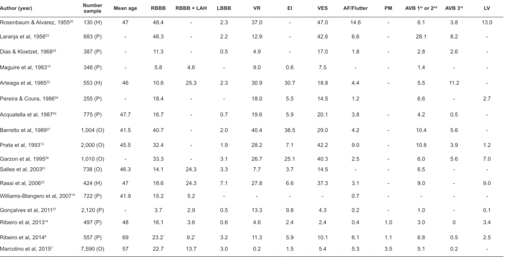
The prevalence of each ECG abnormality depends on the studied population and is shown in Table 1, which describes the results from observational studies that evaluated more than 200 patients with ChD. In Table 1, right bundle branch block (RBBB) associated with left anterior hemiblock (LAH) cases are not included in the RBBB count unless they are signalized. Given that most of the studies are Brazilian and that Rosenbaum20
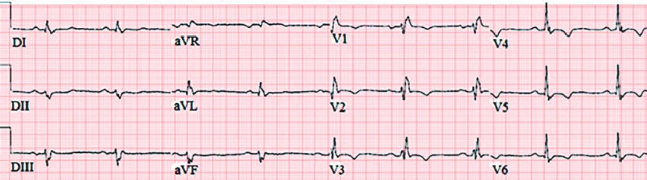
was a pioneer in ChD studies, we chose to include his work in the Table 1 despite the use of a population of less than 200 patients with ChD. Many studies have shown a remarkable prevalence of RBBB7,10,11,16,20-22 and its strong association with ChD7,10. Marcolino et al. found an odds ratio (OR) of 10.73 [95% confidence interval (CI) 10.10 –11.41] that improved when RBBB was combined with LAH (OR: 12.09, 95% CI 11.20-13.04)7. The combination of RBBB and LAH (Figure 1) was more prevalent in ChD than in other cardiomyopathies11,23.
Despite the low frequency in ChD24,left bundle branch
block (LBBB) is markedly associated with heart failure. Patients with ChCM have a longer PR interval10,14,15 and QRS complex duration14,15 compared to seronegative patients.
Moreover, as a consequence of conduction disturbances, ChCM has a strong association with pacemaker (PM) rhythm (OR: 13.29, 95% CI 11.47-15.40), and second degree (OR: 4.05, 95% CI 2.47-6.63) and third degree (OR: 13.29, 95% CI 11.47-5.40) atrioventricular block (AVB)7.
The presence of electrical inactivity (EI), whether isolated or combined with other alterations on the ECG, was associated with
ChCM22,25. Its prevalence ranges from 0.5% to 30% among the
studies in the literature. EIs are often associated with ventricular conduction disturbances and VES10,22, which could indicate
more extensive myocardial damage and a worse survival rat26,27. Ventricular repolarization (VR) abnormalities are quite frequent13,14, and the prevalence ranges from 0.2% to 40%27.
These abnormalities tend to occur during the early course of the disease28 before the occurrence of other abnormalities and are
not related to a worse prognosis28,29. Prata et al. considered VR abnormalities nonspecific13, since they could be a consequence
of diffuse myocarditis13,28, autonomic dysfunction, or even
malnourishment13. Although VR alterations and EIs are usually
associated with coronary artery disease, the patients with ChCM had normal coronary arteries30,31. Moreover, the ischemia shown in
the patients’ scintigraphy was not associated with ECG alterations, thoracic pain30,31, or severe wall-motion abnormalities at rest31.
Atrial fibrillation (AF) is often associated with ChD (OR: 3.15, 95% CI 2.83-3.51)7, and its prevalence is higher in the elderly7,13 and in men7. The prevalence of AF in ChCM is similar to the other cardiomyopathies23. Thus, this arrhythmia is more indicative of a worse prognosis than of a specific alteration14.
VES is a high-frequency abnormality13,22,24 and is associated
with a worse prognosis17. However, the 12-lead ECG does not recognize the transitory character of this ventricular arrhythmia nor does it allow for the evaluation of its severity26. This
as Med
T
rop
51(5):570-577, Sep
-Oct, 2018
TABLE 1: Prevalence of electrocardiographic alterations in Chagas disease patients in the studies.
Author (year) Number
sample Mean age RBBB RBBB + LAH LBBB VR EI VES AF/Flutter PM AVB 1st or 2nd AVB 3rd LV
Rosenbaum & Alvarez, 195520 130 (H) 47 48.4 - 2.3 37.0 - 47.0 14.6 - 6.1 3.8 13.0
Laranja et al, 195653 683 (P) - 48.3 - 2.2 12.9 - 42.6 6.6 - 28.1 8.2
-Dias & Kloetzel, 196829 387 (P) - 11.3 - 0.5 4.9 - 17.0 1.8 - 2.8 2.6
-Maguire et al, 198310 346 (P) - 5.8 4.6 - 9.0 0.6 7.5 - - 1.4 -
-Arteaga et al, 198522 553 (H) 46 10.6 25.3 2.3 30.9 30.7 18.8 4.4 - 5.5 11.2
-Pereira & Coura, 198654 255 (P) - 18.4 - - 18.0 5.5 14.5 1.2 6.6 - 2.7
Acquatella et al, 198755 775 (P) 47.7 16.7 - 0.7 19.6 5.9 20.1 3.8 - 4.2 0.5
-Barretto et al, 198927 1,004 (O) 41.5 40.7 - 2.0 40.4 38.5 29.0 4.2 - 10.4 5.6
-Prata et al, 199313 2,000 (O) 45.5 32.4 - 1.9 28.2 7.1 42.2 9.0 - 10.8 3.9 1.2
Garzon et al, 199526 1,010 (O) - 33.3 - 3.1 26.7 25.1 40.3 2.5 - 6.0 5.6 7.0
Salles et al, 200351 738 (O) 46.3 14.1 24.3 3.3 7.7 3.7 14.5 - - 6.5 -
-Rassi et al, 200652 424 (H) 47 18.6 24.3 7.1 27.8 6.6 37.3 3.1 - 9.0 - 9.0
Williams-Blangero et al, 200715 722 (P) 41.9 15.2 5.2 - - - - 0.7 - - -
-Gonçalves et al, 201157 2,120 (P) - 3.7 2.9 0.5 13.3 9.8 4.3 0.2 - 1.0 - 0.1
Ribeiro et al, 201314 497 (P) 48 16.1 3.6 0.6 4.6 2.4 2.4 0.4 1.0 3.0 0 3.4
Ribeiro et al, 20148 557 (P) 69 23.2* 9.2* 3.2 11.3 5.9 10.1 6.1 1.1 6.8 0.5 2.5
573
ELECTROCARDIOGRAPHIC ALTERATIONS RELATED TO HEART FAILURE
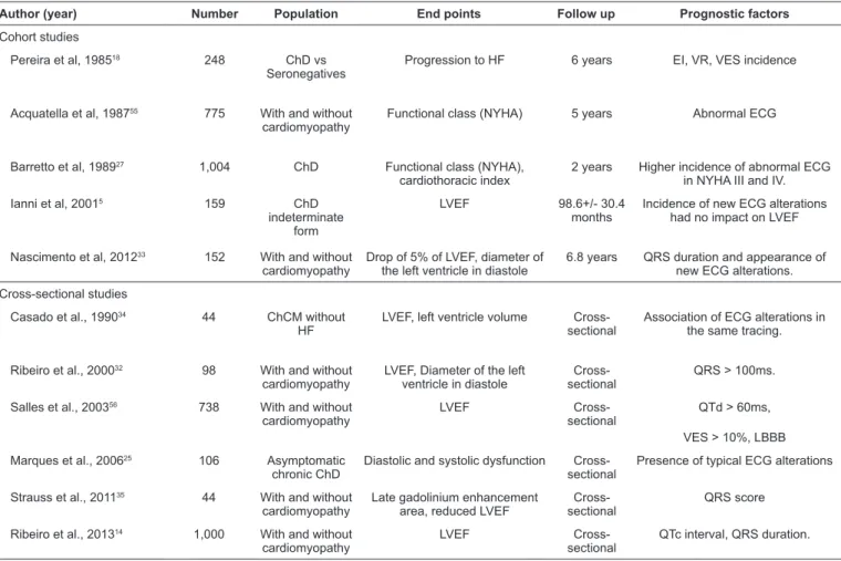
The ECG of patients with ChD can show valuable information about a patient’s evolution to heart failure (HF). The main studies that evaluated this aspect are described in
Table 2. In patients with ChD, there is a significant correlation
between a QRS duration > 100ms and a reduced left ventricle ejection fraction (LVEF) and increased dimensions of the left ventricle in diastole32. However, QRS duration does not
correlate to regional abnormalities of left ventricle contraction or the presence of apical aneurisms; hence, QRS cannot predict a normal left ventricle32. The importance of QRS duration in
this cross-sectional study is corroborated by an 8-year follow-up cohort that identified QRS duration as the only isolated electrocardiographic variable that correlated with a drop of 5% or more in the LVEF and an increase in the diameter of the left ventricle in diastole33. The appearance of new ECG
abnormalities also correlated to a drop in the LVEF33.
Ribeiro et al. reinforced this finding in 2013 when they reported that a QRS duration > 120ms and a QT interval > 440ms can predict with moderate accuracy a reduced LVEF in patients with ChD14. This same study also identified the
abnormalities most frequently associated with LVEF in ChD14:
frequent supraventricular premature beats, VES, AF, RBBB, possible old myocardial infarction, and major isolated ST-T wave abnormalities14. These results corroborate with the findings of Barreto et al. who identified a higher incidence of ECG abnormalities in ChCM populations in heart-failure classes III and IV (New York Heart Association), including VES (p < 0.001), ventricular conduction disturbances (p < 0.001), EI (p < 0.001), and VR alterations (p < 0.001)27. The combination of ventricular conduction disturbances with VES or with sinus bradycardia was associated with both, reduced LVEF and increased left ventricle diameter34.
The QRS score estimates the fibrosis area by considering the alterations of amplitude, duration, and morphology of Q, R, and S waves. Each point corresponds to an area of 3% fibrosis in the left ventricle35. A QRS score > 2 points had the highest
accuracy for predicting the presence of any late gadolinium enhancement and reduced LVEF in cardiac resonance35.
FIGURE 1: An electrocardiogram showing the typical features of Chagas cardiomyopathy. It displays right bundle branch block associated with left anterior hemiblock
ELECTROCARDIOGRAPHIC ALTERATIONS RELATED TO STROKE RISK IN CHAGAS DISEASE
ChD is an independent risk factor for stroke incidence36-38, even
when compared to a high-risk population for this outcome44 and an OR of 7.17 (95% CI 1.50-34.19)37 may be associated with it.
Furthermore, elderly patients with ChD who have had a stroke have a higher risk of death than the seronegative patients39. An evaluation of death due to a stroke in ChD using the Cox model identified that AF is a variable with a higher hazard ratio (HR): 3.87 (95% CI 1.26-11.91), followed by B-type natriuretic peptide39. Although
AF is an important risk factor in the genesis of ischemic stroke related to ChD, one study showed that the occurrence of AF was not associated with stroke in patients with ChCM40, while there was an
association with the presence of LV thrombus and apical aneurysm. These results could be a consequence of the study’s cross-sectional character, and the protection provided by anticoagulation.
Sousa et al.41 elaborated on a score to evaluate thromboembolic risk in ChD. These authors identified several independent risk variables: left ventricle (LV) systolic dysfunction (HR: 13.21, 95% CI 4.72-37), apical aneurysm (HR: 2.32, 95% CI 1.09-4.95), and VR alterations (HR: 2.62, 95% CI 1.20-5.7) on the 12-lead ECG41. Another study illustrated that the incidence of
stroke is higher in patients with mild LV dysfunction (mean LVEF of 48%) compared to those with severe dysfunction (mean LVEF of 36%)42, and there was no association with the
presence of thrombus in the left atrium. This reinforces the role of ECG abnormalities as predictors of stroke.
ELECTROCARDIOGRAPHIC ALTERATIONS RELATED TO DEATH RISK
IN CHAGAS DISEASE
The main causes of death in ChD are HF, sudden death, and stroke, with a predominance of HF28,43,44. Although the
majority of patients show clinical evidence of HF before sudden death, almost one-third of these events occur in asymptomatic individuals who seldom have normal clinical and radiographic exams and who rarely have normal ECGs45,46. ECG carries
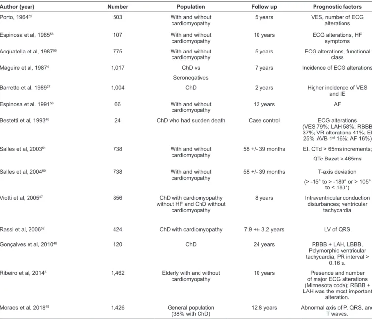
important information about mortality that must be analyzed in the clinical exam. Table 3 summarizes the studies that showed ECG alterations related to the risk of death.
TABLE 2: Electrocardiographic alterations related to heart failure in Chagas disease patients.
Author (year) Number Population End points Follow up Prognostic factors
Cohort studies
Pereira et al, 198518 248 ChD vs
Seronegatives
Progression to HF 6 years EI, VR, VES incidence
Acquatella et al, 198755 775 With and without cardiomyopathy
Functional class (NYHA) 5 years Abnormal ECG
Barretto et al, 198927 1,004 ChD Functional class (NYHA),
cardiothoracic index
2 years Higher incidence of abnormal ECG in NYHA III and IV.
Ianni et al, 20015 159 ChD
indeterminate form
LVEF 98.6+/- 30.4
months
Incidence of new ECG alterations had no impact on LVEF
Nascimento et al, 201233 152 With and without cardiomyopathy
Drop of 5% of LVEF, diameter of the left ventricle in diastole
6.8 years QRS duration and appearance of new ECG alterations.
Cross-sectional studies
Casado et al., 199034 44 ChCM without
HF
LVEF, left ventricle volume Cross-sectional
Association of ECG alterations in the same tracing.
Ribeiro et al., 200032 98 With and without cardiomyopathy
LVEF, Diameter of the left ventricle in diastole
Cross-sectional
QRS > 100ms.
Salles et al., 200356 738 With and without cardiomyopathy
LVEF
Cross-sectional
QTd > 60ms,
VES > 10%, LBBB
Marques et al., 200625 106 Asymptomatic
chronic ChD
Diastolic and systolic dysfunction Cross-sectional
Presence of typical ECG alterations
Strauss et al., 201135 44 With and without cardiomyopathy
Late gadolinium enhancement area, reduced LVEF
Cross-sectional
QRS score
Ribeiro et al., 201314 1,000 With and without cardiomyopathy
LVEF
Cross-sectional
QTc interval, QRS duration.
ChD: Chagas disease; HF: heart failure; EI: electrical inactivity; VR: ventricular repolarization; VES: ventricular extra systoles; NYHA: New York Heart Association; ECG: electrocardiographic; LVEF: left ventricular ejection fraction; ChCM: Chagas cardiomyopathy; QTd: QT dispersion ; QTc:.corrected QT interval.
Patients with normal ECGs have a life expectancy compatible with their gender and age4,43, while those with ECG
abnormalities have a higher mortality rate4,43 even if there is
no other sign of HF50. The mortality rate increases when an
individual with an altered ECG develops HF.
Patients with combined ECG alterations have a higher mortality rate28, and the presence of three or more alterations
indicates a poor prognosis. There has been a preponderance of sudden death in patients who had VES with RBBB or primary T-wave alterations. However, when RBBB is associated with VR alterations, a death caused by HF was more common28.
The number of alterations in the ECG was also a predictor of death in one cohort of patients with ChCM8. In this cohort, the
combination of RBBB and LAH was most heavily related to death8, which corroborates with other studies27,46-48.
Alterations of P, QRS, and T-axes represent a risk of death: (HR: 1.48, 95% CI 1.16-1.88), (HR: 1.34, 95% CI 1.04-1.73), and (HR: 1.35, 95% CI 1.07-1.71), respectively49. T-wave axis deviations (> -15° to > -180° or > 105° to < 180°) were also associated with death in another study50. A wider QT interval
was related to death and was possibly a determining factor of sudden arrhythmic death51. This same study identified that EI
was a prognostic variable51, which corroborated with the findings
of a previous study27.
The analysis of the only cohort comprised solely of patients with ChCM was published in 200652. The final model indicated that only one 12-lead ECG variable, low QRS voltage (LV) (HR: 1.87, 95% CI 1.03-3.37), increased the risk of death. It must be highlighted that LV did not predict adverse outcomes in other cohorts. It is possible that the ECG alterations important to the prognosis of patients with ChD as a whole, do not have the same prognostic value in those with established cardiomyopathy.
CONCLUSIONS
Electrocardiographic abnormalities are frequent in ChD and indicate the presence of cardiomyopathy. However, they are not specific for ChD and, particularly among the elderly, can be caused by other simultaneous cardiomyopathies.
575
Conflict of interest
The authors declare that there is no conflict of interest.
Financial support
Dr. A.L.P. Ribeiro was supported in part by CNPq (grant 310679/2016-8, and Instituto de Avaliação de Tecnologias em Saúde – IATS, grant 465518/2014-1) and by FAPEMIG (Programa Pesquisador Mineiro, PPM-00428-17).
REFERENCES
1. Chagas C, Villela E. Cardiac form of American Trypanosomiasis. Mem Inst Oswaldo Cruz. 1922;14(1):5-61.
2. Dias JC, Ramos Jr AN, Gontijo ED, Luquetti A, Shikanai-Yasuda MA, Coura JR, et al. 2nd Brazilian Consensus on Chagas Disease,
2015. Rev Soc Bras Med Trop. 2016;49(Suppl 1):3-60.
TABLE 3: Electrocardiographic alterations related to death in Chagas disease patients.
Author (year) Number Population Follow up Prognostic factors
Porto, 196428 503 With and without
cardiomyopathy
5 years VES, number of ECG
alterations
Espinosa et al, 198558 107 With and without
cardiomyopathy
10 years ECG alterations, HF
symptoms
Acquatella et al, 198755 775 With and without
cardiomyopathy
5 years ECG alterations, functional class
Maguire et al, 19874 1,017 ChD vs
Seronegatives
7 years Incidence of ECG alterations
Barretto et al, 198927 1,004 ChD 2 years Higher incidence of VES
and IE
Espinosa et al, 199158 66 With and without
cardiomyopathy
12 years AF
Bestetti et al, 199346 24 ChD who had sudden death Case control ECG alterations
(VES 79%; LAH 58%; RBBB 37%; VR alterations 41%; EI 25%, AVB 1st 16%; AF 16%)
Salles et al, 200351 738 With and without
cardiomyopathy
58 +/- 39 months EI, QTd > 65ms increments;
QTc Bazet > 465ms
Salles et al, 200450 738 With and without
cardiomyopathy
58 +/- 39 months T-axis deviation
(> -15° to > -180° or > 105° to < 180°)
Viotti et al, 200547 856 ChD with cardiomyopathy
without HF and ChD without cardiomyopathy
8 years Intraventricular conduction disturbances; ventricular
tachycardia
Rassi et al, 200652 424 ChD with cardiomyopathy 7.9 +/- 3.2 years LV of QRS
Gonçalves et al, 201048 120 ChD 24 years RBBB + LAH, LBBB,
Polymorphic ventricular tachycardia, PR interval >
0.16 s.
Ribeiro et al, 20148 1,462 Elderly with and without
cardiomyopathy
10 years Presence and number
of major ECG alterations (Minnesota code); RBBB + LAH was the most important
alteration.
Moraes et al, 201849 1,426 General population
(38% with ChD)
12.8 years Abnormal axis of P, QRS, and T waves.
VES: ventricular extra systoles; ECG: electrocardiogram; HF: heart failure; ChD: Chagas disease; AF: atrial fibrilation; LAH: left anterior hemiblock; RBBB: right bundle branch block; VR: ventricular repolarization ; EI: electrical inactivity; AVB: atrioventricular block; QTd: : QT interval dispersion ; QTc: corrected QT interval ; LV: low voltage; QRS: QRS wave; LBBB: Left bundle branch block; PR: PR interval; P: P wave.
3. Ribeiro AL, Nunes MP, Teixeira MM, Rocha MO. Diagnosis and management of Chagas disease and cardiomyopathy. Nat Rev
Cardiol. 2012;9(10):576-89.
4. Maguire JH, Hoff R, Sherlock I, Guimaraes AC, Sleigh AC, Ramos NB, et al. Cardiac morbidity and mortality due to Chagas' disease: prospective electrocardiographic study of a Brazilian community.
Circulation. 1987;75(6):1140-5.
5. Ianni BM, Arteaga E, Frimm CC, Pereira Barretto AC, Mady C. Chagas' heart disease: evolutive evaluation of electrocardiographic and echocardiographic parameters in patients with the indeterminate
form. Arq Bras Cardiol. 2001;77(1):59-62.
6. Sabino EC, Ribeiro AL, Salemi VM, Di Lorenzo Oliveira C, Antunes AP, Menezes MM, et al. Ten-year incidence of Chagas cardiomyopathy among asymptomatic Trypanosoma cruzi
-seropositive former blood donors. Circulation. 2013;127(10):
1105-15.
7. Marcolino MS, Palhares DM, Ferreira LR, Ribeiro AL. Electrocardiogram and Chagas disease: a large population database
of primary care patients. Glob Heart. 2015;10(3):167-72.
8. Ribeiro AL, Marcolino MS, Prineas RJ, Lima-Costa MF. Electrocardiographic abnormalities in elderly Chagas disease patients: 10-year follow-up of the Bambui Cohort Study of Aging. J Am Heart Assoc. 2014;3(1):e000632.
9. Silva EM, Rocha MOC Silva RC, Paixao GC, Buzzati H, Santos AN, et al. Clinic and epidemiological study on Chagas disease in the Serra Azul district of Mateus Leme, central-western region of the State of Minas Gerais, Brazil. Rev Soc Bras Med Trop.
2010;43(2):178-81.
10. Maguire JH, Mott KE, Lehman JS, Hoff R, Muniz TM, Guimaraes AC, et al. Relationship of electrocardiographic abnormalities and seropositivity to Trypanosoma cruzi within a rural community in
northeast Brazil. Am Heart J. 1983;105(2):287-94.
11. Rodriguez MV, Hernandez WY, Garcia AN, Colato CM, Cardoza PG, Cardozo LM. ELISA seroprevalence of Trypanosoma cruzi in a
cohort of heart disease patients. J Infect Dev Ctries. 2013;7(4):348-54.
12. Goldbaum M, Ajimura FY, Litvoc J, Carvalho SA, Eluf-Neto J. American trypanosomiasis and electrocardiographic alterations among industrial workers in Sao Paulo, Brazil. Rev Inst Med Trop São Paulo. 2004;46(6):299-302.
13. Prata SP, da Cunha DF, da Cunha SF, Prata SC, Nogueira N. Prevalence of electrocardiographic abnormalities in 2,000 aged and
non-aged chagasic patients. Arq Bras Cardiol. 1993;60(6):369-72.
14. Ribeiro AL, Sabino EC, Marcolino MS, Salemi VM, Ianni BM, Fernandes F, et al. Electrocardiographic abnormalities in
Trypanosoma cruzi seropositive and seronegative former blood
donors. PLoS Negl Trop Dis. 2013;7(2):e2078.
15. Williams-Blangero S, Magalhaes T, Rainwater E, Blangero J, Correa-Oliveira R, Vandeberg JL. Electrocardiographic characteristics in a population with high rates of seropositivity for Trypanosoma cruzi
infection. Am J Trop Med Hyg. 2007;77(3):495-9.
16. da Silva MA, Costa JM, Barbosa JM, Cabral F, Fragata Filho AA, Correa EB, et al. Chronic phase of Chagas disease. Clinical aspects and course of the disease. Arq Bras Cardiol. 1994;63(4):281-5.
17. Barretto AC, Mady C, Ianni BM, Arteaga E, Cardoso RH, da Luz PL, et al. Relationship between ventricular arrhythmia and cardiac function in Chagas disease. Arq Bras Cardiol. 1995;64(6):533-5. 18. Pereira JB, Willcox HP, Coura JR. Morbidity in Chagas' disease. III.
Longitudinal study of 6 years, in Virgem da Lapa, MG, Brazil. Mem
Inst Oswaldo Cruz. 1985;80(1):63-71.
19. Coura JR, de Abreu LL, Pereira JB, Willcox HP. Morbidity in Chagas' disease. IV. Longitudinal study of 10 years in Pains and Iguatama, Minas Gerais, Brazil. Mem Inst Oswaldo Cruz.
1985;80(1):73-80.
20. Rosenbaum MB, Alvarez AJ. The electrocardiogram in chronic
chagasic myocarditis. Am Heart J. 1955;50(4):492-527.
21. Valerio L, Roure S, Sabria M, Balanzo X, Valles X, Seres L. Clinical, electrocardiographic and echocardiographic abnormalities in Latin American migrants with newly diagnosed Chagas disease
2005-2009, Barcelona, Spain. Euro Surveill. 2011;16(38). pii: 19971.
22. Arteaga-Fernandez E, Barretto AC, Mady C, Ianni BM, Bellotti G, Pileggi F. The electrocardiogram in patients with positive serological reactions for Chagas' disease. Study of 600 cases. Arq
Bras Cardiol. 1985;44(5):333-7.
23. Bestetti RB, Muccillo G. Clinical course of Chagas' heart disease: a comparison with dilated cardiomyopathy. Int J Cardiol.
1997;60(2):187-93.
24. Rodrigues N, Ferreira EP, Dias JP. The electrocardiogram in chronic Chagas disease. Study of 100 cases. Arq Bras Cardiol. 1966;19(3):225-34.
25. Marques DS, Canesin MF, Barutta Jr F, Fuganti CJ, Barretto AC. Evaluation of asymptomatic patients with chronic Chagas disease through ambulatory electrocardiogram, echocardiogram and B-Type
natriuretic peptide analyses. Arq Bras Cardiol. 2006;87(3):336-43.
26. Garzon SA, Lorga AM, Nicolau JC. Electrocardiography in Chagas' heart disease. Sao Paulo Med J. 1995;113(2):802-13.
27. Barretto AC, Bellotti G, Deperon SD, Arteaga-Fernandez E, Mady C, Ianni BM, et al. The value of the electrocardiogram in evaluating myocardial function in patients with Chagas' disease. Arq Bras
Cardiol. 1989;52(2):69-73.
28. Porto CC. O eletrocardiograma no prognóstico e evolução da doença
de chagas. Arq Bras Cardiol. 1964;17:313-46.
29. Dias JC, Kloetzel K. The prognostic value of the electrocardiographic features of chronic Chagas' disease. Rev Instit Med Trop Sao Paulo. 1968;10(3):158-62.
30. Marin-Neto JA, Simoes MV, Ayres-Neto EM, Attab-Santos JL, Gallo Jr L, Amorim DS, et al. Studies of the coronary circulation in Chagas' heart disease. Sao Paulo Med J. 1995;113(2):826-34. 31. Marin-Neto JA, Marzullo P, Marcassa C, Gallo Jr L, Maciel BC,
Bellina CR, et al. Myocardial perfusion abnormalities in chronic Chagas' disease as detected by thallium-201 scintigraphy. Am J
Cardiol. 1992;69(8):780-4.
32. Ribeiro AL, Rocha MO, Barros MV, Rodrigues AR, Machado FS.
A narrow QRS does not predict a normal left ventricular function
in Chagas' disease. Pacing Clin Electrophysiol. 2000;23(11 Pt 2):
2014-7.
33. Nascimento BR, Araujo CG, Rocha MO, Domingues JD,
Rodrigues AB, Barros MV, et al. The prognostic significance of
electrocardiographic changes in Chagas disease. J Electrocardiol. 2012;45(1):43-8.
34. Casado J, Davila DF, Donis JH, Torres A, Payares A, Colmenares R, et al. Electrocardiographic abnormalities and left ventricular systolic
function in Chagas' heart disease. Int J Cardiol. 1990;27(1):55-62.
35. Strauss DG, Cardoso S, Lima JA, Rochitte CE, Wu KC. ECG scar
quantification correlates with cardiac magnetic resonance scar size and prognostic factors in Chagas' disease. Heart. 2011;97(5):357-61.
36. Cardoso RN, Macedo FY, Garcia MN, Garcia DC, Benjo AM, Aguilar D, et al. Chagas cardiomyopathy is associated with higher incidence of stroke: a meta-analysis of observational studies. J Card Fail. 2014;20(12):931-8.
37. Paixão LC, Ribeiro AL, Valacio RA, Teixeira AL. Chagas disease: independent risk factor for stroke. Stroke. 2009;40(12):3691-4. 38. Oliveira-Filho J, Viana LC, Vieira-de-Melo RM, Faical F, Torreao
JA, Villar FA, et al. Chagas disease is an independent risk factor for stroke: baseline characteristics of a Chagas Disease cohort. Stroke.
2005;36(9):2015-7.
39. Lima-Costa MF, Matos DL, Ribeiro AL. Chagas disease predicts 10-year stroke mortality in community-dwelling elderly: the
Bambui cohort study of aging. Stroke. 2010;41(11):2477-82.
40. Nunes MC, Kreuser LJ, Ribeiro AL, Sousa GR, Costa HS, Botoni FA, et al. Prevalence and risk factors of embolic cerebrovascular events associated with Chagas heart disease. Glob Heart.
2015;10(3):151-7.
577
42. Nunes MCP, Barbosa MM, Rocha MOC. Peculiar aspects of cardiogenic embolism in patients with Chagas' cardiomyopathy: a transthoracic and transesophageal echocardiographic study. J Am
Soc Echocardiogr. 2005;18(7):761-7.
43. Espinosa R, Carrasco HA, Belandria F, Fuenmayor AM, Molina C, Gonzalez R, et al. Life expectancy analysis in patients with Chagas'
disease: prognosis after one decade (1973-1983). Int J Cardiol.
1985;8(1):45-56.
44. Ayub-Ferreira SM, Mangini S, Issa VS, Cruz FD, Bacal F, Guimaraes GV, et al. Mode of death on Chagas heart disease: comparison with other etiologies. a subanalysis of the REMADHE
prospective trial. PLoS Negl Trop Dis. 2013;7(4):e2176.
45. Ribeiro ALP, Rocha MOC. Forma indeterminada da doença de Chagas: consideracoes acerca do diagnostico e do prognostico. Rev Soc Bras Med Trop. 1998;31(3):301-14.
46. Bestetti RB, Freitas OC, Muccillo G, Oliveira JS. Clinical and morphological characteristics associated with sudden cardiac death in patients with Chagas' disease. Eur Heart J. 1993;14(12):1610-4.
47. Viotti R, Vigliano C, Lococo B, Petti M, Bertocchi G, Alvarez MG, et al. Clinical predictors of chronic chagasic myocarditis
progression. Rev Esp Cardiol. 2005;58(9):1037-44.
48. Goncalves JGF, Dias Silva VJ, Calzada Borges MC, Prata A, Correia D. Mortality indicators among chronic Chagas patients living in an endemic area. Int J Cardiol. 2010;143(3):235-42.
49. Moraes DNM, Nascimento BR, Beaton AZ, Soliman EZ, Lima-Costa MF, dos Reis RCP, et al. Value of the electrocardiographic
(P Wave, T Wave, QRS) axis as a predictor of mortality in 14 years
in a population with a high prevalence of Chagas disease from the Bambui cohort study of aging. Am J Cardiol. 2018;121(3):364-9.
50. Salles GF, Xavier SS, Sousa AS, Hasslocher-Moreno A, Cardoso CR. T-wave axis deviation as an independent predictor of mortality in chronic Chagas' disease. Am J Cardiol. 2004;93(9):1136-40. 51. Salles G, Xavier S, Sousa A, Hasslocher-Moreno A, Cardoso C.
Prognostic value of QT interval parameters for mortality risk stratification in Chagas' disease: results of a long-term follow-up
study. Circulation. 2003;108(3):305-12.
52. Rassi Jr A, Rassi A, Little WC, Xavier SS, Rassi SG, Rassi AG, et al. Development and validation of a risk score for predicting death in
Chagas' heart disease. N Engl J Med. 2006;355(8):799-808.
53. Dias E, Laranja FS, Miranda A, Nobrega G. Chagas' disease; a clinical, epidemiologic, and pathologic study. Circulation. 1956;14(6):1035-60.
54. Pereira JB, Coura JR. Morbidade da doenca de Chagas. Estudo seccional em uma area endemica, Virgem da Lapa, Minas Gerais. Rev Soc Bras Med Trop. 1986;19(3):139-48.
55. Acquatella H, Catalioti F, Gomez-Mancebo JR, Davalos V, Villalobos L. Long-term control of Chagas disease in Venezuela:
effects on serologic findings, electrocardiographic abnormalities, and clinical outcome. Circulation. 1987;76(3):556-62.
56. Salles GF, Cardoso CR, Xavier SS, Sousa AS, Hasslocher-Moreno A. Electrocardiographic ventricular repolarization parameters in chronic Chagas' disease as predictors of asymptomatic left ventricular systolic dysfunction. Pacing Clin Electrophysiol. 2003;26(6):1326-35.
57. Goncalves JG, Prata A, Dias JC, Macedo V. O inquerito
eletrocardiografico. Rev Soc Bras Med Trop. 2011;44(Suppl 2):40-6.
58. Espinosa RA, Pericchi LR, Carrasco HA, Escalante A, Martinez O, Gonzalez R. Prognostic indicators of chronic chagasic cardiopathy. Int J Cardiol. 1991;30(2):195-202.